Neuroendocrine tumors: An epidemiological study of 250 cases
at a tertiary hospital
FELIPE SILVEIRA1, MAÍRA LEITE BASILE1, FÁBIO SEIJI KUGA1, JOSÉ DONATO PRÓSPERO1, ROBERTO ANTONIO PINTO PAES1,
FABÍOLA DEL CARLO BERNARDI1*
1Faculdade de Ciências Médicas da Santa Casa de São Paulo, São Paulo, SP, Brazil
S
UMMARYStudy conducted at Faculdade
de Ciências Médicas da Santa Casa de São Paulo, São Paulo, SP, Brazil
Article received: 2/9/2017
Accepted for publication: 3/12/2017
*Correspondence:
Faculdade de Ciências Médicas
da Santa Casa de São Paulo Address: Rua Dr. Cesário Motta Jr., 61
São Paulo, SP – Brazil Postal code: 01221-020
http://dx.doi.org/10.1590/1806-9282.63.10.856
Objective: To compare the frequency of neuroendocrine tumors in our service with that reported in the literature considering age, gender, location, degree of differentiation and increase in incidence by means of a retrospective study.
Method: Search of variables from a database of neuroendocrine tumor cases diagnosed at the Department of Pathological Sciences, Hospital da Santa Casa de São Paulo over the past 10 years, relating them to epidemiological data such as gender, age, distribution across organs, most-used immunohistochemical markers and presence or absence of either lymph node or distant metastases.
Results: In all, 250 cases were reviewed, 133 involving females, predominantly in the 61-70 age range. The lung was the most frequent site, followed by the stomach. CD56, synaptophysin and chromogranin were the immunohistochemical markers used most often and to a lesser extent Ki67, a marker of cell proliferation that indicates a higher or lower degree of histological malignancy. Metastases, either in lymph nodes and/or distant sites, were found in 44 cases (17.6%).
Conclusion: The results were largely consistent with those in the literature, including age group, gender and location. Most metastases originated from high-grade tumors, with high Ki67 levels and greater impairment of the liver. However, only 36.4% of the cases had Ki67 index. Reevaluation of the Ki67 proliferative index using image analysis in doubtful cases will allow for a correlation between progression and prognosis.
Keywords: neuroendocrine tumors/epidemiology, neoplasms, epidemiologic studies.
INTRODUCTION
Neuroendocrine tumors (NETs) can be located in any organ and constitute a group of primitive neoplasms originating from endodermal cells that proliferate from epithelia or other tissue structures with or without endocrine action.1-3
Most publications agree that the sites where NETs appear most frequently are the gastrointestinal tract and pancreas (70%) followed by the bronco-pulmonary system (20-30%). Other sites such as head, neck, thymus, genital and urinary system and skin are very rare (< 10%).1 Various
studies published in the literature from centers all around the world confirmed such organ distribution, affecting males and females alike, with an age peak in the sixth and seventh decades of life.1,2,4-6
In Brazil, Younes et al.,7 in a study using the database
of the GETNE – Grupo de Estudo de Tumores
Neuro-endócrinos (Neuroendocrine Tumors Study Group) in-volving 32 centers, which comprised 1,000 patients since 1985, found that the sites most frequently affected were the thoracic cavity (71.6%) and gastro-enteric-pancreatic tract (20.2%).
The behavior and local aggressiveness of neuroendo-crine tumors vary, which in turn is related to tumor size or secretion and histological grade of malignancy. In some cases there are no well-defined histopathological criteria for classifying neuroendocrine tumors in terms of prog-nosis or progression factors.3,8 Irrespective of neoplasm
grade, metastases may occur, more frequently to the liver, and are often present by the time of primary diagnosis in 45-95% of cases.9,10
improve-ment in the diagnosis and histological classification of neuroendocrine tumors. Mitotic counts and the Ki67 protein-based cell proliferation index are critical to assess the classification and the possible disease prognosis.11,12
Under the 2010 World Health Organization (WHO) classification scheme, NETs are classified based on bio-logical behavior as grade 1 (NET G1), 2 (G2 NET) and neuroendocrine carcinomas (NEC): carcinomas are sub-divided into large cell neuroendocrine carcinoma (LCNEC) and small cell neuroendocrine carcinoma (SCNEC), and mixed adenoneuroendocrine carcinomas. Histologic grades are dependent on mitotic counts and the Ki-67 labeling index: when the Ki67 index is low (< 3%) or the mitotic count is less than 2/10 HPFs, it is classified as G1 NET. G2 NET has the Ki67 index values between 3 and 20% and mitotic count (2-20/10 HPFs). Neuroendocrine carcinomas (NEC) have > 20% (Ki67 index) and > 20 mi-totic count in 10 HPFs. However, the classification of NETs of the lung is different, being divided into low- (typical and atypical carcinoid) and high- (large cell and small cell neuroendocrine carcinoma) grade.6,11-13
We intend to describe the distribution of neuroen-docrine tumors and analyze the epidemiological profile of cases from a single institution using data collection that included a distribution by gender, age and primary site, as well as the existence or absence of metastases (whenever possible) and the frequency of use of immu-nohistochemistry for making definitive diagnoses and defining prognosis.
METHOD
A computerized search was performed for a retrospective assessment of cases with diagnosis of neuroendocrine neoplasm from the electronic files of the Department of Pathological Sciences, Santa Casa de Misericórdia de São Paulo between May 2004 and May 2014. The lack of a code for Merkel cell carcinoma prevented the inclusion of cases with this diagnosis.
Patients with neuroendocrine tumors were cataloged and, based on pathology reports, analyzed and distrib-uted according to gender, age, diagnosis, tumor location, primary site (either known or indeterminate), presence or absence of metastases, and immunohistochemical assay results with antigenic markers. There was no slide review by the authors. Cases without pathology reports were not included in the manuscript and we did not review any medical records to assess the patients’ clinical evolution.
We grouped the tumors into ten categories taking into account their site of origin. The descriptive statisti-cal analysis of the variables was included.
The cases were classified as follows: neuroendocrine tumor grade 1 (NET G1), neuroendocrine tumor grade 2 (NET G2) and neuroendocrine carcinoma (NEC)-large cell and small cell.11 We established that cases previously
designated as low-grade neuroendocrine neoplasm, low proliferative index neuroendocrine neoplasm, carcinoid tumor and other similar tumors were included in our study under the nomenclature of neuroendocrine tumors grade 1 (NET G1).
RESULTS
Two hundred and fifty (250) cases of neuroendocrine tumors were reviewed for this ten-year period. The num-ber of cases increased over the years from 2004-2014, with 110 between 2004 and 2009, and 140 cases from 2010 to 2014, a 27.3% increase in the number of cases. Of the 250 patients, 133 (53%) were females and 117 (47%) were males. Regarding age, the median was 64 years (range 9 to 87), while the mean age was 54 years. Sixty-three (63/26%) cases were in the most frequent age group, from 61 to 70 years old. In 38 cases (15.2%), examination was based on biopsy and surgical specimens; in other 69 cases (27.6%), it was based only on surgical specimens; whereas in 143 cases (57.2%), it was performed on biopsy alone.
Of 250 tumors, 242 (97%) had their primary site identified. With respect to metastases, the liver was the most frequently affected site (50%). The other cases pre-sented with metastases to the chest wall, cervical spine, soft tissues, and bone (one case each). In two cases (0.9%), the diagnosis was made in the metastatic sites and the primary sites were identified with the help of immuno-histochemistry assays. One of them involved lymph node and liver biopsies with a diagnosis of metastatic medul-lary carcinoma of the thyroid, whereas, in the other, a bone biopsy proved to be useful to diagnose a meta-static pheochromocytoma.
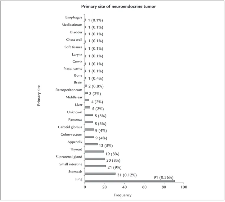
The most frequent primary site was the lung (36% of NETs, 91 cases). The stomach was the second most fre-quent site, with 31 cases (12.5%), followed by the small intestine, with 21 cases (9%). Gastrointestinal tumors (esophagus, stomach, small and large intestines, and cecal appendix) totaled 74 (30%) of cases (Figure 1).
Immunohistochemical assays were used to identify the site of origin in 176 cases. The most commonly used mark-ers were chromogranin in 144 cases (81.8%), with 88.8% positive results; synaptophysin in 115 (65.3%), with 93.9% positive results; and CD56 in 56 cases (31.8%), with 94.6% positive results, as shown in Table 1.
pri-TABLE 1 Distribution of the neuroendocrine markers chromogranin, synaptophysin and CD56 by main primary sites.
Site Total IHC Positive/synaptophysin (%) Positive/chromogranin (%) Positive/CD56 (%)
Lung 91 74 35/40 (87.5%) 40/52 (76.9%) 36/38 (94.7%)
Stomach 31 26 18/19 (94.7%) 24/26 (92.3%) 4/4 (100%)
Small intestine 21 15 13/13 (100%) 13/14 (92.8%) 3/3 (100%)
Appendix 13 3 2/2 (100%) 3/3 (100%) 1/1 (100%)
Colon-rectum 8 6 5/5 (100%) 5/5 (100%) 2/3 (80%)
Pancreas 8 8 7/7 (100%) 8/8 (100%) 3/3 (100%)
Thyroid 19 16 8/8 (100%) 10/10 (100%) 2/2 (100%)
Suprarenal gland 20 8 6/6 (100%) 7/7 (100%) 1/1 (100%) Paraganglioma 25 20 14/15 (93.3%) 18/19 (94.7%) 1/1 (100%)
Total 236 176 108/115 (93.9%) 128/144 (88.8%) 53/56 (94.6%)
IHC: immunohistochemistry.
Note: There are other markers not listed in the table.
FIGURE 1 Distribution of neuroendocrine tumor by primary site.
Primary site of neuroendocrine tumor
Primar
y sit
e
Frequency 1 (0.1%)
1 (0.1%) 1 (0.1%) 1 (0.1%) 1 (0.1%) 1 (0.1%) 1 (0.1%) 1 (0.1%) 1 (0.4%) 2 (0.8%) 3 (2%)
4 (2%) 5 (2%)
8 (3%) 8 (3%) 9 (4%) 9 (4%)
13 (5%)
19 (8%) 20 (8%)
21 (9%) 31 (0.12%)
0 20 40 60 80 100 91 (0.36%)
Esophagus
Mediastinum
Bladder
Chest wall
Soft tissues
Larynx
Cervix
Nasal cavity
Bone
Brain
Retroperitoneum
Middle ear
Liver
Unknown
Pancreas
Carotid glomus
Colon-rectum
Appendix
Thyroid
Suprarenal gland
Small intestine
Stomach
mary site. TTF1 was used in 71 cases (28.4%), with 48% pos-itives; AE1/AE3 in 103 cases (41.2%), with 80% positive re-sults; enolase in 16 cases (6.4%), with 87.5% positive rere-sults; S100 protein in 41 cases (16.4%), with 44% positive re-sults; calcitonin in 18 cases (7.2%), with 100% positive results; as well as other markers, such as thyroglobulin, CK7, CK20, 34BE12 and 35bH11. For assaying islets of Langerhans tumors, specific markers were used such as insulin, glucagon, gastrin, and somatostatin.
The proliferation index was evaluated based on the Ki67 marker in 91 cases (36.4%), with low index (NET G1) results in 38 cases, moderate index (NET G2) in 34 cases, and high-grade (NEC) in 19 cases, especially in tumors originating in the lung with 8 in 32 cases analyzed; two cases involving the stomach in 15 analyzed; two cases of the pancreas in eight and the remaining seven cases in-volving the cervix, bladder, nasal cavity, liver, esophagus, colon/rectum and chest/abdominal wall (one case each).
In the respiratory tract, small-cell carcinoma pre-dominated, with 55 cases (60.4%). In four cases (7.4%) of small-cell carcinoma, biopsy was performed in the me-tastases, the most frequent found in the brain (three cases), liver (one case) and bone (one case), with one patient presenting metastases in both the brain and bone. Of the 28 cases of carcinoid lung tumors, two (7.1%) had lymph node metastases in 17 cases assessed.
Of the 31 cases involving the stomach, 28 (90%) cor-responded to NET G1 and three (9.7%) to NEC. Metas-tases were observed in none of the NET G1. There were lymph node metastases in two cases of gastric NEC and liver metastasis in one case.
Of 21 cases of neuroendocrine tumors in the small intestine, NET G1 was the most frequent, with 13 cases (62%), followed by four cases of NEC (19%), two cases of paraganglioma (10%), one case of NET G2 (5%), and one case of gastrin-secreting neuroendocrine tumor (NET) (5%). Metastases to the lymph nodes were present in five cases, two of which originated from NET G1 and three from NEC. Distant metastasis was positive in one case of NET G1 (diagnostic of carcinoid tumor before 2010). In two cases of NEC, the Ki67 proliferative index was < 2%.
Thirteen (13) cases of NETs of the cecal appendix were diagnosed as NET G1 without metastases.
NETs of the colorectal segment totaled eight cases, of which six were NET G1 and two were NEC. Lymph node metastases occurred in two cases, of which one case was a NET G1 and the other was NEC with a Ki67 index of 90%, indicative of a high-grade tumor.
NETs of the pancreas totaled eight cases, most of which (four cases; 50%) were diagnosed as NEC, two
cases (25%) NET G1, one case somatostatinoma-produc-ing NET, and one case as insulinoma-producsomatostatinoma-produc-ing NET. In two cases, lymph node metastasis was observed. Metas-tases to the liver occurred in three cases, two of which were NEC and one was NET G1, with metastasis also to the duodenum.
The diagnosis of insulinoma and somatostatinoma was based on specific markers used in immunohistochem-istry assays.
NETs of the thyroid amounted to 19 cases, all of which were medullary carcinomas. Positive results for thyro-globulin were found in 31% of cases. Lymph node metas-tasis was positive in 11 of 19 cases (58%), whereas distant metastases were found in two cases (10%).
Diagnoses of adrenal gland pheochromocytoma totaled 21 cases, one of which involved metastasis to the bone.
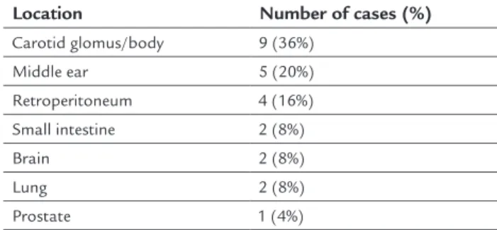
In terms of the whole sample, there were 25 cases (10%) of paraganglioma, summarized in Table 2.
TABLE 2 Topographic distribution of paragangliomas.
Location Number of cases (%)
Carotid glomus/body 9 (36%) Middle ear 5 (20%) Retroperitoneum 4 (16%) Small intestine 2 (8%)
Brain 2 (8%)
Lung 2 (8%)
Prostate 1 (4%)
Metastases were found in 44 cases (17.6%), being negative in 49 cases (19.6%) and not reported in 157 (62.8%). Either lymph node or distant metastases were found in 24 cases each (54.5%), while four cases involved (10%) both types simultaneously. As for metastases to the lymph nodes, medullary thyroid carcinoma was strik-ingly found in 11 cases (46%); followed by the small in-testine with five (21%) cases; colon/rectum two (8%); stomach two (8%); pancreas two (8%); lung two (8%); and larynx with one (4%) case.
DISCUSSION
Of the 250 neuroendocrine tumors in our study, the location of the primary tumor was not known in eight cases (3%) only. In the work conducted by Alsina et al.,1
4.7% of the cases involved an unknown primary site, while the work by Taal and Visser5 presented no case with an
unknown site. The liver was the most affected site, with an unknown primary site, consistent with the data in the literature.8,13
Regarding the most frequent primary site, our data coincide with those reported by Alsina et al.1 as well as
Calderella et al.,2 with the lung and stomach being the
most frequently affected organs and the former alternat-ing its rank position with the latter, we followed the latest WHO classification of lung neoplasms, where small-cell carcinoma is classified into the group of neuroendocrine tumors.12 We believe this to be the reason why, in some
studies, the highest incidence occurs in the gastrointes-tinal tract; if we excluded the small-cell lung carcinoma, the frequency of pulmonary neuroendocrine tumors would be 11.7%. In the study carried out by Taal and Visser,5 the cecal appendix was the most frequent
pri-mary site (27%).
As already described in many articles, neuroendocrine tumors can affect any organ, with case reports involving the pancreas, bladder, esophagus, larynx, retroperitoneum, cervix, ear, liver, also present in our survey.1-3,8,13
Regarding immunohistochemistry results, CD56 was more sensitive than synaptophysin, followed by chromo-granin, in the diagnosis of neuroendocrine tumors; nev-ertheless, it was used in only 24% of cases.
The distribution of lung neuroendocrine tumors was similar to that in the literature, with small-cell carcinomas being the most frequent tumor, and the brain being the site most frequently affected by distant metastasis.12,15
The assessment of metastases was hampered because, in most cases of small-cell carcinoma following diagnosis, the patients received chemotherapeutic treatment with no biopsies indicating probable metastases.
In two out of 21 cases (10%) of typical lung carcinoid tumors, there was lymph node metastasis. According to the WHO, 10-15% of carcinoid tumors can lead to lymph node metastasis.12
With regard to gastric NETs, the diagnostic diversity encountered in this group is due to a change in nomen-clature, and the subjectivity of each pathologist as well as particularities in writing histopathological reports, which could lead to bias, especially regarding data collec-tion. Classification has changed over the years. In 2000, the WHO classified them as well-differentiated endocrine tumors (secretory or not); well- and poorly differentiated
endocrine carcinoma; and tumor-like lesions (hyperplasia and dysplasia). In the current classification (2010), the nomenclatures well-differentiated neuroendocrine tumor (NET G1 and NET G2) (secretory or not) grade 1 (low grade, synonymous with carcinoid) and grade 2 (well--differentiated endocrine carcinoma, intermediate), and poorly differentiated neuroendocrine carcinoma (NEC) were subdivided into small- and large-cell carcinomas. An example of how classification has changed is the fact that low-grade neuroendocrine neoplasm used to be inter-preted as a low proliferative index neuroendocrine tumor and, according to the old classification system, the term used was a carcinoid.8,11,13 No cases of mixed
adenoneu-roendocrine tumors (MANETs) and carcinomas (MANECs) were observed in this database.
In the case of tumors of the small intestine, NET G1 was the most frequent type, consistent with the literature.11,13
As for tumors of the cecal appendix, the most frequent diagnosis was that of carcinoid tumors (nomenclature observed in the reports pathological), none of which pre-sented with metastases,5,11,13 therefore also consistent with
findings reported in the literature. According to them, neuroendocrine tumors of the cecal appendix usually have a good prognosis, and are classified as carcinoid tumors (NET G1or NET G2) or neuroendocrine carcinoma. The most frequently affected group is that below 50 years of age, Ki67 > 3% and mitotic index > 2 mitoses/mm2.8
Of all NETs of the pancreas, neuroendocrine carci-noma was the most frequently encountered, and some cases presented as functional tumors. According to the literature, these pancreatic tumors develop in the islets of Langerhans and are generally well- or moderately dif-ferentiated and classified according to the type of hormone or peptide secretion. They may secrete insulin (insulino-ma), gastrin (gastrino(insulino-ma), glucagon (glucagono(insulino-ma), va-soactive intestinal polypeptide (VIP – VIPoma) or soma-tostatin (somasoma-tostatinoma). The diagnosis of those that have no detectable hormonal secretion must be made exclusively on an anatomic-pathological basis.8,11,13
In the thyroid, medullary carcinomas correspond to 5-10% of all neoplasms affecting this gland, 80% of cases are sporadic and 20% are related to syndromes (MEN2A and MEN2B, with MEN2 standing for Multiple Endocrine Neoplasia type 2). Lymph node metastasis is present in 10-20% of cases and distant metastasis in less than 5% of cases. For immunohistochemistry, calcitonin is a more specific marker than other markers such as chromogranin and synaptophysin, which are also generally positive.8 In
nodes can be verified given that all patients underwent surgical resection with lymph nodes dissection, whereas the treatment of other affected sites following diagnosis, was often only chemotherapy, as in the case of small-cell lung carcinoma.
Some cases classified as NET G1 and NET G2 pre-sented with lymph node or distant metastases, even when they had a low proliferative index. One limitation, how-ever, is that the proliferation index was only carried out in 36.4% of cases.
CONCLUSION
Our results were largely consistent with those in the lit-erature, especially regarding age group, gender and loca-tion. Most metastases originated from high-grade tumors, with high Ki67 levels and greater impairment of the liver.
Our work was limited, as we did not correlate with the survival curve and the histological classification. In addition, we did not assess the patients’ medical records and clinical data in details. In the future, we propose to conduct a more detailed study of the proliferative index (Ki67) of these tumors, by using a quantitative score for image analysis of scanned slides, geared towards a better assessment of this prognostic marker, and to compare this data with the mitotic count, in addition to surveying medical records and imaging studies, especially with regard to the presence or absence of metastases and dis-ease progression.
RESUMO
Tumores neuroendócrinos: estudo epidemiológico de 250 casos em um hospital terciário
Objetivo: Comparar a frequência de tumores neuroen-dócrinos em nosso serviço com a literatura em relação idade, sexo, localização, grau de diferenciação e aumento da incidência por meio de um estudo retrospectivo.
Método: Levantamento em banco de dados de casos de tumores neuroendócrinos diagnosticados no Serviço de Anatomia Patológica do Hospital da Santa Casa de São Paulo nos últimos 10 anos, relacionando com os dados epidemiológicos, como sexo, idade, distribuição pelos diversos órgãos, marcadores imuno-histoquímicos mais utilizados e presença ou não de metástase em linfonodos ou a distância.
Resultados: Foram revistos 250 casos, 133 femininos, com faixa etária predominante entre 61 e 70 anos. O pulmão foi o local com maior frequência, seguido do estômago. Os marcadores imuno-histoquímicos mais utilizados
foram CD56, sinaptofisina e cromogranina, às vezes com-plementados pelo Ki67, que permite avaliar o grau de proliferação celular, indicativo de maior ou menor grau de malignidade histológica. Metástases em linfonodos e/ ou a distância foram constatadas em 44 casos (17,6%).
Conclusão: Os resultados foram em grande parte concor-dantes com os dados da literatura, como idade, sexo e localização. A maioria das metástases se originou de neo-plasias de alto grau, com alto índice do Ki67, com maior comprometimento do fígado. No entanto, o índice proli-ferativo do Ki67 foi feito em apenas 36,4% dos casos. A reavaliação dos índices proliferativos do Ki67 por meio de análise de imagem, de casos duvidosos, permitirão rela-cionar com a evolução e o prognóstico dos pacientes.
Palavras-chave: tumores neuroendócrinos/epidemiolo-gia, neoplasias, estudos epidemiológicos.
REFERENCES
1. Alsina M, Marcos-Gragera R, Capdevila J, Buxó M, Ortiz RM, Barretina P, et al. Neuroendocrine tumors: a population-based study of incidence and survival in Girona Province, 1994-2004. Cancer Epidemiol. 2011; 35(6):e49-54. 2. Caldarella A, Crocetti E, Paci E. Distribution, incidence and prognosis in
neuroendocrine tumors: a population based study from a cancer registry. Pathol Oncol Res. 2001; 17(3):759-63.
3. Yao JC, Hassan M, Phan A, Dagohoy C, Leary C, Mares JE, et al. One hundred years after “carcinoid”: epidemiology of and prognostic factors for neuroendocrine tumors in 35,825 cases in the United States. J Clin Oncol. 2008; 26(18):3063-72.
4. Modlin IM, Lye KD, Kidd M. A 5-decade analysis of 13,715 carcinoid tumors. Cancer. 2003; 97(4):934-59.
5. Taal BG, Visser O. Epidemiology of neuroendocrine tumors. Neuroendocrinology. 2004; 80(Suppl 1):3-7.
6. Kim JY, Hong SM. Recent updates on neuroendocrine tumors from the gastrointestinal and pancreatobiliary tracts. Arch Pathol Lab Med. 2016; 140(5):437-48.
7. Younes RN; GETNE (Grupo de Estudo de Tumores Neuroendócrinos). Neuroendocrine tumors: a registry of 1,000 patients. Rev Assoc Med Bras. 2008; 54(4):305-7.
8. Faggiano A, Mansueto G, Ferolla P, Milone F, del Basso de Caro ML, Lombardi G, et al. Diagnostic and prognostic implications of the World Health Organization classification of neuroendocrine tumors. J Endocrinol Invest. 2008; 31(3):216-23.
9. Saeed A, Buell JF, Kandil E. Surgical treatment of liver metastases in patients with neuroendocrine tumors. Ann Transl Med. 2013; 1(1):6.
10. Kandil E, Saeed A, Buell J. Surgical approaches for liver metastases in carcinoid tumors. Gland Surg. 2015; 4(5):442-6.
11. Bosman FT, Carneiro F, Hruban RH, Theise ND. WHO Classification of tumors of the digestive system. 4. ed. Geneva: World Health Organization Publisher; 2010.
12. Travis WD, Brambilla E, Muller-Hermelink HK, Harris CC. Pathology & genetics: tumors of the lung, pleura, thymus and heart. Lyon: IARC Press; 2014.
13. Younes RN. Tumores neuroendócrinos. São Paulo: MBC Marketing e Propaganda; 2012.
14. Korse CM, Taal BG, van Velthuysen ML, Visser O. Incidence and survival of neuroendocrine tumours in the Netherlands according to histological grade: experience of two decades of cancer registry. Eur J Cancer. 2013; 49(8):1975-83.