Mo rpho lo gic and bio che m ical change s
in m ale rat lung afte r surgical and
pharm aco lo gical castratio n
Departamento de Q uímica Biológica, Facultad de Q uímica, Bioquímica y Farmacia, Universidad Nacional de San Luis, San Luis, Argentina
M.S. O jeda, N.N. Gómez, E. Gil, L. Scardapane and M.S. Gimenez
Abstract
The morphology of the rat lung was studied by light microscopy in different situations: after surgical and pharmacological castration and after administration of testosterone to the castrated rat to determine if the androgen is required to maintain the normal morphology of the lung. We also determined the effect of flutamide on the phospholipid composition of both the surfactant and microsomes of the lung. Rats were separated into five groups: I control noncastrated rats, II -castrated rats sacrificed 21 days after castration, III - -castrated rats that received testosterone daily from day 2 to day 21 after castration, IV -castrated rats that received testosterone from day 15 to day 21 after castration, and V - control rats injected with flutamide for 7 days. The amount of different phospholipids in the surfactant and microsomes of the lung was measured in group I and V rats. At the light microscopy level, the surgical and pharmacological castration provoked alter-ations in the morphology of the lung, similar to that observed in human lung emphysema. The compositions of surfactant and microsomes of the lung were similar to those previously reported by us for the surgically castrated rats. These results indicate that androgens are necessary for the normal morphology as well as for some metabolic aspects of the lung.
Co rre spo nde nce
M.S. Gimenez
Departamento de Q uímica Biológica Facultad de Q uímica, Bioquímica y Farmacia
Universidad Nacional de San Luis Avenida Ejército de los Andes, 950 5700 San Luis
Argentina Fax: + 54-652-30224 E-mail: mgimenez@ unsl.edu.ar
Research supported by the Consejo Nacional de Investigaciones Científicas y Técnicas and Universidad Nacional de San Luis, Argentina (Project No. 8104). M.S. Gimenez is a member of the Career of Scientific Investigations, Consejo Nacional de Investigaciones Científicas y Técnicas (CO NICET), Argentina.
Received July 28, 1999 Accepted January 12, 2000
Ke y wo rds ·Lung
·Antiandrogens ·Phospholipids ·Flutamide ·Lung morphology
Intro ductio n
The alveolar surfaces of the lungs are lined with a lipid-protein complex called pulmonary surfactant. This material consists of approximately 90% lipids and 5-10% pro-teins. The surfactant component is synthe-sized by type II pneumocytes. The surfactant lipids spread as a monolayer at the air-liquid interface. Serious surfactant deficiency may compromise the structural and functional in-tegrity of the alveoli and become life
threat-ening. This situation may occur in the prema-ture newborn (1). An abnormal surfactant also appears to be an important characteristic of the adult respiratory distress syndrome (2).
regu-lating the circulation of liquid throughout the TLU. The film of bubbles is not only formed on the alveolar epithelium and on the interalveolar septa but also across the open-ings of the alveoli, forming a network through which liquid channels connect the intersti-tial spaces, the surfactant and the surface liquids. The foam maintains the surface ten-sion near zero, thus avoiding the collapse of the alveoli (3).
Normal rat lungs contain receptors for both androgens and estrogens, and the num-ber of these receptors varies with age, sex and hormonal conditions of the animal (4). We have recently shown that 21 days after surgical castration, the phospholipid content in the adult male rat lung is increased com-pared to the non-castrated rats (5).
The effect of androgens on the target tissue can be studied not only by surgical but also by pharmacological castration, i.e., by administering antiandrogens such as fluta-mide (6,7). These substances recognize re-gions of the ligand-binding receptor domain that result in their dimerization and DNA binding, but leave the C-terminus of the ligand-binding domain in a form appropriate for protease and antibody recognition. As a result, the repressor function is not removed and the receptor is not able to induce tran-scription (8,9). The aim of the present study was to determine whether a) surgical castra-tion produces morphologic changes in the lung, b) testosterone administration at dif-ferent times can prevent the morphologic changes observed in the lung of castrated rats, and c) the antiandrogen flutamide ad-ministered to control rats produces the same morphologic and biochemical changes in the lung as those observed in the surgically cas-trated rat. In this way, we were able to obtain information about whether the androgen regu-lates some metabolic aspects that could alter the morphology of the lung. To our knowl-edge this is the first report that shows mor-phologic changes in the lung of androgen-deprived male rats.
Mate rial and Me tho ds
Che micals
All reagents were of analytical grade. Phospholipid standards and flutamide were purchased from Sigma Chemical Co. (St. Louis, MO, USA).
Anim als, fe e ding pro ce dure
Four-month-old adult male Wistar rats weighing 250 g were kept in a light- (lights on 6:00-20:00 h) and temperature- (22-24oC)
controlled room; rat chow (Cargill, Buenos Aires, Argentina) and tap water were avail-able ad libitum. Rats were housed individu-ally and divided into five groups. Group I was sham operated and used as control, group II was subjected to castration by simple chidectomy, group III was subjected to or-chidectomy and received testosterone (100 µg/kg body weight) daily from day 2 until day 21 after castration by intramuscular (im) injection, and group IV was subjected to orchidectomy and received testosterone daily from day 15 to day 21 after castration. Group V consisted of sham-operated animals which received flutamide im (5 mg/250 g body weight) dissolved in 5% ethanol and mixed with gelatin twice a day (at 8:00 a.m. and 8:00 p.m.) from day 15 to day 21 after sur-gery (10). All animals were killed 21 days after surgery.
Iso latio n o f m icro so m e s and e xtrace lullar
surfactant
ice-cold 0.9% saline. The combined lavages were centrifuged at 4oC and 580 g for 10 min
to sediment macrophages. The resulting su-pernatant was centrifuged at 198,000 g for 30 min in a Beckman ultracentrifuge LS 65 B using a 65 Ty rotor to obtain the surfactant pellet. After extraction of the extracellular surfactant, the lungs were quickly removed, washed with ice-cold 0.9% saline solution, and weighed. They were then homogenized in 0.32 M sucrose (1 g lung tissue/6 ml). The crude homogenate was centrifuged at 1000 g
for 5 min and the supernatant was filtered and centrifuged at 7700 g for 20 min. The microsomes in the supernatant were isolated as a pellet after centrifugation at 100,000 g
for 1 h in 0.8 M sucrose/Tris/NaCl buffer, pH 7.4 (11).
Analysis o f pho spho lipids
Lipids were extracted from microsomes and the extract was resuspended in 2:1 chloroform:methanol (v/v). Phospholipids were separated into component species by thin-layer chromatography using silica gel H plates and chloroform, methanol, and water at a ratio of 65:25:4 (v/v/v), respectively, as solvent (12). Exposing the plates to iodine vapors identified the location of individual phospholipids, which were scraped off and quantified (13). The position of each phos-pholipid was determined using the respec-tive standard, and the results were expressed as the percentage of total phospholipid phos-phorus.
Light micro sco py - histo lo gical te chnique s
The lung was perfused with 0.9% saline solution and then with Bouins liquid through the pulmonary artery. Subsequently, sections of lung tissue were cut, which were sub-merged in the same fixing liquid for 5 h. All sections were obtained from the same region of the lung for effective comparison. The samples were embedded in paraffin and
con-tiguous 5-6-µm thick sections were taken using a Reichter-Jung H 40 microtome and stained with hematoxylin-eosin. The photo-micrographs were taken with a Leitz Dialux microscope equipped with a Leica camera.
Statistical analysis
Results are reported as means ± standard deviation. Data were analyzed statistically by ANOVA and the Tukey test.
Re sults
Bo dy and lung we ights
Body weight gains were similar for surgi-cally and pharmacologisurgi-cally castrated rats, but lower for both groups in comparison to controls. The lung weight of surgically and pharmacologically castrated rats was similar and was higher compared to control (Table 1). When testosterone was administered to castrated rats the lung weight was similar to that of the control, as previously reported by us (5).
Analysis o f pho spho lipids
The phospholipid content of microsomes of rats that received flutamide was increased compared to that of sham-operated controls (0.92 ± 0.2 and 0.14 ± 0.05 µmol/g wet tissue, respectively, P<0.001).
Table 1 - Body and lung w eights of rats after surgical and pharmacological castration.
Data are reported as means ± SD, N = 8 for each case. Across a row , values w ith different superscript letters indicate significant differences by analysis of variance and the Tukey test. a,b,c,dP<0.01.
Control Surgical castration Pharmacological castration
Body w eight (g)
Initial 268 ± 12 272 ± 9 285 ± 15
Final 312 ± 18 293 ± 7 305 ± 8
Gain 40 ± 12a 18 ± 5b 16 ± 7b
On a percentage basis, lysophosphatidyl-choline (LPC), phosphatidylinositol + phos-phatidylserine (PI + PS) and phosphatidyl-ethanolamine (PE) were decreased and phos-phatidylcholine (PC) was increased, but the amounts of sphingomyelin (Sph) and phosphatidylglycerol (PG) were unchanged in rats that received flutamide compared to sham-operated controls. In the extracellular surfactant of flutamide-treated rats the con-centration of phospholipids increased in com-parison with that of the control (21.82 ± 2.5 and 14.89 ± 1.2 µmol/g wet tissue, respec-tively, P<0.01). On a percentage basis, LPC, Sph, PI + PS and PG were increased, PC was decreased and PE showed no change compared to sham-operated control rats (Table 2).
Light m icro sco py
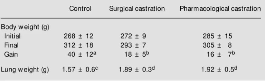
The lung parenchyma of a control rat is shown in Figure 1A. Significant morpho-logical changes in lung parenchyma were observed in surgically castrated rats com-pared to controls (Figure 1B).
The TLU showed a significantly altered appearance. We observed large spaces formed by the fusion of the alveolar cavities as the interalveolar septa disappeared. The alveolar epithelium was not structurally af-fected, the pneumocytes appeared normal, and the alveolar surface was continuous. The interstitial tissue exhibited an evident hypertrophy due to the increase in the num-ber of connective finum-bers and macrophagic invasion. The interstitial spaces contained an exceptionally large number of blood cells. Significant damage to the lung paren-chyma was observed in rats injected with flutamide. The alveolar structure was signif-icantly altered, the thin histological weft dis-appeared and large spaces were formed by the collapse of the interalveolar septa. In the other regions of the lung, the amount of fibrous connective tissue increased sig-nificantly and large numbers of
macrophag-Table 2 - Phospholipid composition of microsomes and extracellular surfactant from male rat lung after administration of flutamide.
Co: Control; F: control + flutamide; LPC: lysophosphatidylcholine; Sph: sphingomyelin; PC: phosphatidylcholine; PI + PS: phosphatidylinositol + phosphatidylserine; PE: phos-phatidylethanolamine; PG: phosphatidylglycerol. Data are reported as means ± SD for 8 rats in each group, expressed as percent of total phospholipids. a,bP<0.001, c,dP<0.01
by analysis of variance and the Tukey test.
M icrosomes Extracellular surfactant
Co F Co F
LPC 1.65 ± 0.1a 0.82 ± 0.08b 0.42 ± 0.01a 0.80 ± 0.02a
Sph 8.90 ± 0.9 8.74 ± 1.1 0.89 ± 0.05c 1.14 ± 0.10d
PC 67.50 ± 2.3a 76.09 ± 4.2b 78.70 ± 4.40a 68.70 ± 3.20b
PI + PS 10.7 ± 0.3a 8.24 ± 0.3b 2.80 ± 0.20a 10.50 ± 2.70b
PE 9.80 ± 0.0a 4.09 ± 1.6b 6.20 ± 0.60 5.40 ± 1.10
PG 3.2 ± 0.2 3.71 ± 0.5 9.70 ± 0.10a 14.20 ± 2.90b
Figure 1 - Effect of surgical and pharm acological castration on male rat lung. H-E staining, X200.
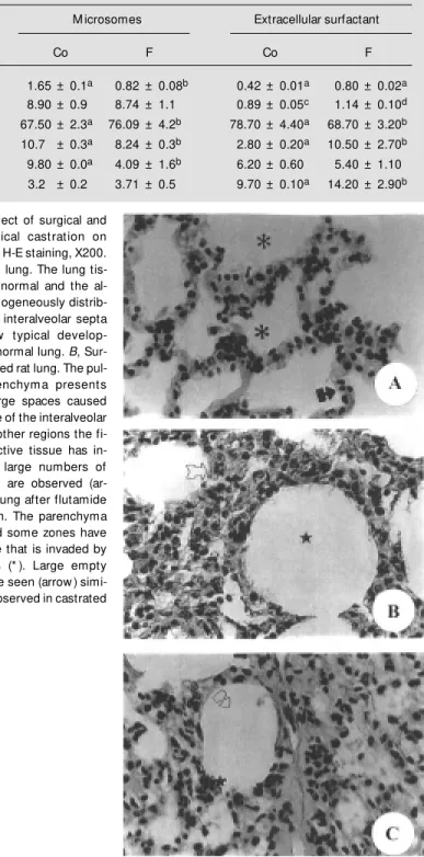
es invaded these zones compared to controls (Figure 1C). The lungs of castrated rats that received testosterone on day 2 did not show morphological changes (Figure 2A). On the other hand, when androgen was adminis-tered on day 15 the lung showed the same structural alterations as in the castrated rat lung. This indicates that testosterone did not reverse the effect of castration when admin-istered at an advanced stage of lung damage (Figure 2B).
D iscussio n
The present results showed that pharma-cological castration by the administration of flutamide modifies the composition of the phospholipids in the microsomes and in the extracellular surfactant of the male rat lung. These changes are similar to those previ-ously observed in our laboratory using surgi-cally castrated rats (14). Androgen suppres-sion by surgical or pharmacological meth-ods provokes significant alterations in the lung. These degenerative changes may be related, at least in part, to alterations in the chemical composition of cell membranes and lung surfactants. The alterations observed could be a consequence of the alteration in the composition of the phospholipids that make up the alveolar surface. This would provoke a disorganization of the gas-liquid interface with consequent damage to the film of bubbles, which keeps the surface tension near zero, thus allowing the TLU to maintain its physical integrity. The loss of the bubble film affects the network, primarily the zone corresponding to the alveolar septa, in which the amount of interstitial tissue is dimin-ished, thereby providing little structural sup-port. We observed that the septa break up and contiguous alveolar cavities fuse into one. The damage incurred cannot be due to the histological techniques used, since the same technique was employed in all cases.
The action of androgens on the lung has been studied primarily in the fetal lung. The
synthesis of surfactant glycerophospholipids and proteins is under multifactorial control and is regulated by a number of hormones and factors, including glucocorticoids, pro-lactin, insulin, growth factors, estrogens, androgens, thyroid hormones and catechola-mines acting through beta-adrenergic recep-tors, and cAMP (15).
Dihydrotestosterone (DHT) inhibits pul-monary surfactant production in fetal rab-bits, and flutamide administration to preg-nant rabbits eliminates the sex difference in the saturated phosphatidylcholine/sphingo-sine ratio in lung lavage by increasing the male ratios up to that observed in the females (16-18). Many observations support the view that surfactant deficiency is indeed the major cause of neonatal respiratory distress syn-drome (19).
Male sex hormone (androgen) has an inhibitory effect on antenatal lung develop-ment. Androgen would decrease antenatal lung glucocorticoid receptor (GR). Further-more, antenatal administration of DHT re-duced tissue levels of GR mRNA and pro-tein, consistent with androgenic inhibition
of antenatal lung development by decreasing GR. Glucocorticoids and androgens exert opposite effects on fetal lung GR (20).
When explanted midgestation human fe-tal lung tissue is maintained in serum-free medium in the presence of dexamethasone, increased synthesis of saturated phosphati-dylcholine (SPC) occurs. Addition of an equimolar concentration of DHT to the me-dium delays the spontaneous and dexameth-asone-stimulated increases in SPC synthesis by 24 h after exposure. The antiandrogen flutamide neutralizes the effect of DHT, in-dicating that it acts through the androgen receptor to block the glucocorticoid (21). On the other hand, it is known that rats with pulmonary fibrosis present a decrease in the concentration of plasma corticosterone and testosterone and a lower content of pulmo-nary cytosol corticosteroid, androgen and estrogen receptors (22).
In the present study, in animals injected with flutamide, the parenchyma was enlarged by connective fibers. This could prevent the rupture of the alveolar structure, permitting
the synthesis of cicatricial tissue. Contrary to what has been observed in castrated rats, blood in the alveolar space and lung paren-chyma was absent. The morphology of the lung of castrated rats which were treated with testosterone on the second day after castration was similar to that observed in the control rat lung, possibly due to the fact that serum androgen concentration was similar to control (data not shown). When the cellu-lar damage is produced 15 days after castra-tion, the administration of testosterone does not reverse the structural alterations observed after castration, although the biochemical changes are reversed (14).
Our data demonstrate that the phospho-lipid composition of microsomes and extra-cellular surfactant in the male rat lung is highly dependent on androgenic stimulation and that the damage in lung parenchyma takes place in the absence of testosterone. Our data suggest that the lung should be regarded as an organ, which can be affected by gonadal steroid imbalances.
Re fe re nce s
1. Jobe A (1988). The role of surfactant in neonatal adaptation. Perinatology, 12: 123-133.
2. Hallman M , Spragg R, Harrell JH & M oser KM (1982). Evidence of lung surfactant abnormality in respiratory failure. Journal of Clinical Investigation, 70: 673-683. 3. Scarpelli EM (1998). The alveolar surface
netw ork. A new anatomy and its physi-ological significance. Anatomical Record, 251: 491-527.
4. M orishige E & Uetake CA (1978). Recep-tors for androgen and estrogen in the rat lung. Endocrinology,102: 1827-1837. 5. Ojeda M S, Gomez NN & Gimenez M S
(1997). Androgen regulation of lung lipids in the male rat. Lipids,12: 57-62. 6. Simard J, Luthy Y, Belanger A & Labrie F
(1986). Characteristics of interaction of the antiandrogen Flutamide w ith andro-gen receptor in various target tissues.
M olecular and Cellular Endocrinology,44: 261-270.
7. Kallio PJ, Janne OA & Palvino JJ (1994). Agonist but not antagonists alter the con-formation of the hormone-binding domain of the androgen receptor. Endocrinology, 134: 998-1001.
8. Tsai M J & O’M alley BW (1994). M olecu-lar mechanisms of action of steroid/thy-roid receptor superfamily members. An-nual Review of Genetics, 25: 89-123. 9. M ulder E & Kuil CW (1996).
Deoxyribo-nucleic acid-binding ability of androgen receptors in w hole cells: Implications for the action of androgens and antiandro-gens. Endocrinology,137: 1870-1877. 10. Sardanons M L, de las Heras M A, Calandra
RS, Solano AR & Podesta EJ (1989). Ef-fect of the antiandrogen flutamide on pi-tuitary LH content and release. Neuroen-docrinology, 50: 211-216.
11. Adachi H, Hayashi H, Sato H, Dempo K & Akino T (1989). Characterization of phos-pholipids accumulated in pulmonary sur-factant compartments of rats
intratrache-ally exposed to silica. Biochemical Jour-nal, 262: 781-786.
12. Kuroki Y & Akino T (1991). Pulmonary sur-factant protein A (SP-A) specially binds dipalmitoylphosphatidylcholine. Journal of Biological Chemistry, 266: 3068-3073. 13. Rouser G, Fluster S & Yam am oto A
(1970). Tw o dimensional thin-layer chro-matographic separation of polar lipid and determination of phospholipids analysis of spots. Lipids,5: 494-496.
14. Ojeda M S (1995). Lipids of male rat lung. Effects of the androgens suppressed. Doctoral thesis, National University of San Luis, Argentina.
15. M endelson CR & Boggaram V (1991). Hor-monal control of the surfactant system in fetal lung. Annual Review of Physiology, 53: 415-440.
16. Nielsen HC, Zinman HM & Torday JS (1982). Dihydrotestosterone inhibits fetal rabbit pulmonary surfactant production.
611-616.
17. Nielsen HC (1985). Androgen receptors influence the production of pulmonary surfactant in the testicular feminization mouse fetus. Journal of Clinical Investiga-tion, 76: 177-181.
18. Klain JM & Nielsen HC (1993). Androgen regulation of epidermal grow th factor re-ceptor binding activity during fetal rabbit lung development. Journal of Clinical
In-vestigation, 91: 425-431.
19. Jobe A & Ikegami M (1987). Surfactant for the treatment of respiratory distress syndrome. American Review of Respira-tory Disease, 136: 1256-1275.
20. Sw eezey NB, Ghibu F, Gagnon S, Schotman E & Hamid Q (1998). Glucocor-ticoid receptor mRNA and protein in fetal rat lung in vivo: modulation by glucocorti-coid and androgen. American Journal of
Physiology,275: L103-L109.
21. Torday JS (1990). Androgens delay hu-man fetal lung maturation in vitro. Endo-crinology,126: 3240-3244.