ISSN 0100-879X
BIOMEDICAL SCIENCES
AND
CLINICAL INVESTIGATION
www.bjournal.com.br
www.bjournal.com.br
Volume 44 (12) 1194-1298 December 2011
Institutional Sponsors
The Brazilian Journal of Medical and Biological Research is partially financed by
Faculdade de Medicina de Ribeirão Preto Campus
Ribeirão Preto
Ex plor e H igh - Pe r for m a n ce M S Or bit r a p Te ch n ology I n Pr ot e om ics & M e t a bolom ics
analit icaw eb.com .br S C I E N T I F I C
Braz J Med Biol Res, December 2011, Volume 44(12) 1202-1208
doi: 10.1590/S0100-879X2011007500138
The
Streptococcus mutans
GlnR protein exhibits an increased
affinity for the glnRA operon promoter when bound to GlnK
The
Streptococcus mutans
GlnR protein
exhibits an increased affinity for the
glnRA
operon promoter when bound to GlnK
P. Castellen
1, F.G.M. Rego
2, M.E.G. Portugal
1and E.M. Benelli
11Departamento de Bioquímica e Biologia Molecular, 2Departamento de Patologia Médica,
Universidade Federal do Paraná, Curitiba, PR, Brasil
Abstract
The control of nitrogen metabolism in pathogenic Gram-positive bacteria has been studied in a variety of species and is in -volved with the expression of virulence factors. To date, no data have been reported regarding nitrogen metabolism in the odontopathogenic species Streptococcus mutans. GlnR, which controls nitrogen assimilation in the related bacterial species,
Bacillus subtilis, was assessed in S. mutans for its DNA and protein binding activity. Electrophoretic mobility shift assay of the S. mutans GlnR protein indicated that GlnR binds to promoter regions of the glnRA and amtB-glnK operons. Cross-linking and
pull-down assays demonstrated that GlnR interacts with GlnK, a signal transduction protein that coordinates the regulation of nitrogen metabolism. Upon formation of this stable complex, GlnK enhances the affinity of GlnR for the glnRA operon promoter. These results support an involvement of GlnR in transcriptional regulation of nitrogen metabolism-related genes and indicate
that GlnK relays information regarding ammonium availability to GlnR.
Key words: Streptococcus mutans; GlnR; Transcriptional regulator; GlnK; PII-type protein
Introduction
Correspondence: E.M. Benelli, Departamento de Bioquímica e Biologia Molecular, Universidade Federal do Paraná, Caixa Postal 19046, 81531-990 Curitiba, PR, Brasil. Fax: +55-41-3266-2042. E-mail: [email protected] or [email protected]
Received April 28, 2011. Accepted September 5, 2011. Available online October 21, 2011. Published November 28, 2011.
Among odontopathogens, Streptococcus mutans is
re-sponsible for most of human dental decay (1). The primary
S. mutans virulence factors associated with cariogenicity
include adhesion, acidogenicity, and acid tolerance (2). The influence of nitrogen metabolism on virulence has been
described for the Gram-positive bacteria S. pyogenes,
S. pneumoniae, Bacillus anthracis,and Staphylococcus aureus (3-7). A recent study suggested a similar associa -tion between putative proteins of nitrogen metabolism and
virulence in S. mutans (8).
Most bacteria regulate nitrogen uptake via AmtB, a well-conserved ammonium transport membrane protein
(9). Intracellularly, nitrogen is assimilated by the glutamine synthetase/glutamate synthetase (GS/GOGAT) pathway or the glutamate dehydrogenase (GDH) pathway (9). The regulation of nitrogen metabolism is coordinated by the PII-type signal transduction proteins, GlnB and GlnK (formerly NrgB), which control the activities of enzymes,
membrane transport proteins, and transcription factors
(9,10). Although the GS/GOGAT assimilatory enzymes and PII signal transduction proteins are widely conserved in
Gram-positive bacteria, other modes of regulating nitrogen metabolism also exist (11).
The regulation of nitrogen metabolism in S. mutans is
poorly understood. The best characterized Gram-positive
bacterium is Bacillus subtilis, which is used as a model
organism for low G+C Gram-positive bacteria. B. subtilis
expresses two DNA-binding proteins of the MerR family,
GlnR and TnrA, which cooperate in the transcriptional re-pression or activation of nitrogen metabolism genes (12,13).
GlnR represses the glnRA and ureABC operons, which
encode GlnR and glutamine synthetase and urea utilization enzymes, respectively, and the tnrA gene (14-16). TnrA
activates the transcription of the amtB-glnK (ammonium
transport), ureABC, nasBC and nasDEF (nitrate and nitrite
utilization) operons and represses glnRA and gltAB, which
encodes glutamate synthase (17-20). Feedback-inhibited GS regulates the activities of both proteins by stabilizing
the GlnR-DNA complex and preventing TnrA from binding
to DNA (21,22). A study of the B. subtilis GlnK protein
In vitro characterization of Streptococcus mutans GlnR protein 1203
Studies of nitrogen metabolism in S. mutans may provide
insight into the virulence mechanisms of this pathogen. In
this report, we demonstrate that S. mutans GlnR binds to
the promoter regions of amtB-glnK and glnRA operons.
In addition, we show for the first time a stable interaction
between the GlnR and GlnK proteins. GlnK within this
complex enhances the affinity of GlnR for DNA.
Material and Methods
Bacterial strains and growth conditions
Escherichia coli BL21-AI was grown at 37°C in Luria broth (LB) or Luria agar (LA) medium supplemented with
kanamycin (50 µg/mL) as appropriate (Table 1).
Cloning, expression and purification of recombinant
GlnR
The S. mutans UA159 glnR gene was amplified by PCR
with primers N-GlnR and C-GlnR (Table 2) using S. mutans
chromosomal DNA as a template. PCR products were
digested with NdeI and BamHI and cloned into pET28b+
(Novagen, EMD4 Biosciences, USA) to produce the plasmid
pET28R, which expresses GlnR-(His)6. The same digested
PCR products were cloned into pET29b+ (Novagen, EMD4 Biosciences) to produce the plasmid pET29R, which ex-presses native GlnR (Table 1). These plasmids were then
transformed into E. coli BL21-AI and transformants were
selected on LA supplemented with kanamycin.
Bacteria transformed with pET29R were cultured on 1 L LB medium at 16°C for 16 h after induction with 0.2%
ara-binose. Cells were pelleted by centrifugation, resuspended
in bufferA (50 mM Tris-HCl, pH 8.0, 200 mM NaCl, 0.1 mM
EDTA, 10% glycerol), and lysed by sonication. The soluble fraction of the cell extract was purified by ion exchange chromatography on a Q-Sepharose column (GE Healthcare
Ltd., UK), and proteins were eluted with an NaCl gradient (0.2-1 M). Fractions containing GlnR were selected after SDS-PAGE visualization. These fractions were mixed and
diluted in 50 mM Tris-HCl, pH 8.0, and 10% glycerol to a final concentration of 0.2 M NaCl and loaded onto a heparin column (GE Healthcare Ltd.). Proteins then were eluted
through a second NaCl gradient (0.2-2 M).
GlnR fused to an N-terminal hexahistidyl tag was over
-expressed in E. coli BL21-AI transformed with pET28R
and grown on LA at 30°C. Cells were harvested by cen
-trifugation, resuspended in lysis buffer(50 mM Tris-HCl, pH 8.0, 200 mM NaCl, 40 mM imidazole, 10% glycerol), and disrupted by sonication. The GlnR-(His)6 protein was purified from the soluble cell fraction by chromatography on an immobilized nickel ion affinity HisTrap HP column (GE Healthcare Ltd.). Proteins were eluted with an imidazole gradient (40 µM to 1 M).
Protein purity was analyzed by 12% SDS-PAGE (25). Apparently homogeneous fractions were dialyzed in two
steps: 1) against the equilibration buffer DA (50 mM
Tris-HCl, pH 8.0, 500 mM NaCl, 20% glycerol), and 2) against the buffer DB (50 mM Tris-HCl, pH 8.0, 200 mM NaCl, 20% glycerol). GlnR and GlnR-(His)6 proteins were stored
in buffer DB, and protein concentration was determined
by the Bradford method (26). Protein concentrations were
calculated as proteins in monomeric state.
S. mutans GlnK and GlnK-(His)6 proteins were ex-pressed and purified as described by Portugal et al. (27).
Polyclonal antibody production
Three Wistar albino rats (140 ± 20 g) were immunized
subcutaneously with 20 µg GlnR or GlnK, both in native
form, in complete Freund’s adjuvant. Three booster injec-tions were given at 4-week intervals and rats were bled 4 weeks after the last injection. Whole blood was centrifuged
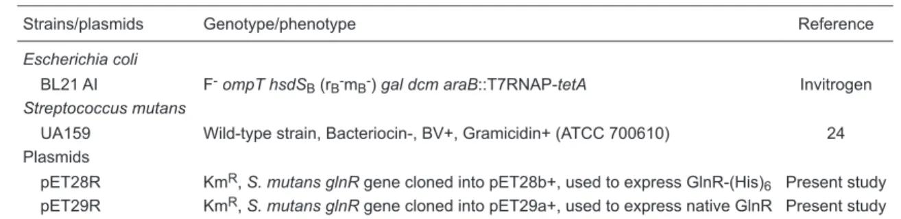
Table 1. Bacterial strains and plasmids.
Strains/plasmids Genotype/phenotype Reference
Escherichia coli
BL21 AI F-ompThsdS
B (rB-mB-) gal dcmaraB::T7RNAP-tetA Invitrogen Streptococcus mutans
UA159 Wild-type strain, Bacteriocin-, BV+, Gramicidin+ (ATCC 700610) 24 Plasmids
pET28R KmR, S. mutansglnR gene cloned into pET28b+, used to express GlnR-(His)6 Present study
pET29R KmR, S. mutansglnR gene cloned into pET29a+, used to express native GlnR Present study
Table 2. Primers used in the present study.
Primer Sequence
N-GlnR 5’ GAAAGGAGGAAACATATGAAAG 3’ C-GlnR 5’ AAAAATTAAGGATCCACGTTACG 3’ PamtBfor 5’ AGCTAAGCTTTAGAGCCCTAG 3’ PamtBrev 5’ GATAATAAATGCTATAGATCC 3’ PglnRfor 5’ GGCCATGAGTAATAAGACGGG 3’ PglnRrev 5’ CCGCCATTGATCGTCGAAGC 3’
and the serum was used in the experiments. The antibody specificity was tested in dot blot experiments.
Electrophoretic mobility shift assay (EMSA)
The S. mutans promoter regions PamtB-glnK and PglnRA were amplified by PCR using primers PamtBfor/
PamtBrev and PglnRfor/PglnRrev, respectively (Table 2). After amplification, PCR products were purified using the QIAquick PCR Purification Kit (Qiagen, Germany). When indicated, probes were labeled with [γ32P]-ATP using T4 polynucleotide kinase (New England Biolabs, USA). For
DNA binding experiments, reaction buffer (10 mM Tris,
pH 8.0, 5 mM MgCl2, 1 mM DTT, 8% [v/v] glycerol, 0.05%
N-octyl glucoside, 100 µg/mL BSA, and 50 µg/mL
poly[dA-dC]) was combined with DNA fragments corresponding
to the PglnRA or PamtB-glnK region. Either unlabeled or
radiolabeled fragments were used, as indicated. GlnR or
GlnR-(His)6 was then added at various concentrations, and
the reactions were incubated at 30°C for 15 min. Samples were subjected to native 6.5% PAGE in TG buffer (50 mM
Tris, 50 mM glycine, pH 8.0). Gels were stained with SYBR
Green (Invitrogen, USA) when unlabeled DNA fragments were used. Radiolabeled DNA fragments in dried gels were
visualized using a PhosphorImager (Molecular Dynamics, USA) system. Band intensities were analyzed using the LabWorks software (UVP, USA).
The effect of GlnK on the binding affinity of GlnR to
PglnRA DNA was determined by adding various concentra -tions of native GlnK to the incubation buffer.
Approximated apparent dissociation constants (K0.5)
were calculated by fitting the data to a double-reciprocal plot as described by Levitzki (28). K0.5 corresponds to the concentration of GlnR necessary for 50% of DNA binding
sites to be occupied.
His tag pull-down assays
GlnR/GlnK complex formation was studied in vitro using
pull-down assays. The proteins GlnR and GlnK-(His)6 were incubated in buffer A (10-mM Tris, pH 8.0, 5 mM MgCl2, 8% glycerol, 0.05% N-octyl glucoside, 80 mM imidazole) in the
presence or absence of 15 nM DNA at room temperature. Ni2+-chelating Sepharose resin (50 µL; GE Healthcare Ltd.) was equilibrated in buffer A, and 5 µM GlnK-(His)6 was added. After 5 min, 2.5 µM GlnR was added. The mixtures were washed three times with 300 µL buffer A with or without DNA. Proteins were eluted with buffer B (10 mM Tris, pH
8.0, 5 mM MgCl2, 8% glycerol, 0.05% N-octyl glucoside, 1
M imidazole). As control, the resin was incubated in buffer
A either with GlnK-His or GlnR proteins. Samples were analyzed by 10% Tricine-SDS-PAGE stained with colloidal
Coomassie blue.
Cross-linking experiments
The interaction of GlnR and GlnK was further analyzed via chemical cross-linking with glutaraldehyde. GlnK and
GlnR, both in native form, were incubated in HGNED buffer (1 mM DTT, 100 mM NaCl, 0.2 M EDTA, 0.05% [v/v] NP-40, 10% glycerol, 25 mM HEPES, pH 8.0) at room temperature for 15 min. Glutaraldehyde was added to a final concentra
-tion of 0.1% (v/v) and the reac-tions were quenched after 7 min by the addition of 0.1 volume of 2 M Tris. Proteins were fractionated by 15% SDS-PAGE (25) and transferred to polyvinylidene difluoride membranes (Millipore, USA)
(29). Membranes were probed with mouse antiserum
against GlnK or GlnR, and proteins were visualized by
chemiluminescence using horseradish peroxidase-labeled
secondary antibodies and luminol (ECL Western Blotting System, GE Healthcare Ltd.).
In these experiments, the Prestained Protein Molecular
Weight Marker (Fermentas, Canada) was used to verify the Western transfer efficiency and to estimate the protein weight through relative mobility (30).
All experiments were performed at least twice.
Results
GlnR binds DNA corresponding to glnK-amtB and
glnRA promoters in vitro
GlnR binding to glnK-amtB and glnRA promoter regions
was examined by EMSA. The K0.5 values for PglnRA and
PamtB-glnK were 50 and 24 nM, respectively (Figure 1A and
B). The PglnRA DNA fragment used in this study includes
a putative palindromic binding site (ATGTNAN7TNACAT)
(8) at -58 bp relative to the possible transcriptional start
point (Figure 1C). The DNA fragment with the PamtB-glnK
region contains the putative GlnR binding site at -18 bp relative to the possible transcriptional start point and a 6-base conserved repeat at -79 bp (Figure 1C). The pres-ence of two band shifts in GlnR concentrations over 50 nM
suggests that these binding sites are recognized by GlnR
in vitro (Figure 1B).
GlnR interacts with GlnK in vitro
The oligomeric states of GlnR and GlnK and complex formation between these proteins were studied using
chemical cross-linking with glutaraldehyde (Figure 2A and B). GlnR existed as a dimer independently of the presence
of glnRA promoter DNA (Figure 2A, lanes 2 and 3). After chemical cross-linking, GlnK protein samples were
visual-ized as five bands with different migration characteristics (Figure 2B, lane 5). Although this result may be explained
as a cross-linking artifact, it is also possible that GlnK exists
as five different oligomers under the tested conditions. The observed bands possibly correspond to many oligomeric forms of GlnK. GlnR interacts directly with different putative
GlnK oligomers (Figure 2A, lane 7).
Complex formation between native GlnR and
GlnK-(His)6 was examined using pull-down assays in the absence
or presence of glnRA promoter DNA (Figure 2C and D).
In vitro characterization of Streptococcus mutans GlnR protein 1205
Figure 1. GlnR-(His)6 binding toDNA. A, Increasing quantitiesof GlnR-(His)6 were incubated in the presence of 0.3 nM of a radio-labeled glnRA promoter DNA fragment (311 bp). Lane 1 = no GlnR; lanes 2-10 = 10, 25, 50, 10, 250, and 500 nM, 1, 2.5, and 5 µM GlnR, respectively. B, Increasing quantities of GlnR-(His)6 were incubated in the presence of 0.6 nM of an amtB-glnK promoter DNA fragment (174 bp). Lane 1 = no GlnR; lanes 2-10 = 10, 25, 50, 10, 250, and 500 nM, 1, 2.5, and 5 µM GlnR, respectively. C, DNA sequence of amtB-glnK and glnRA promoter regions. The putative GlnR binding sites (8) are boxed, the possible transcription start
points are indicated by asterisks and possible -10 and -35 elements are presented in gray boxes. These are the sequences of DNA fragments used in electrophoretic mobility shift assay(EMSA) experiments. D, GlnR binding site consensus sequence with 6-base
inverted repeats spaced by 7 nt (8).
C
of DNA or in the presence of 15 nM glnRA promoter DNA
(Figure 2), implying that DNA is not required for GlnR-GlnK
complex formation.
GlnK enhances DNA-binding activity of GlnR in vitro
The effect of GlnK on the ability of GlnR to bind glnRA
promoter DNA was determined using EMSA (Figure 3).
Various concentrations of GlnK were added to 50 nM GlnR
(protomer). In the presence of 150 nM GlnK (protomer), which corresponds to a 6-fold excess over dimeric GlnR
(25 nM), binding of GlnR to DNA was increased 5-fold as
estimated by band densitometry (Figure 3A, lane 5). This result suggests that two GlnK trimers or, possibly, the hexameric form of GlnK increases the affinity of the GlnR
dimer for DNA.
The EMSA analyses detected the formation of a stable ternary complex between GlnR, GlnK and DNA and con
-firmed the results of the pull-down experiments in the pres
-ence of DNA. Long-term EMSA indicated that the ternary complex exhibited a considerably lower mobility than the
Figure 2. Formation of the GlnR-GlnK complex
in vitro. A and B, Western blots of GlnR and GlnK cross-linking experiments. Cross-linking reactions containing the indicated components
were separated by 15% SDS-PAGE and evalu
-ated by Western blot analysis with anti-GlnR
(A) or anti-GlnK (B) antiserum. Native GlnR, native GlnK, glnRA promoter DNA, and
glutar-aldehyde (Glut) were used at concentrations of 2.5, 2.5, and 0.4 µM, and 0.1% (v/v), respec
-tively. C and D, His-tag pull-down assay. Resin was loaded with 5 µM GlnK-(His)6 (12.1 kDa;
C and D, lanes 1 and 3) and 2.5 µM GlnR (14.3
kDa; C and D, lanes 2 and 3). DNA fragments containing glnRA promoter DNA (15 nM) were added to the reaction mixture (D) Tricine-SDS 10% gel electrophoresis of proteins eluted
from the resin. Arrows indicate the identified
proteins.
Figure 3. The influence of GlnK on GlnR binding
to DNA. A, Native GlnR (50 nM promoter) was incubated with 0.3 nM of a radiolabeled 311-bp glnRA promoter DNA fragment, and increasing quantitiesof native GlnK protomer. Lane 1 = no GlnR; lane 2 = GlnR; lanes 3-5 = GlnR and 25 nM GlnK; GlnR and 75 nM GlnK, and GlnR and 150 nM GlnK, respectively. B, Native GlnR (50 nM) was incubated with 6 nM of the glnRA promoter DNA fragment and 25 nM native GlnK, and elec-trophoresis was performed for twice the duration of the other gels described in this study. Lane 1 =
no GlnR; lane 2 = GlnR; lane 3 = GlnR and GlnK. C, Increasing quantities of native GlnR were in-cubated in the presence of 3 nM glnRA promoter DNA fragments. Lane 1 = no GlnR; lanes 2-7 =
In vitro characterization of Streptococcus mutans GlnR protein 1207
GlnR-DNA complex (Figure 3B, lane 3).
The effect of GlnK on the affinity of GlnR binding to
glnRA promoter DNA was also examined by EMSA (Figure
3C and D). GlnK did not bind DNA fragments under these conditions (Figure 3D, lane 2). In the presence of a 6-fold
excess of GlnK protomer relative to the GlnR dimer, the K0.5
was 13 nM (Figure 3D). When only GlnR was present in
the reaction mixture, the K0.5 was 50 nM (Figures 1A and
3C). These results indicate that GlnK increases GlnR-DNA
binding activity by a factor of approximately 4.
Discussion
We report here the cloning, expression, and purification
of the S. mutans GlnR protein. Our results demonstrate that
S. mutans GlnR binds to glnRA promoter DNA, as described for B. subtilis GlnR (12), and that S. mutans GlnR binds the amtB-glnK promoter. These results agree with the in vivo results obtained by Chen et al. (8), which showed that S. mutans GlnR regulates its own transcription and the
transcription of the amtB-glnK operon.
The present study describes for the first time an inter
-action between S. mutans GlnK and GlnR in vitro (Figure
2). EMSA analyses identified stable GlnR-GlnK-DNA com
-plexes (Figure 3B). In B. subtilis,GlnK binds to the TnrA
protein, which is functionally and structurally analogous to GlnR (23). However, no studies have examined the modula
-tion of TnrA or GlnR activity by GlnK.
Compared to GlnR alone, S. mutans GlnR bound to GlnK
has a 4-fold higher affinity for glnRA promoter DNA (Figure 3C and D), suggesting that GlnK enhances GlnR-mediated
transcriptional repression of the glnRA promoter.
Interactions between GlnK and various transcription
factors have been described for the Gram-positive
Coryne-bacterium glutamicum. In this microorganism, adenylylated
GlnK interacts with AmtR, a transcriptional repressor, lead-ing to de-repression of genes involved in the control of
nitrogen metabolism (31). In cyanobacteria, GlnK regulates gene expression by binding to NtcA and PipX proteins. NtcA
is a global transcriptional activator, and PipX binds to and
enhances the activity of NtcA during nitrogen starvation.
Under nitrogen excess, GlnK interacts with PipX to impair NtcA activation (32-34).
Almost all PII-type proteins are trimeric; however, gel filtration chromatography results suggest that the GlnK pro
-tein of S. mutans has the molecular weight of a hexamer in
solution (27). Supporting this, an S. mutans GlnK structure
deposited in the Protein Data Bank (PDB ID: 3L7P) is
de-scribed as a biological assembly of six domains (Fan X-X, Wang K-T, Su X-D; Protein Data Bank, 2011). Our results
suggest that a 6-fold excess of GlnK promoter increases the
affinity of the GlnR dimer for glnRA operon promoter DNA.
This implies that two trimers of GlnK protein, or possibly its
hexameric form, are responsible for the increase in GlnR
DNA-binding activity (Figure 3A and D).
GlnR-DNA binding was confirmed in vitro for the
amtB-glnK and glnRA promoters. The in vitro formation of a stable
complex between GlnR and GlnK is described here for
the first time. Once bound to GlnK, GlnR exhibits a higher affinity for glnRA promoter DNA. Information regarding
nitrogen availability is likely to be transmitted to GlnR via
GlnK binding in S. mutans.
Future in vivo studies should permit the determination
of the physiological conditions in which the GlnR-GlnK
interaction occurs.
Acknowledgments
We thank Prof. Susan H. Fisher for advice regarding
EMSA parameters, Prof. Dennis G. Cvitkovitch for providing the S. mutans UA159 strain, and Prof. Fábio de Oliveira
Pedrosa, Instituto Nacional de Ciência e Tecnologia - Fixa-ção de Nitrogênio, for access to the equipment. Research
supported by CNPq (#140152/2007-5).
References
1. Loesche WJ. Role of Streptococcus mutans in human dental
decay. Microbiol Rev 1986; 50: 353-380.
2. Banas JA. Virulence properties of Streptococcus mutans.
Front Biosci 2004; 9: 1267-1277.
3. Tamura GS, Nittayajarn A, Schoentag DL. A glutamine transport gene, glnQ, is required for fibronectin adherence
and virulence of group B streptococci. Infect Immun 2002; 70: 2877-2885.
4. Hendriksen WT, Kloosterman TG, Bootsma HJ, Estevao S, de Groot R, Kuipers OP, et al. Site-specific contributions of
glutamine-dependent regulator GlnR and GlnR-regulated genes to virulence of Streptococcus pneumoniae. Infect Immun 2008; 76: 1230-1238.
5. van Schaik W, Chateau A, Dillies MA, Coppee JY, Sonen
-shein AL, Fouet A. The global regulator CodY regulates toxin
gene expression in Bacillus anthracis and is required for full virulence. Infect Immun 2009; 77: 4437-4445.
6. Gustafson J, Strassle A, Hachler H, Kayser FH,
Berger-Bachi B. The femC locus of Staphylococcus aureus required
for methicillin resistance includes the glutamine synthetase
operon. J Bacteriol 1994; 176: 1460-1467.
7. Somerville GA, Proctor RA. At the crossroads of bacterial
metabolism and virulence factor synthesis in Staphylococci. Microbiol Mol Biol Rev 2009; 73: 233-248.
8. Chen PM, Chen YY, Yu SL, Sher S, Lai CH, Chia JS. Role
of GlnR in acid-mediated repression of genes encoding proteins involved in glutamine and glutamate metabolism in Streptococcus mutans. Appl Environ Microbiol 2010; 76: 2478-2486.
Mi-crobiol Rev 1995; 59: 604-622.
10. Arcondeguy T, Jack R, Merrick M. P(II) signal transduction proteins, pivotal players in microbial nitrogen control. Micro-biol Mol Biol Rev 2001; 65: 80-105.
11. Amon J, Titgemeyer F, Burkovski A. Common patterns -
unique features: nitrogen metabolism and regulation in Gram-positive bacteria. FEMS Microbiol Rev 2010; 34: 588-605.
12. Schreier HJ, Brown SW, Hirschi KD, Nomellini JF, Son -enshein AL. Regulation of Bacillus subtilis glutamine syn
-thetase gene expression by the product of the glnR gene. J Mol Biol 1989; 210: 51-63.
13. Wray LV Jr, Ferson AE, Rohrer K, Fisher SH. TnrA, a tran -scription factor required for global nitrogen regulation in
Bacillus subtilis. Proc Natl Acad Sci U S A 1996; 93: 8841-8845.
14. Brown SW, Sonenshein AL. Autogenous regulation of the
Bacillus subtilis glnRA operon. J Bacteriol 1996; 178: 2450-2454.
15. Wray LV Jr, Ferson AE, Fisher SH. Expression of the Bacillus subtilis ureABC operon is controlled by multiple regulatory factors including CodY, GlnR, TnrA, and Spo0H. J Bacteriol
1997; 179: 5494-5501.
16. Zalieckas JM, Wray LV Jr, Fisher SH. Cross-regulation of the Bacillus subtilis glnRA and tnrA genes provides evidence for
DNA binding site discrimination by GlnR and TnrA. J Bacte-riol 2006; 188: 2578-2585.
17. Wray LV Jr, Zalieckas JM, Ferson AE, Fisher SH. Mutational analysis of the TnrA-binding sites in the Bacillus subtilis
nrgAB and gabP promoter regions. J Bacteriol 1998; 180: 2943-2949.
18. Brandenburg JL, Wray LV Jr, Beier L, Jarmer H, Saxild HH, Fisher SH. Roles of PucR, GlnR, and TnrA in regulating ex -pression of the Bacillus subtilis ure P3 promoter. J Bacteriol
2002; 184: 6060-6064.
19. Nakano MM, Hoffmann T, Zhu Y, Jahn D. Nitrogen and oxy -gen regulation of Bacillus subtilis nasDEF encoding NADH-dependent nitrite reductase by TnrA and ResDE. J Bacteriol
1998; 180: 5344-5350.
20. Belitsky BR, Wray LV Jr, Fisher SH, Bohannon DE, Sonen -shein AL. Role of TnrA in nitrogen source-dependent repres-sion of Bacillus subtilis glutamate synthase gene expression. J Bacteriol 2000; 182: 5939-5947.
21. Fisher SH, Wray LV Jr. Novel trans-acting Bacillus subtilis
glnA mutations that derepress glnRA expression. J Bacteriol
2009; 191: 2485-2492.
22. Fisher SH, Wray LV Jr. Bacillus subtilis glutamine synthetase
regulates its own synthesis by acting as a chaperone to
stabilize GlnR-DNA complexes. Proc Natl Acad Sci U S A
2008; 105: 1014-1019.
23. Heinrich A, Woyda K, Brauburger K, Meiss G, Detsch C,
Stulke J, et al. Interaction of the membrane-bound GlnK-AmtB complex with the master regulator of nitrogen me-tabolism TnrA in Bacillus subtilis. J Biol Chem 2006; 281: 34909-34917.
24. Ajdic D, McShan WM, McLaughlin RE, Savic G, Chang J, Carson MB, et al. Genome sequence of Streptococcus mu-tans UA159, a cariogenic dental pathogen. Proc Natl Acad Sci U S A 2002; 99: 14434-14439.
25. Laemmli UK. Cleavage of structural proteins during the
as-sembly of the head of bacteriophage T4. Nature 1970; 227: 680-685.
26. Bradford MM. A rapid and sensitive method for the quantita-tion of microgram quantities of protein utilizing the principle
of protein-dye binding. Anal Biochem 1976; 72: 248-254.
27. Portugal ME, Souza EM, Pedrosa FO, Benelli EM. Strepto-coccus mutans GlnK protein: an unusual PII family member. Braz J Med Biol Res 2011; 44: 394-401.
28. Levitzki A. Ligand binding. In: Creighton TE (Editor), Protein function: a practical approach. 2nd ed. Oxford: IRL Press;
1997. p 119.
29. Sambrook J, Fritsch EF, Maniatis T. Molecular cloning: a laboratory manual. New York: Cold Spring Harbor Labora
-tory Press; 1989.
30. Neville DM Jr. Molecular weight determination of
protein-dodecyl sulfate complexes by gel electrophoresis in a discontinuous buffer system. J Biol Chem 1971; 246: 6328-6334.
31. Beckers G, Strosser J, Hildebrandt U, Kalinowski J, Farwick
M, Kramer R, et al. Regulation of AmtR-controlled gene ex-pression in Corynebacterium glutamicum: mechanism and characterization of the AmtR regulon. Mol Microbiol 2005; 58: 580-595.
32. Espinosa J, Forchhammer K, Burillo S, Contreras A.
Interac-tion network in cyanobacterial nitrogen regulaInterac-tion: PipX, a
protein that interacts in a 2-oxoglutarate dependent manner with PII and NtcA. Mol Microbiol 2006; 61: 457-469. 33. Espinosa J, Forchhammer K, Contreras A. Role of the
Synechococcus PCC 7942 nitrogen regulator protein PipX in NtcA-controlled processes. Microbiology 2007; 153: 711-718.