The Brazilian Journal of
INFECTIOUS DISEASES
w w w . e l s e v i e r . c o m / l o c a t e / b j i d
Original article
Phenotypic and genotypic characterization of
Rhodococcus
equi
isolated from sputum
Paulo da Silva
a,∗, Adolfo Carlos Barreto Santos
b, Daisy Nakamura Sato
a,
Jaqueline Otero Silva
a, Marta Inês Cazentini Medeiros
a, Ana Maria Machado Carneiro
a,
Sergio Roberto de Andrade Leite
a, Clarice Queico Fujimura Leite
baInstituto Adolfo Lutz, Núcleo de Ciências Biomédicas/Centro de Laboratório Regional (IAL-NCB/CLR-VI), Ribeirão Preto, SP, Brazil
bUniversidade Estadual Paulista “Júlio de Mesquita Filho” (UNESP), Laboratório de Micobactérias “Professor. Dr. Hugo David”,
Pharmaceutical Sciences School, Araraquara, SP, Brazil
a r t i c l e
i n f o
Article history:
Received 29 October 2011 Accepted 13 March 2012
Available online 10 September 2012
Keywords:
Polymerase chain reaction Rhodococcus equi
Mycolic acids
a b s t r a c t
Introduction: Rhodococcus equiis an opportunistic pathogen, causing rhodococcosis, a con-dition that can be confused with tuberculosis. Often, without identifyingM. tuberculosis, physicians initiate empiric treatment for tuberculosis.R. equiandM. tuberculosishave dif-ferent susceptibility to drugs. Identification ofR. equiis based on a variety of phenotypic, chromatographic, and genotypic characteristics.
Objective:This study aimed to characterize bacterial isolates from sputum samples sugges-tive ofR. equi.
Methods:The phenotypic identification included biochemical assays; thin-layer chromatog-raphy (TLC) and polymerase chain reaction (PCR) were used for genotypic identification. Results:Among 78 Gram-positive and partially acid-fast bacilli isolated from the sputum of tuberculosis-suspected patients, 51 were phenotypically and genotypically characterized asR. equibased on literature data. Mycolic acid analysis showed that all suspectedR. equi had compounds with a retention factor (Rf) between 0.4-0.5. Genotypic characterization indicated the presence of thechoEgene 959 bp fragments in 51 isolates CAMP test positive. Twenty-two CAMP test negative isolates were negative for thechoEgene. Five isolates pre-sumptively identified asR. equi, CAMP test positive, werechoEgene negative, and probably belonged to other bacterial species.
Conclusions: The phenotypic and molecular techniques used constitute a good methodologi-cal tool to identifyR. equi.
© 2012 Elsevier Editora Ltda. All rights reserved.
Introduction
Rhodococcus equi is considered a bacterial agent of medical
importance in the group of mycolic-acid-containingbacteria
∗ Corresponding author at:Núcleo de Ciências Biomédicas - Instituto Adolfo Lutz - Centro de Laboratório Regional (IAL-CLR-VI), Rua Minas,
877, Campos Elíseos, 14085-410, Ribeirão Preto, São Paulo, Brazil. E-mail address:[email protected](P. da Silva).
or aerobic actinomycetes. R. equi is the main species of the genusRhodococcuscausing infections (rhodococcosis) in animals and humans. This bacterial agent has emerged as a significant opportunistic pathogen in immunocompro-mised people, particularly, human immunodeficiency virus
1413-8670/$ – see front matter © 2012 Elsevier Editora Ltda. All rights reserved.
(HIV)-infected patients. Rhodococcosis affects mainly the lungs, with clinical and pathological features similar to pul-monary tuberculosis.1,2
TheR. equicell wall includes mycolic-acids of carbon length 30-54 atoms, resulting in a highly variable acid-fast staining. The organism presents the shape of partially acid-fast bacilli (PAFB). Because of its morphologic characteristics,R. equican easily be mistaken for mycobacteria, and may be associated with invasive or systemic infections.2,3
Diagnosis of pulmonary rhodococcosis may be obtained from analysis of clinical specimens such as sputum, bronchial-alveolar lavage (BAL), and biopsy. Smears of clinical specimens stained by modified Kinyon or Ziehl-Neelsen methods may show PAFB.4Culture and biochemical identification are,
how-ever, the gold standard methods for confirming the presence of the pathogen. The colonies grow satisfactorily in nonse-lective culture media, including those used in the isolation of mycobacteria and fungi.5
The phenotypic identification of R. equi is obtained by classical morphological and biochemical tests. Initially, a presumptive identification is determined by theequifactor production, shown by a positive CAMP test reaction.6
Bio-chemically,R. equi is non-reactive and does not oxidize or ferment carbohydrates in standard Gordon media, and does not use acetate, citrate, or malonate as unique carbon sources. It produces catalase and lipase.4,6
R. equi may also be presumptively identified through
thin layer chromatography (TLC) by detection of compounds (mycolic acids), with a retention factor (Rf) between 0.4 and 0.5.7,8Identification ofR. equi by polymerase chain reaction
(PCR), a widely used molecular method, is possible through detection of thevapA gene present in an 85 kb plasmid asso-ciated with virulence. However, this plasmid is not always present in isolates of human origin.9–12
Another effective PCR assay is that described by Ladrón et al.13for species-specificR. equiidentification, which is based
on the amplification of a 959 bp fragment of thechoEgene. This is a chromosomal locus encoding cholesterol oxidase (COX), a secreted enzyme considered as aR. equivirulence factor.1,13COX is the cytolytic factor responsible for the
syner-gistic hemolysis that occurs in the CAMP test reaction, elicited
byR. equiin the presence of sphingomyelinase C-producing
bacteria, such asListeria ivanovii,Bacillus cereus, and Staphylo-coccus aureus.1
The similarity of the clinical courses of rhodococcosis and tuberculosis and the high prevalence of tuberculosis in Brazil14
make it difficult to detectR. equiinfection and establish an appropriate treatment. Considering the clinical importance of R. equi, this study aimed to identify the species in bacterial isolates recovered from sputum samples. A set of techniques, including TLC, is proposed for phenotypic identification, and PCR to characterize genotype, thus establishing an algorithm for the identification ofR. equi.
Material and methods
Bacterial isolates and microbiological procedures
Seventy-eight bacterial isolates (PAFB) suspected of Rhodococ-cus spp. and the reference strain R. equi ATCC 6939 were
included in this study. The bacterial isolates were recov-ered in cultures of sputum samples obtained from patients suspected to have pulmonary tuberculosis, attending pub-lic health units in Ribeirão Preto, SP, Brazil. The phenotypic characterization was performed by the Laboratory of Bacteri-ology of the Instituto Adolfo Lutz, and analysis of mycolic acid and molecular identification was performed by the “Profes-sor. Dr. Hugo David” Mycobacteria Laboratory, Pharmaceutical Sciences School, Universidade Estadual Paulista (UNESP) – Araraquara, SP, Brazil.
R. equi phenotypic identification
The bacterial isolates were cultured on Müeller-Hinton agar (MH) plates and isolated colonies were transferred to tubes with MH agar slants. Standardized techniques, including Gram- and modified acid-fast staining plus additional tests were used to characterize Rhodococcus species in bacterial isolates.15,16The isolates were additionally tested by the CAMP
test, performed on sheep blood agar plates with MH base medium (Dfico) andListeria ivanoviiATCC 19119 as the indica-tor strain. As previously described, theR. equireference strain ATCC 6939 was included as a control.17
Analysis of mycolic acid by TLC
The analysis of mycolic acid was performed according to the technique recommended by Miyaji et al.,8with some
modi-fications. Bacterial isolates were plated on MH agar and the cultures were kept at room temperature until satisfactory growth was observed (up to seven days). The extraction of mycolate methyl-ester was performed in screw cap glass tubes (13 x 100 mm) containing the bacterial suspensions (±30 mg of bacteria) in one mL of methanol/toluene/sulfuric acid (30/15/1). After incubation at 75◦C from 10 to 12 hours to extract the methyl-ester mycolate, the tubes containing the extract received one mL of hexane at room temperature, and were vortexed and left to stand for the separation of the two phases. The supernatants containing methyl-ester mycolate were transferred (0.5 mL) to other tubes, which were kept at 45◦C to evaporate the hexane. To perform TLC, dry residues of methyl-ester mycolate were dissolved in 40 mL of hexane and applied by capillary tubes on silica gel plates (Merck®, Silica gel 60F254). The plates were placed in a glass chamber (22 x 22 x 10 cm) containing 100 mL of hexane/diethyl ether (8/2) for the chromatographic separation of the components. Sprays of 10% ethanolic solution of rhodamine were used to analyze results. The presence of mycolic-acids spots was determined by the Rf. Reference strains (Corynebacterium pseudodiphther-icumIAL 0104,C. xerosisIAL 0105,Nocardia asteroidesIAL 2125, N. brasiliensisIAL 2126,N. brevicatenaIAL 2123,Rhodococcus equi ATCC 33701, R. equiATCC 33702,R. equiATCC 33703,R. equi ATCC 6939,Mycobacterium aurumATCC 23366,M. vaccaeATCC 15483 andM. smegmatisATCC14468) were also tested for the presence of mycolic-acids.
Identification of R. equi by PCR
DNA was obtained according to Telenti et al.18. Briefly, a
loopful of bacteria grown on solid medium (MH agar) was suspended in 300L of TE buffer (10 mM Tris, 1 mM EDTA [pH 8]) in 2.5 mL tubes, boiled for 10 minutes and then frozen (-70◦C) for 20 minutes. The boiling and freezing procedures were repeated twice. After stabilization at room temperature, the tube contents were submitted to the process of extrac-tion and purificaextrac-tion of DNA using the procedures established by Van Soolingen et al.19, with modifications. Briefly, 600 mL
of phenol-chloroform-isoamyl alcohol (25:24:1, vol/vol) were added to the tubes, and the mixture was vortexed for 10 s. After centrifugation (10,000 g at 4◦C) for 20 minutes, the tubes were left to stand until phase separation. The aqueous top phase containing DNA was transferred to other tubes, and mixed with 600 mL of chloroform-isoamyl alcohol (24:1, vol/vol). This was followed by vortexing for 10 s, centrifugation (10,000 g at room temperature) for five minutes, and standing for the separation of the phases formed. The aqueous top phase was transferred to another tube, and the procedure was repeated once more. Precipitation of DNA in the final aqueous phase was performed by addition of absolute ethanol (300 mL) and centrifugation (12,000 g at 4◦C) for 20 minutes. The super-natant was discarded and the pellet was resuspended in 500 mL of 70% ethanol (previously kept at -20◦C, for at least 18 hours) and centrifuged (12,000 g at 4◦C) for 20 minutes. The pellet was separated from the discarded supernatant (carefully pouring from the tube). The DNA precipitation pro-cedures were repeated twice. The tubes were kept inverted (on paper towel) to dry completely. The air-dried pellet was resus-pended in 50 mL of TE buffer (10 mM Tris, 1 mM EDTA [pH 8]), carefully rubbing the pipette tip on the tube wall to release the DNA, which was kept at -20◦C until use.
choE gene amplification
choE gene amplification followed the protocol of Ladrón et al.13. A mixture of 2.5 mL of 10 X PCR buffer; 0.75 mL
of 10 mM deoxynucleoside triphosphate (DNTP); 0.75 mL of 50 mM MgCl2; 0.5 mL of Taq DNA polymerase; 1 mL (40 pmol) of each primer; 10 mL of DNA template; and 8.5 mL milli Q water (final volume of 25 mL) was used for PCR. In a thermal cycler (PTC-100, MJ Research), the DNA was first denatured at 95◦C for five minutes and then subjected to 30 amplification cycles under the following conditions: denaturation at 95◦C for one minute, annealing at 55◦C for one minute, and exten-sion at 72◦C for one minute. After the final cycle, the reactions were terminated by an extra run at 72◦C for 10 minutes. Elec-trophoresis on horizontal 2% agarose gels was performed on 25 mL of the amplified product and with 0.5 mg of ethidium bromide per mL for two hours using TAE buffer (40 mM Tris-acetate, 2 mMNa2 EDTA2H2O) and an electric current of 100 V. Fragments corresponding to the 959 pb product ofchoEgene amplification, indicative ofR. equi, were photographed using AlphaImager (Alpha Innotech ®).
Results
Among the 78 bacterial isolates showing morphologic charac-teristics ofRhodococcusspp., 57 were presumptively identified
Rf = 0,4 - 0,5
1 2 3 4 5 6 7 8 9 10 11 12 13 14 15
Fig. 1 – Representative TLC chromatograms on plates of silica gel showing compounds with Rfs corresponding to mycolic-acids separated by dichloromethane. Samples 1-15 are suspected isolates ofRhodococcus equi.
as R. equi by a positive CAMP test reaction. However, the
presence of the genechoE, indicated by the 959 bp fragment amplification product, was observed in only 51 of these iso-lates, as well as in the reference strain R. equiATCC 6939. Thus, six isolates CAMP test positive and 21 isolates CAMP test negative were also negative for the presence of thechoEgene. Phenotypic characteristics assigned forR. equiwere shown by all 51 isolates CAMP test-positive having a confirmed pres-ence of the gene choE. Accordingly, these isolates did not utilize carbohydrates, acetate, citrate, or malonate as a sole carbon source, but produced catalase and lipase. In addition, all isolates showed negative results in tests for amylase, beta-galactosidase (ONPG), casease, DNase, gelatinase, esculinase, H2S, indole, oxidase, and motility. A variable behavior was demonstrated in the production of nitrate reductase, urease, and reduction of hippurate. While adenine was completely transformed, hypoxanthine, tyrosine, and xanthine were not. Other features consistent withR. equi included the optimal development of cultures in a strictly aerobic environment at temperatures between 25◦C and 37◦C. All these features were confirmed by the reference strainR. equiATCC 693914,16.
Fig. 1shows representative results of mycolic-acid analy-sis by TLC. Spots corresponding to products having Rfaround 0.4-0.5 were observed in all suspected isolates of R. equi and were comparable with the results from standardR. equi ATCC 6939. Other reference strains tested simultaneously for mycolic-acids yielded Rf values between 0.2-0.6, as follows: Corynebacterium pseudodiphthericumIAL 0104 andC. xerosisIAL 0105 - Rf 0.2; Nocardia asteroidesIAL 2125,N. brasiliensisIAL 2126,N. brevicatenaIAL 2123,Rhodococcus equiATCC 33701,R. equiATCC 33702,R. equiATCC 33703 andR. equiATCC 6939 -Rfbetween 0.4 and 0.5;Mycobacterium aurumATCC 23366,M.
vaccaeATCC 15483 andM. smegmatisATCC14468 - Rf≥0.6.
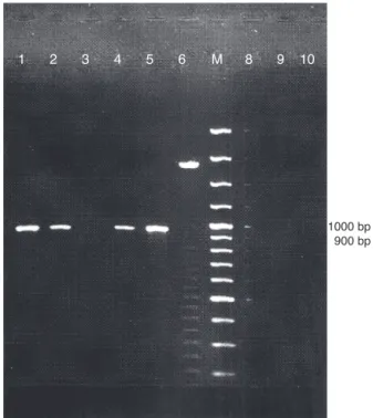
1 2 3 4 5 6 M 8 9 10
1000 bp 900 bp
Fig. 2 – Representative eletroforetic pattern of PCR amplified products of choE in genomic DNA samples of suspected
Rhodococcus equi. Lanes 3, 6, 8, 9 and 10 (isolates CAMP test
negative); Lanes 1, 2 and 4 (isolates CAMP test positive); Lane 5 reference strainRhodococcus equiATCC 6939; M = DNA size marker (100 bp).
five isolates CAMP test-positive were negative for the presence of thechoEgene.
Discussion
R. equiis recognized as an opportunistic pathogen in humans
and has emerged as an important cause of morbidity and mor-tality among immunocompromised patients, especially those infected with HIV. The increased awareness of rhodococcosis improves chances for rapid and correct diagnosis. Basic and clinical analysis is necessary for an appropriate treatment.
Seventy eight bacterial isolates suspected to beRhodococcus spp. were investigated using phenotypic and genotypic tests, including TLC and PCR. A total of 51 isolates were confirmed
asR. equi. The phenotypic identification was based on
mor-phology, cultivation, nutrition, and biochemical features. The 51 isolates ofR. equihad characteristics very similar to those described in the literature.4–6,8,20–22
All 51 isolates grew well when incubated in aerobic con-ditions at 37◦C in a non-selective medium, MH agar. After incubation for 24 hours, colonies measuring about one to two mm in diameter were not distinct. Colonies had their appar-ent features developed in 48 hours with appar-entire edges, shaped like irregular round teardrops, smooth, semitransparent, glis-tening, coalescent, and mucoid.
The characteristics described by Prescott6forR. equiwere
investigated in the confirmed isolates. Colony variation was observed in cultures with the predominant classical type of coalescent viscous mucoid colonies, but less mucoid forms
were also present. Pigment production was not observed in cultures less than four days old, but in up to seven days colonies developed a delicate salmon-pink color. Cultures maintained in slant medium for prolonged periods without sub-culture often became rough, dry, and orange-red, but reverted to the classic colony type when returned to sub-cultures. Morphologically, typical colonies of R. equi ATCC 6939 appeared smaller, smooth, dry rather than mucoid, and appeared to be more pigmented than the colonies of the sus-pected isolates ofR. equi.
Regarding the microscopic morphology and staining prop-erties, all suspect R. equi were Gram-positive pleomorphic coccobacillus, varying from distinctively coccoid to bacil-lary depending on growth conditions. Coccoid forms were usual on solid media (MH agar), but in liquid media (brain-heart infusion), particularly in young cultures, there were forms of long rods or short filaments, which sometimes showed rudimentary branching. When stained by the Ziehl-Neelsen Kinyoun-modified method, the isolates showed partial resistance to acid-alcohol, maintaining basically the same morphology observed in smears stained by the Gram method. All these characteristics are exactly as those related
toR. equi by Prescott,6 and most isolates proved to be
par-tially acid-fast. According to the author, this is an inconsistent feature of the microorganism.
Several features found in this study can be routinely used for identification ofR. equi.All isolates produced theequi fac-tor, emphasizing the importance of the CAMP test.This test was used as a phenotypic marker for the rapid presumptive identification of R. equi, but it may miss strains in isolates not expressing COX despite having the choE gene, or give false-positive results for other extra cellular COX-producing actinomycetes. As well asR. equi, other Gram-positive mycolic acid-containing bacteria, such asMycobacteriumspp.,R. ery-thropolis,R. rhodochrous, andDietziaspp. have COX23,24activity
and the possibility of presenting a positive result in the CAMP test. Nevertheless, the assay proved to be an important tool to presumptively identifyR. equi, since the 51 isolates were CAMP test-positive. According to Prescott,6the test is distinctive for
this organism and must always be used in presumptive iden-tification, since noR. equiisolate has been described asequi factor negative.
TLC, the method chosen to investigate mycolic-acids and recommended by Miyaji et al.,8is the easiest to perform and
is inexpensive. The usual revelation reagent was substituted in this study by 10% rhodamine in ethyl alcohol, which does not interfere with the detection of methyl-esters present. In the 51 isolates suspected to beR. equiand in the reference strainR. equiATCC 6939, spots corresponding to mycolic acids were detected, with Rfs between 0.4 to 0.5, in agreement with the values obtained by Amat.7Other mycolic acid-containing
bacteria belong to the generaCorynebacterium,Dietzia, Gordo-nia, Millisia, Nocardia,Segniliparus, Skermania, Smaragdicoccus, Tomitella,Tsukamurella, andWilliamsia. Mycolic-acids are con-sidered very important chemotaxonomic markers, and their identification by TLC is a useful tool, especially in the screen-ing of members of some genera.25–30
According to Miyaji et al.,8 mycolic-acids based
values≥0.6; group ii- Gordonia, Nocardia, and Rhodococcus, which present Rf between 0.2 - 0.6; and group iii- Corynebac-terium, which presents Rf ≤0.2. According to these criteria, the results for mycolic-acids analysis in this study are com-patible withR. equiidentification and indicate that the TLC methodology may be standardized in this laboratory.
In the PCR assay used to detect thevapAgene for rapid molecular identification ofR. equi,9,10,12,17 the sequences of
16S rDNA are generally accepted as a method for the dif-ferentiation of species, but heterogeneity may exist between different isolates of the same species.24,31A study of the 16S
rDNA sequences of several representative isolates has shown variations of above 4%, indicating thatR. equiis a very het-erogeneous taxon.32Conversely, closely related species may
have identical or nearly identical sequences of 16S rDNA.33,34
Therefore, other species-specific targets are needed, such as thechoEgene, to undertake a reliable identification of the bac-terial species. Taylor et al.34developed a method of PCR-RFLP
targeting a heat shock protein (hsp)gene of 65-kDa, which had previously been used for identification of mycobacteria, and was shown to be useful to discriminate isolates ofR. equi. However, according to Ladrón et al.,13 the test is very
labo-rious because of the conservative nature of this gene. The amplification product is of the same size for all species of actinomycetes, andR. equican be discriminated by restriction analysis of amplicons.
The PCR method proposed by Ladrón et al.13was chosen to
confirm the identifiedR. equiin bacterial isolates that express the activity of COX through the CAMP Test. The results, indi-cated that sequences complementary to the primers COX were present in 51 isolates suspected ofR. equi, suggesting that they are preserved in this bacterial species. In all 51 isolates, PCR results were consistent with the production of COX activ-ity of the extracellular-derivedchoEgene that was expressed phenotypically by a positive reaction in the CAMP test. The choEgene was not detected in any of the 21 bacterial iso-lates that tested negative for the CAMP test. Moreover, among the 57 bacterial isolates that showed detectable activity in the CAMP test, six were negative for thechoEgene, indicating that they probably belong to other species of actinomycetes.32The
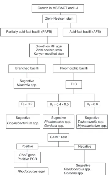
data strengthen the validity of the method and the use of COX molecular primers to identifyR. equi, especially when the CAMP test is positive. Thus, as suggested by Ladrón et al., the presence of thechoEgene identified by a specific PCR method should be considered in the rapid and correct identification of R. equi, differentiating it from other pathogenic actinomycetes. Results of the investigation of the 78 isolates from sputum suggest a simplified identification scheme forR. equi, as a first step in the search for partially acid-fast bacilli based on the determination of mycolic-acids by TLC technique, followed by the CAMP test and PCR using the COX primers that amplify thechoEgene (Fig. 3).
According to the literature, R. equi is a frequent cause of cavitary infections in immunocompromised patients, but extra-pulmonary infections in immunocompetent patients have also occurred. Clinical symptoms may be insidious and resemble those of mycobacterial infections, fungi, and other actinomycetes.R. equishould always be suspected when cul-tures, especially those for tuberculosis, show colonies with distinct morphology and microscopic observations after Gram
Growth in MB/BACT and LJ
Ziehl-Neelsen stain
Partially acid-fast bacilli (PAFB) Acid-fast bacilli (AFB)
Pleomorphic bacilli Growth on MH agar
Ziehl-neelsen stain Kunyon-modified stain
TLC
Rf< 0.2 Rf> 0.6
CAMP Test
Positive Negative
Sugestive Rhodococcus spp.
Gordona spp.
Tsukamurella spp. Mycobacterium spp. Corynebacterium spp.
ChoE gene Positive PCR
Rhodococcus equi Rhodococcus spp.
Gordona spp. Rf = 0.4 - 0.5 Sugestive
Nocardia spp. Branched bacilli
Sugestive Sugestive
Sugestive
Fig. 3 – Flowchart ofRhodococcus equiidentification.
stain indicates the presence of Gram-positive pleomorphic coccobacilli. Staining of culture smears by the Ziehl-Neelsen Kinyoun-modified method and microscopic observations of partially acid-fast microorganisms strengthen a positive iden-tification.
In many instances, identification ofM. tuberculosisin rou-tine clinical laboratory procedures can be faulty and lead the physician to start empirical treatment of tuberculosis, leaving a patient with untreated rhodococcosis. This study could be an alert to healthcare professionals about the importance of identifyingR. equi, even presumptively, in human isolates.
It is concluded that the identification ofR. equi can be achieved by combining the three methods in an optimized algorithm (Fig. 3). Cultures and phenotypic identification including TLC should be considered as the gold standard method to detect the presence of this bacterial agent in the investigated clinical sample. Phenotypic tests and TLC should be performed on primary cultures to suggest presence of
Rhodococcusspp., and implementation of the CAMP test should
be used for presumptive R. equi identification. PCR should be considered an alternative or supplement method to pro-vide highly specific and sensitive results that can be obtained faster. More rapid and accurate identification of the organ-ism provides for correct treatment and faster recovery of the patient.
Conflict of interest
All authors declare to have no conflict of interest.
r e f e r e n c e s
1. Navas J, Gonzalez-Zorn B, Ladrón N, et al. Identification and mutagenesis by allelic exchange of ChoE, encoding a cholesterol oxidase from the intracellular pathogen Rhodococcus equi. J Bacteriol. 2001;183:4796–805.
2. Kedlaya I.Rhodococcus equi. [cited 12 Mar 2009]. Available from:http://emedicine.medscape.com/article/235466 -Updated: Sep 12, 2008.
3. Weinstock DM, Brown AE.Rhodococcus equi: an emerging pathogen. Clin Infect Dis. 2002;34:1379–85.
4. McNeil MM, Brown JM. The medically important aerobic actinomycetes: epidemiology and microbiology. Clin Microbiol Rev. 1994;7:357–417.
5. Brown JM, McNeil MM, Desmond EP.Nocardia,Rhodococcus, Gordona,Actinomadura,Streptomyces, and other actinomycetes of medical importance. In: Murray PR, Baron EJ, Pfaller MA, Tenover FC, Yolken RH, editors. Manual of Clinical microbiology. 7th ed. Washington, DC: ASM Press; 1999. p. 370–98.
6. Prescott JF.Rhodococcus equi: an animal and human pathogen. Clin Microbiol Rev. 1991;4:20–34.
7. Amat GC. Detección y caracterización por métodos fenotípicos y moleculares de mycolata formadores de espumas en estaciones depuradoras de aguas residuales domésticas con sistemas de fangos activos. PhD Thesis. Valencia: Departamento de Biotecnología, Universidad Politécnica de Valencia; 2004.
8. Miyaji M, Yamamoto K, Moretti-Branchini ML, et al. Actinomicetos. In: Fungos patogênicos – guia prático de laboratório. v. 1. Campinas-SP. Brasil: JICA/UNICAMP; 2002, 65–75.
9. Takai S, Morishita T, Nishio Y, et al. Evaluation of a monoclonal antibody-based colony blot test for rapid identification of virulentRhodococcus equi. J Vet Med Sci. 1994;56:681–4.
10. Takai S, Ikeda T, Sasaki Y, et al. Identification of virulent Rhodococcus equiby amplification of gene coding for 15- to 17-kilodalton antigens. J Clin Microbiol. 1995;33:1624–7. 11. Takai S, Imai Y, Fukunaga N, et al. Identification of
virulence-associated antigens and plasmids inRhodococcus equifrom patients with AIDS. J Infect Dis. 1995;172:1306–11.
12. Takai S, Hines SA, Sekizaki T, et al. DNA sequence and comparison of virulence plasmids fromRhodococcus equiATCC 33701 and 103. Infect Immun. 2000;68:6840–7.
13. Ladrón N, Fernández M, Agüero J, et al. Rapid identification of Rhodococcus equiby a PCR assay targeting the ChoE gene. J Clin Microbiol. 2003;41:3241–5.
14. Silva P, Miyata M, Sato DN, et al.Rhodococcus equiisolation from sputum of patients with suspected tuberculosis. Mem Inst Oswaldo Cruz. 2010;105:199–202.
15. Balows A, Hausler Jr WJ, Herrmann KL, Isenberg HD, Shadomy HJ. Manual of clinical microbiology. 5th ed Washington DC: ASM Press; 1991.
16. Silva P. Valorizac¸ão do diagnóstico laboratorial, na identificac¸ão deRhodococcus equiisolado do escarro de pacientes suspeitos de tuberculose. PhD Thesis. Faculdade de Ciências Farmacêuticas De Araraquara, Universidade Estadual Paulista “Julio De Mesquita Filho”: São Paulo; 2009. 17. Bille J, Doyle MP. Listeria and Erysipelothrix. In: Balows A,
Hausler Jr WJ, Herrmann KL, Isenberg HD, Shadomy HJ, editors. Manual of clinical microbiology. 5th ed. Washington DC: ASM Press; 1991. p. 287–95.
18. Telenti A, Marchesi F, Balz M, et al. Rapid identification of mycobacteria to the species level by polymerase chain reaction and restriction enzyme analysis. J Clin Microbiol. 1993;31:175–8.
19. Van Soolingen D, Hermans PWM, De Haas PEW, et al. Occurrence and stability of insertion sequences in
Mycobacterium tuberculosiscomplex strains: evaluation of an insertion sequence-dependent DNA polymorphism as a tool in the epidemiology of tuberculosis. J Clin Microbiology. 1991;29:2578–86.
20. Bizet C, Barreau C, Harmant C, et al. Identification of Rhodococcus,GordonaandDietziaspecies using carbon source utilization tests (“biotype-lOO” strips). Res Microbiol. 1997;148:799–809.
21. Fiss E, Brooks GF. Use of a siderophore detection medium, ethylene glycol degradation, and 3-galactosidase activity in the early presumptive differentiation ofNocardia,Rhodococcus, Streptomyces, and rapidly growingMycobacteriumSpecies. J Clin Microbiol. 1991;29:1533–5.
22. Flores M, Ford EG, Janda JM. Value of the O – nitrophenyl – p – D -galactopyranoside test to differentiate among the aerobic actinomycetes. J Clin Microbiol. 1990;28:2142–4.
23. Mac Lachlan J, Wotherspoon AT, Ansell RO, et al. Cholesterol oxidase: sources, physical properties and analytical applications. J Steroid Biochem Mol Biol. 2000;72: 169–95.
24. Kirschner P, Bottger E. Micro-heterogeneity within RNA of Mycobacterium gordonae. J Clin Microbiol. 1992;30:
1049–50.
25. Butler WR, Floyd MM, Brown JM, et al. Novel mycolic acid-containing bacteria in the family Segniliparaceae fam. nov., including the genusSegniliparusgen. nov., with descriptions ofSegniliparus rotundussp. nov. andSegniliparus rugosussp. nov. Int J Syst Evol Microbiol. 2005;55:
1615–24.
26. Goodfellow M, Maldonado LA. The families Dietziaceae, Gordoniaceae, Nocardiaceae and Tsukamurellaceae. In: Dworkin M, Falkow S, Rosenberg E, Schleifer KH, Stackebrandt E, editors. The prokaryotes. A handbook on the biology of bacteria, 3, 3rd ed. New York: Springer; 2006. p. 843–88. 27. Soddell JA, Stainsby FM, Eales KL, et al.Millisia brevisgen.
nov., sp. nov., an actinomycete isolated from activated sludge foam. Intern J System Evolut Microbiol. 2006;56:739–44. 28. Adachi K, Katsuta A, Matsuda S, et al.Smaragdicoccus
niigatensisgen. nov., sp. nov., a novel member of the suborder Corynebacterineae. Int J Syst Evol Microbiol. 2007;57:
29. Katayama T, Kato T, Tanaka M, et al.Tomitella biformatagen. nov., sp. nov., a new member of the suborderCorynebacterineae isolated from a permafrost ice wedge. Int J Syst Evol
Microbiol. 2010;60:2803–7.
30. Brandão PFB, Clapp JP, Bull AT. Discrimination and taxonomy of geographically diverse strains of nitrile-metabolizing actinomycetes using chemometric and molecular sequencing techniques. Environ Microbiol. 2002;4:262–76.
31. McMinn EJ, Alderson G, Dodson HI, et al. Genomic and phenomic differentiation ofRhodococcus equiand related strains. Anton Leeuw Int J G. 2000;78:331–40.
32. Fox GE, Wisotzkey JD, Jurtshuk P. How close is close: 16S rRNA sequence identity may not be sufficient to guarantee species identity. Int J Syst Bacteriol. 1992;42:166–70.
33. Patel R, Piper KE, Rouse MS, et al. Determination of 16S rRNA sequences of enterococci and application to species identification of nonmotileEnterococcus gallinarumisolates. J Clin Microbiol. 1998;36:3399–407.