Orthopaedics and Traumatology Department, Hospital Regional do Paraná, State University of Londrina – Londrina/PA, Brazil.
Orthopaedics and Traumatology Institute, Hospital das Clinicas, São Paulo University Medical School – São Paulo/SP, Brazil.
Email: [email protected]
Received for publication on June 09, 2005. Accepted for publication on August 25, 2005.
ORIGINAL RESEARCH
ACCURACY OF MAGNETIC RESONANCE IN
IDENTIFYING TRAUMATIC INTRAARTICULAR KNEE
LESIONS
Carlos Eduardo Sanches Vaz, Olavo Pires de Camargo, Paulo José de Santana, and Antonio Carlos Valezi
Vaz CES, Camargo OP de, Santana PJ de, Valezi AC. Accuracy of magnetic resonance in identifying traumatic intraarticular knee lesions. Clinics. 2005; 60(6):445-50.
PURPOSE: To evaluate the diagnostic accuracy of magnetic resonance imaging of the knee in identifying traumatic intraarticular knee lesions.
METHOD: 300 patients with a clinical diagnosis of traumatic intraarticular knee lesions underwent prearthoscopic magnetic resonance imaging. The sensitivity, specificity, positive predictive value, negative predictive value, likelihood ratio for a positive test, likelihood ratio for a negative test, and accuracy of magnetic resonance imaging were calculated relative to the findings during arthroscopy in the studied structures of the knee (medial meniscus, lateral meniscus, anterior cruciate ligament, posterior cruciate ligament, and articular cartilage).
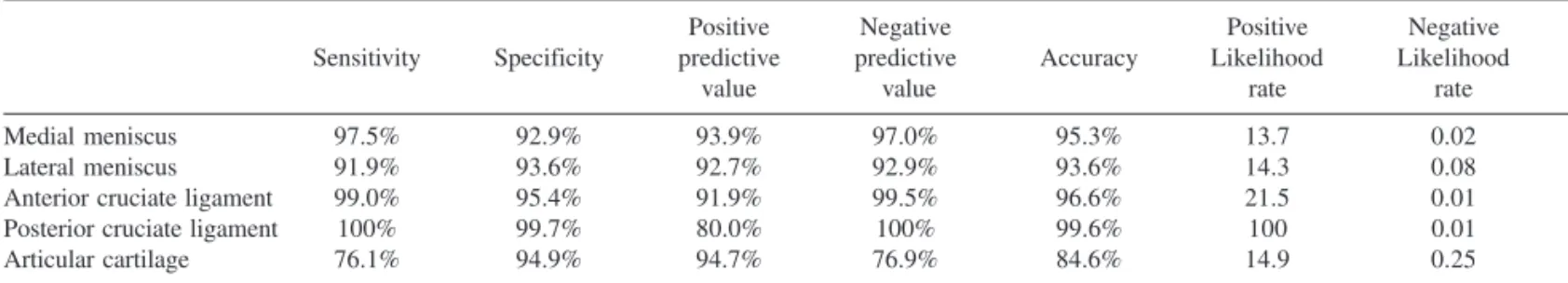
RESULTS: Magnetic resonance imaging produced the following results regarding detection of lesions: medial meniscus: sensitivity 97.5%, specificity 92.9%, positive predictive value 93.9%, positive negative value 97%, likelihood positive ratio 13.7, likelihood negative ratio 0.02, and accuracy 95.3%; lateral meniscus: sensitivity 91.9%, specificity 93.6%, positive predictive value 92.7%, positive negative value 92.9%, likelihood positive ratio 14.3, likelihood negative ratio 0.08, and accuracy 93.6%; anterior cruciate ligament: sensitivity 99.0%, specificity 95.9%, positive predictive value 91.9%, positive negative value 99.5%, likelihood positive ratio 21.5, likelihood negative ratio 0.01, and accuracy 96.6%; posterior cruciate ligament: sensitivity 100%, specificity 99%, positive predictive value 80.0%, positive negative value 100%, likelihood positive ratio 100, likelihood negative ratio 0.01, and accuracy 99.6%; articularcartilage: sensitivity 76.1%, specificity 94.9%, positive predictive value 94.7%, positive negative value 76.9%, likelihood positive ratio 14.9, likelihood negative ratio 0.25, and accuracy 84.6%.
CONCLUSION: Magnetic resonance imaging is a satisfactory diagnostic tool for evaluating meniscal and ligamentous lesions of the knee, but it is unable to clearly identify articular cartilage lesions.
KEYWORDS: Magnetic resonance. Knee. Arthroscopy. Traumatology.
Due to its anatomical configuration and because of its being the biggest joint of the human body, the knee is fre-quently subjected to direct trauma that can result in inju-ries of variable gravity.1–5.
Intraarticular knee lesions are associated with
signifi-cant morbidity and frequently need surgical treatment and extensive rest. Although they are common, their correct di-agnosis still is a challenge.6–8
Clinical tests may be confusing, and delay in diagno-sis can result in social and economic problems and some-times in a worse prognosis.8–10 Therefore, complementary diagnostic tools are often necessary,11–14 mainly when sus-picion of multiple lesions exists.15,16
hospitalization and regional or general anesthesia, thus pre-senting all the potential complications of an open surgical procedure.21–27
During the last decade, magnetic resonance imaging has been confirmed as the ideal approach for primary diagno-sis of traumatic intraarticular knee lesions.28–31 It is noninvasive, fast, can be done on an outpatient basis, and is free of complications. Despite this, magnetic resonance imaging is a new technology, 32 which means that clinical experience is still lacking. Additionally, there are doubts about the accuracy of magnetic resonance imaging and the clinical advantages of this test, since it is still an expen-sive procedure.33–36
With the purpose of investigating the accuracy of mag-netic resonance imaging in patients with clinical signs of traumatic intraarticular knee lesions, we compared its find-ings with those obtained from the subsequent arthroscopies.
METHODS
Design: Diagnostic test evaluation.
Setting: Department of Orthopedics and Traumatology, University of Londrina, Londrina, Brazil.
Participants: A population of 300 consecutive patients with clinical signs of traumatic intraarticular knee lesions examined from August 1998 through March 2002 who un-derwent prearthoscopic magnetic resonance imaging where included in the study. Patients with previous knee injuries and knee surgery where excluded.
Procedures:
Magnetic Resonance:
All the exams were performed in the same diagnostic imaging center with blinded interpretation by 3 radiology specialists in magnetic resonance imaging. A Phillips de-vice model NT5 with magnetic-field strength of 0.5 TESLA was used, along with a special knee bobbin. The magnetic resonance imaging sequences were TT2; coronal SE-T1, TSE-T2, SPIR, and TSE-T2 oblique; and coronal and sagittal GRE-T2 (special sequences for the meniscus).
Arthroscopy:
The arthroscopies were all done in a hospital environ-ment, with complete preoperative care, with most them be-ing outpatient surgery. A Stryker 3 CCD video camera was used with a 4 mm Karl-Storz arthroscope with a 30-degree angle. Standard arthroscopic portals were used: the superomedial portal for fluid outflow, the inferolateral por-tal for the arthroscope, and the inferomedial porpor-tal for
in-strumentation. During arthroscopy, a systematic examina-tion of the knee was performed with a complete evalua-tion of the joint. All arthroscopies where done by the same surgeon and filmed on appropriate tapes.
Data Analysis:
The intraarticular structures included in the study were the medial meniscus, the lateral meniscus, the anterior and posterior cruciate ligaments, and the articular cartilage. All these structures were evaluated to identify lesions, and the results of the magnetic resonance imaging were compared with the arthroscopies (considered the gold standard for di-agnosis). The following were calculated as defined in Fig-ure 1 using Epi Info 6, V 6.04 software: sensitivity, specificity, accuracy, positive predictive value, negative pre-dictive value, likelihood ratio for a positive test, likelihood ratio for a negative test, and the 95% confidence intervals.
RESULTS
Results for all the measured parameters are presented in Table 1. It can be seen that MRI has very high levels of sensitivity, specificity, positive predictive value, positive-negative value and accuracy for meniscal and ligamentous lesions. Likelihood of positive and negative ratio was also excellent. For articular cartilage lesions, results were not nearly as precise.
DISCUSSION
While clinical data remain the most important tool for identifying intraarticular knee lesions, sometimes it is in-sufficient to elucidate the final diagnosis.
Until the last decade, diagnostic arthroscopy was the only possible way to clarify a doubtful diagnosis. Unfor-tunately, it is an invasive and expensive procedure, and its overuse has produced unnecessary complications, such as infection, neurovascular lesions, damaged intraarticular materials, amongst others.
With the evolution of the materials and surgical tech-niques, arthroscopy has become more of a surgical method than a diagnostic tool, and magnetic resonance is fast be-coming the favorite diagnostic method for many of sur-geons52–54.
Table 1 - Results of the data analysis: sensitivity, specificity, positive predictive value, negative predictive value, positive likelihood rate, negative likelihood rate, and accuracy of magnetic resonance imaging to evaluate lesions of the medial meniscus, lateral, meniscus, anterior cruciate ligament, posterior cruciate ligament, and articular cartilage
Positive Negative Positive Negative
Sensitivity Specificity predictive predictive Accuracy Likelihood Likelihood
value value rate rate
Medial meniscus 97.5% 92.9% 93.9% 97.0% 95.3% 13.7 0.02
Lateral meniscus 91.9% 93.6% 92.7% 92.9% 93.6% 14.3 0.08
Anterior cruciate ligament 99.0% 95.4% 91.9% 99.5% 96.6% 21.5 0.01
Posterior cruciate ligament 100% 99.7% 80.0% 100% 99.6% 100 0.01
Articular cartilage 76.1% 94.9% 94.7% 76.9% 84.6% 14.9 0.25
A = number of true positive results; B= number of false positive results C = number of false negative results; D= number of true negative results
S = Sensitivity; E = Specificity
Willians49 performed a study in which magnetic reso-nance imaging scans were performed on 69 patients wait-ing for knee arthroscopy. All patients had a clinical diag-nosis of traumatic intraarticular knee lesion. Of the patients scanned, magnetic resonance imaging ruled out lesions in 24 patients, who were removed from the waiting list. Af-ter 9 months, only 1 of them had been re-listed for thera-peutic arthroscopy because of continued symptoms.
Since magnetic resonance imaging results in a fast and accurate diagnosis, it allows the surgeon time to plan the surgical procedure prior to surgery for treatment, whereas diagnostic arthroscopy necessitates immediate treatment, without previous study.
Although knee magnetic resonance is still considered an expensive tool, with costs ranging from US $250 to US $500, the total cost of arthroscopy is far greater, ranging from US $1500 to US $3000. Weinstabl et al, 48 studying the cost-benefit of knee magnetic resonance, evaluated 201patients with clinical signs of knee meniscal lesions who later had undergone arthroscopy. They report that 30% of the diagnoses were false-positives, and that 30% fewer arthroscopies would have resulted in an economy of US $723,600 dollars. The total cost of knee magnetic resonance was US $160,800, and magnetic resonance offered a com-parative accuracy of 96% against 78% for clinical exami-nation.
Bui-Mansfield et al46 performed a study to ascertain
whether there would be a significant economy if magnetic resonance to complement the clinical examination was done in all cases for which a diagnostic arthroscopy was indicated, using a value of US $1000 dollars for each mag-netic resonance procedure. Of 50 diagnostic arthroscopies, 42% had been unnecessary (false-positive results). They observed that if the results of the magnetic resonance had been taken into account before the performance of the arthroscopy, there would have been an economy of US $680 dollars for each case.
Our study demonstrates that magnetic resonance does not appear to have a satisfactory accuracy for diagnosing knee articular cartilage lesions, since it was associated with a great number of false negative results (low sensitivity). It has been proposed that enhancement of the magnetic resonance imaging accuracy with articular cartilage lesions is obtained by the introduction of a special contrast in the knee.50 This procedure is called resonance (or arthro-MRI), and recent studies have demonstrated this innova-tion to have good accuracy.51
CONCLUSION
Magnetic resonance imaging has high accuracy to for diagnosing knee meniscal and cruciate ligament lesions, but does not have satisfactory accuracy in detecting articular cartilage lesions.
RESUMO
Vaz CES, Camargo OP de, Santana PJ de, Valezi AC. Acurácia da ressonância magnética para identificar lesões traumáticas intra-articulares do joelho. Clinics. 2005; 60(6):445-50.
OBJETIVO: Avaliar a validade da ressonância magnética do joelho no diagnóstico das lesões intra-articulares traumáticas do joelho.
MÉTODO: População de 300 pacientes, com quadro clínico sugestivo de lesões intra-articulares traumáticas do joelho, que tiveram seus laudos de ressonância magnética comparados com os resultados obtidos nas artroscopias realizadas posteriormente. Foram calculados a sensi-bilidade, especificidade, valor preditivo positivo, valor preditivo negativo, razão de verossimilhança positiva, razão de verossimilhança negativa e acurácia da ressonância magnética do joelho para o diagnóstico de lesões em cada estrutura intra-articular estudada do joelho (menisco me-dial, menisco lateral, ligamento cruzado anterior, ligamento
cruzado posterior e cartilagem articular).
100%, a especificidade de 99%, o valor preditivo positivo de 80%, o valor preditivo negativo de 100%, a razão de verossimilhança positiva de 100, a razão de verossimilhança negativa de 0.01 e a acurácia de 99.6%. Para as lesões condrais a sensibilidade da ressonância magnética foi de 76.1%, a especificidade de 94.9%, o valor preditivo positivo de 94.7%, o valor preditivo negativo de 76.9%, a razão de verossimilhança positiva de 14.9, a razão de
verossimi-lhança negativa de 0.25 e a acurácia de 84.6%.
CONCLUSÃO: A ressonância magnética apresenta alta acurácia para identificar as lesões meniscais e ligamentares do joelho, mas é insatisfatória para diagnosticar as lesões da cartilagem articular.
PALAVRAS-CHAVE: Ressonância magnética. Joelho. Artroscopia. Traumatologia.
REFERENCES
1. Bruns W, Maffulli N. Lower limb injuries in children in sports. Clin Sports M ed. 2000;19(4):1-18.
7. Cohen M, Abdallla RJ, Ejnisman B, Amaro JT. Lesões ortopédicas no futebol. Rev Bras Ortop. 1997;32(12):940-4.
8. Hernandez AJ, Vieira EA. Função dos ligamentos na estabilização do joelho. In: Camanho GL, ed. Patologia do joelho. São Paulo: Sarvier; 1996. p.2-4.
9. Insall JN. Examination of the knee. In: Insall JN, editor. Surgery of the knee. 2nd ed. New York: Churchill Livingstone; 1984. p.63.
10. Kannus P, Jarvinen M. Conservatively treated tears of the anterior cruciate ligament. Long-term result. J Bone Joint Surg Am. 1987;69:1007-12.
6. Adalberth T, Roos H, Lauren M, Akeson P, Sloth M, Jonsson K, et al. Magnetic resonance imaging, scintilography, and arthroscopy evaluation of traumatic hemarthrosis of the knee. Am J Sports Med. 1997;25(2):231-7.
7. Camanho GL, Viegas AC. Avaliação da reconstrução do ligamento cruzado anterior em doentes com idade acima de 45 anos. Rev Bras Ortop. 2001;36(1-2):37-40.
8. Oberlander MA, Shalvoy RM, Hughston JC. The accuracy of the clinical knee examination documented by arthroscopy. A prospective study. Am J Sports Med. 1993;21(6):773-8.
9. O’ Shea KJ, Murphy KP, Heekin RD, Herzwurm PJ. The diagnostic accuracy of history, physical examination, and radiographs in the evaluation of traumatic knee disorders. Am J Sports Med. 1996;24(2):164-8.
10. Passariello R, Trecco F, de Paulis F, Bonanni G, Masciocchio C. Computed tomography of the knee joint: clinical results. J Comput Assist Tomogr. 1983;7:1043-9.
11. Manco LG, Berlow ME, Czajka J, Alfred R. Diagnosis of meniscal tears using high-resolution computed tomography. J Bone Joint Surg Am. 1987;69:498-502.
12. Verdonk R, Meire D, Van de Velde C, de Meulemeester C, Van Eteetvelde G, Claessens H. CT scan of the knee: correlation with clinical and arthroscopic findings. Acta Orthop Scand. 1991;57(Supl):49-55. 13. White LM, Schweitzer ME, Deely DM. The effect of training and
experience on the magnetic resonance imaging interpretation of meniscal tears. Arthoscopy. 1997;13:224-8.
14. Yazaki CM, Assis JR, Cundari AMMV. Estudo comparativo entre tomografia computadorizada e artroscopia nas lesões meniscais do joelho. Rev Bras Ortop. 1995;30(6):409-16.
15. Gray SD, Kaplan PA, Dussault RG. Imaging of the knee. Orthop Clin North Am. 1997;28(4):643-57.
16. Severino NR, Camargo OPA, Aihara T, Cury RPL, Oliveira VM, Vaz CES, et al. Comparação entre a ressonância magnética e a artroscopia no diagnóstico das lesões do joelho. Rev Bras Ortop. 1997;32(4):275-8.
17. Dandy DJ, Jackson RW. Diagnosis of internal derangements of the knee. The role of arthroscopy. J Bone Joint Surg Br. 1975;57:346-8. 18. De Haven KE, Collins HR. Diagnosis of internal derangements of the
knee. J Bone Joint Surg Am. 1975;57:802-10.
19. Fischer SP, Fox JM, Pizzo W, Friedman MJ, Snyder SJ, Ferkel RD. Accuracy of diagnosis from magnetic resonance imaging of the knee. J Bone Joint Surg Am. 1991;73(1):2-10.
20. Halbrecht JL, Jackson DW. Office arthroscopy: a diagnostic alternative. Arthroscopy. 1992;8:320-6.
21. Affonseca A. Artroscopias desnecessárias. Rev Bras Ortop. 1997;32(4):255.
22. Carmichael IW, Macleod AM, Travlos J. MRI can prevent unnecessary arthroscopy. J Bone Joint Surg Br. 1997;79(4):624-5.
23. Gillies H, Seligson D. Precision in the diagnosis of meniscal lesions: a comparison of clinical evolution, arthrography, and arthroscopy. J Bone Joint Surg Am. 1979;61:343-6.
24. McGinty JB. Complications of arthroscopy and arthroscopic surgery. In: Parisien JS, editor. Arthroscopic surgery. New York: MacGray-Hill; 1988. p. 221-30.
25. Patel D. Complications in arthroscopic surgery. In: Sherman OH, Minkoff J, editors. Current management of complications in orthopedics: arthroscopic surgery. Baltimore: Williams and Wilkins; 1990. p. 104-113.
26. Sherman OH, Fox JM, Snyder SJ. Arthroscopy - “no problem surgery”. An analysis of complications in two thousand six hundred and forty cases. J Bone Joint Surg Am. 1986;68:256-65.
28. Rappeport ED, Wieslander SB, Stephensen S, Launten GS, Thomsen HS. MRI preferable to diagnostic arthroscopy in knee joint injuries. A double-blind comparation of 47 patients. Acta Orthop Scand. 1997;68(3):277-81.
29. Reicher MA, Rauschning W, Gold RH, Bassett LW, Lufkin RB, Glen WJ. High-resolution magnetic resonance imaging of the knee joint: normal anatomy. Am J Roentgenol. 1985;145:895-902.
30. Spiers AS, Meagher T, Ostlere SJ, Wilson DJ, Dodd CA. Can MRI of the knee affect arthroscopy practice? A prospective study of 58 patients. J Bone Joint Surg Br. 1993;75(1):49-52.
31. Stanitiski CL. Correlation of arthroscopic and clinical examinations with magnetic resonance imaging findings of injured knees in children and adolescents. Am J Sports Med. 1998;26(1):2-7.
32. Riel KA, Reinisch M, Kersting-Sommerhoff B, Holf N, Merl T. 0.2-Tesla magnetic resonance imaging of internal lesions of the knee joint: a prospective arthroscopically controlled clinical study. Knee Surg Sports Traumatol Arthrosc. 1999;7(1):37-41.
33. Alioto RJ. The influence of MRI on treatment decisions regarding knee injuries. Am J Knee Surg. 1999;12(2):91-7.
34. Gelb HJ, Glasgow SG, Sapega AA, Torg JS. Magnetic resonance imaging of knee disorders: clinical value and cost-effectiveness in a sports medicine practice. Am J Sports Med. 1996;24(1):99-104.
35. Khanna AJ, Cosgarea AJ, Mont MA, Andres BM, Domb BG, Evans PJ, et al. Magnetic resonance imaging of the knee. Current techniques and spectrum of disease. J Bone Joint Surg Am. 2001;83(Suppl 2, Pt 2):128-41.
36. Mackenzie R, Palmer CR, Lomas DJ, Dixon AK. Magnetic resonance imaging of the knee: diagnostic performance statistics. Clin Radiol. 1996;51(4):251-7.
37. Crabtree SD, Bedford AF, Edgar MA. The value of arthrography and arthroscopy in association with a sports injuries clinic: a prospective and comparative study of 182 patients. Injury. 1981;13:220-6. 38. De Smet AA, Norris MA, Yandow DR, Graf BK, Keene JS. Diagnosis
of meniscal tears of the knee with MR imaging: effect of observer variation and sample size on sensitivity and specificity. Am J Roentgenol. 1993;160(3):555-9.
39. Munshi M, Davidson M, MacDonald PB, Froese W, Sutherland K. The efficacy of magnetic resonance imaging in acute knee injuries. Clin J Sports Med. 2000;10(1):34-9.
40. Sherman PM, Penrod BJ, Lane MJ, Ward JA. Comparison of knee magnetic resonance imaging findings in patients referred by orthopaedic surgeons versus nonorthopaedic practitioners. Arthroscopy. 2002;18(2):201-5.
41. Elvenes J, Jerome CP, Reikeras O, Johansen O. Magnetic resonance imaging as a screening procedure to avoid arthroscopy for meniscal tears. Arch Orthop Trauma Surg. 2000;120(1-2):14-6.
42. Rangger C, Klestil T, Kathrein A, Inderster A, Hamid L. Influence of magnetic resonance imaging on indications for arthroscopy of the knee. Clin Orthop. 1996;330:133-42.
43. Rappeport ED, Mehta S, Wieslander SB, Schwartz LG, Thomsen HS. MR imaging before arthroscopy in knee joint disorders? Acta Radiol. 1996;37(5):602-9.
44. Trieshmann HWJ. The impact of magnetic resonance imaging of the knee on surgical decision making. Arthroscopy. 1996;12(5):550-5. 45. Bryan S, Weatherburn G, Bungay H, Hatrick C, Salas C, Parry D, et al.
The cost-effectiveness of magnetic resonance imaging for investigation of the knee joint. Health Technol Assess. 2001;5(20):1-5.
46. Bui-Mansfield LT. Potential cost savings of MR imaging obtained before arthroscopy of the knee: evolution of 50 consecutive patients. Am J Roentgenol. 1997;168(4):913-8.
47. Suarez-Almazor ME, Kaul P, Kendall CJ, Saunders LD, Johnston DWC. The cost effectiveness of magnetic resonance imaging for patients with internal derangement of the knee. Int J Technol Assess Health Care. 1999;15(2):392-405.
48. Weinstabl R, Muellner T, Vecsei V, Kainberger F, Kramer M. Economic considerations for the diagnosis and therapy of meniscal lesions: can magnetic resonance help reduce the expense? World J Surg. 1997;21(4):363-8.
49. Williams RL, Williams LA, Watura R, Fairclough JA. Impact of MRI on a knee arthroscopy waiting list. Ann R Coll Surg Engl. 1996;78(5):450-2.
50. Munk B, Lundorf E, Staunstrup H, Staunstrup H, Schmidt SA, Bolvig L, et al. Clinical magnetic resonance imaging and arthroscopic findings in knees: a comparative prospective study of meniscus anterior cruciate ligament and cartilage lesions. Arthroscopy. 1998;14(2):171-5. 51. Zairul-Nizan ZF, Hyzan MY, Gobinder S, Razak MA. The role of
preoperative magnetic resonance imaging in internal derangement of the knee. Med J Malaysia. 2000;55(4): 433-8.
52. Santos-Machado TM, Oliveira CRM, Croci AT, Fernandes A, Abadi, Baptista MDAM, Camargo OP. Parosteal osteosarcoma with myocardial metastasis 13 years after follow-up. Rev Hosp Clin Fac Med S Paulo. 2003;58:113-118.
53. Etchebehere M, Camargo OP, Croci AT Oliveira CRCM, Batista AM. Relationship between surgical procedure and outcome for patients with grade I chondrosarcomas. Clinics. 2005;60:121-126.