Total phenolic,
fl
avonoid, alkaloid and iridoid content
and preventive effect of Lider-7-tang on
lipopolysaccharide-induced acute lung injury in rats
Ch. Erdenechimeg
1,3, A. Guiqide
2, B. Dejidmaa
1, Ch. Chimedragchaa
1and S. Purevsuren
3 1Institute of Traditional Medicine and Technology, Ulaanbaatar, Mongolia
2
The Inner Mongolia Autonomous Region International Mongolian Hospital, HuhHot, Inner Mongolia, China
3
School of Pharmacy, Mongolian National University of Medical Sciences, Ulaanbaatar, Mongolia
Abstract
Lider-7-tang, a medicine used for the treatment of respiratory diseases especially pneumonia and fever in Mongolian Traditional Medicine, was selected for this phytochemical and pharmacological study. The objectives of the study were to determine total biological active substances and analyze the effects of Lider-7-tang treatment in rats with acute lung injury (ALI). Quantitative determination of the total active constituents (phenolic,flavonoid, iridoid and alkaloid) of the methanol extract of Lider-7-tang was performed using Folin-Ciocalteu reagent, aluminum chloride reagent, Trim-Hill reagent, and Bromocresol green reagent, respectively. A total offifty 8–10-week-old male Wistar rats (200–240 g) were randomized into three groups: control group,
lipopolysaccharide (LPS) group (7.5 mg/kg) and LPS+Lider-7 group (90 mg/kg Lider-7-tang before LPS administration). The total content of alkaloids was 0.2±0.043%, total phenols 7.8±0.67%,flavonoids 3.12±0.206%, and iridoids 0.308±0.0095%.
This study also evaluated the effects of Lider-7 on levels of inflammatory mediators by observing histopathological features associated with LPS-induced ALI. The rats pretreated with Lider-7 had significantly lower levels of IL-6 (at 3 and 6 h), and TNF-a
(at 3, 6, 9, and 12 h). The current study showed that Lider-7 exerted a preventive effect against LPS-induced ALI, which appeared to be mediated by inhibiting the release of pro-inflammatory cytokines.
Key words: Traditional medicine; Lider-7-tang; Phenolic; Acute lung injury; Lipopolysaccharide
Introduction
Acute lung injury (ALI) is an acute inflammatory disease, characterized by excess production of inflammatory factors in lung tissue, and followed by non-cardiogenic dyspnea, severe hypoxemia, and pulmonary edema, thus leading to both high morbidity and mortality (1,2). A major cause of the development of ALI is sepsis, wherein Gram-negative bacteria are a prominent cause (3). The intraperitoneal injection of lipopolysaccharide (LPS), a component of the outer cell wall of most Gram-negative bacteria, mimics human Gram-negative ALI and is one of the most com-monly accepted models for ALI (4). Lipopolysaccharide, binding to its receptor, toll-like receptor 4, provokes the activation of a key pro-inflammatory transcription factor,
nuclear factorkB, which induces the expression of various
pro-inflammatory cytokines and chemokines, such as tumor
necrosis factor-a(TNF-a), interleukin-1b, and macrophage
inflammatory protein-2 (5). As a consequence of the strong inflammatory response, alveolar structures change,
endothelial and alveolar permeability increase and alveolar fluid clearance decreases, thus critically impairing lung function (3,6).
Lider-7-tang is one of the traditional Mongolian herbal
medicines consisting of seven herbs, Radix Sophoroe
alopecuroides, Radix Inulae helenium, Fructus
Garden-iae, Fructus Terminaliae billericae, Fructus T. chebulae,
Herba Gentianae barbatae and Herba Lagotis
integrifo-liae. Lider-7-tang has been used to treat cold and flu
symptoms such as nasal congestion, headache, body ache, fever, sore throat pain, and cough for a long time in Traditional Mongolian Medicine (7,8). Lider-7-tang has a light green color, has an odor, and tastes bitter, smooth, fatty and soft.
S. alopecuroidesL. shows a wide spectrum of pharma-cological activities, including detoxification, anti-bacterial, inflammatory, pain killing, asthma cough, and
anti-tumor, among others (9–12). There are many chemical
Correspondence: Ch.Erdenechimeg:<[email protected]>|<[email protected]>
constituents inS. alopecuroidesL., and the main bioactive
components of this plant are alkaloids, flavones, volatile
oils, and quinones. In the 1980’s, there were more than 20 kinds of alkaloids isolated and identified from S. alopecuroidesL., such as sophocarpine, matrine,
oxyma-trine, sophoridine, sophoramine etc. (13,14). S.
alopecur-oides L. contains quercetin, rutoside, isobavachin, glabol, trifolirhizin, ammthamnidin, vexibinol and vexibidin (14).
Three new flavonostilbenes (alopecurones M–O) were
isolated from the root bark ofS. alopecuroidesL. together
with 21 known compounds. All isolates were evaluated for their potential to inhibit LPS-induced nitric oxide
produc-tion in RAW 264.7 cells (15). S. alopecuroides L. has a
great effect as an anti-inflammatory. The main effective substances associated with anti-inflammatory activity are
considered the alkaloids ofS. alopecuroidesL. (11).
I. helenium L. has been investigated for pharmaco-logical benefits including antioxidant and anti-inflammatory activities, hepatoprotective characteristics, cytotoxicity,
and antimicrobial properties (16–18). Chemical analysis
of the rhizome and roots showed thatI. heleniumcontains
many bioactive compounds including polysaccharide inulin (up to 44%), essential oil with eudesmane-type sesquiterpene (up to 5%), lactones (mainly alantolactone and isoalantolactone), thymol derivatives, terpenes, and sterols (19,20).
Flavonoids are a group of polyphenolic compounds and exhibit several biological effects such as anti-hepatotoxic, anti-inflammatory and anti-ulcer activity. All
ingredients of Lider-7-tang containflavonoids and
pheno-lic compounds. For example, 5,7,3’,4’
-tetrahydroxyfla-vone, doismetin, apigenin, chrysoeriol, tilianin and luteolin,
etc. have been isolated from G. barbatae L (14). There
are iridoid glycosides Gardenia jasminoides Ells and
L. integrifolia.G. jasminoidesextracts and their main active phytoconstituents geniposide, genipin, crocin, crocetin have been reported for a wide range of pharmacological activities such as anti-hyperglycemic, anti-atherosclerotic, anti-inflam-matory, anti-arthritis, and anti-cancer etc. (21,22).
Gallic acid is a polyphenolic compound with anti-oxidant property. Gallic acid, a major constituent of T. bellirica(Barur),T. chebula(Arur), is useful for common colds and fever and has diuretic, laxative, liver tonic, refrigerant, stomachic, restorative, alterative, antipyretic, and anti-inflammatory effects (23,24).
Therefore, we postulated that Lider-7-tang could protect against LPS-induced lung injury. In the present study, we tested this hypothesis using a rat model of LPS-induced ALI.
Material and Methods
Plant materials
The crude herbal medicines from S. alopecuroides,
I. helenium, T. chebula, T. bellerica, G. jasminoideswere purchased from Traditional Drug Factory at the Institute of
Traditional Medicine and Technology (Mongolia).G. barbatae
andL. integrifoliawere collected from Khuvsgul, Mongolia in 2015. The origin of each herbal medicine was taxonomically confirmed by Prof. Ganbold E (Ulaanbaatar University, Ulaanbaatar, Mongolia).
Ethics statement
All experimental procedures performed in this study were in accordance with the Guide for the Care and Use of Laboratory Animals, proposed by the Institute of Tradi-tional Medicine and Technology. The study protocol was approved by the Biomedical Ethics Subcommittee of Mongolian National University of Medical Sciences, Mongolia.
Experimental animals
A total of fifty 8–10-week-old male Wistar rats (200–
240 g) were used in this study. All experimental animals were obtained from the Experiment Animal House, Institute of Traditional Medicine and Technology. The rats were housed in cages and maintained at room tempera-ture with a 12-h light/dark cycle. They were fed with
standard pellet diet and tap waterad libitum.
Reagent
Standards of gallic acid, rutin, oxymatrine and aucubin were obtained from Sigma-Aldrich (USA). Folin
Ciocal-teu’s phenol reagent and aluminum chloride (AlCl3) of
Sangon (China) were used in the study. All other solvents and chemicals were of analytical grade.
Escherichia coli055:B5 endotoxin from Sigma-Aldrich and the cytokine immunoassay kits from Shanghai MLBIO Biotechnology Co. Ltd. (China) were used in the study.
Chemical analysis
Sample preparation. Powdered medicine was pre-cisely weighed (1.0 g), extracted with 50 mL of 70%
ethanol in reflux for 30 min, andfiltrated. The supernatant
was used as the test solution.
Estimation of total flavonoid contents. The solution
was treated with 1 mL of 5% NaNO2, 1 mL of 10%
Al(NO3)3and 10 mL of 4% NaOH solution, and absorbance
values were determined using a spectrophotometer (UNICO UV-2102 C, China) at 500 nm. The content of flavonoids in extracts is reported as rutin equivalent (mg of RU/g of extract) (25).
Estimation of total polyphenolic compounds. The amount of total phenolics was determined using the Folin-Ciocalteu assay. The Folin-Folin-Ciocalteu reagent (diluted 1:10
in water) and aqueous Na2CO3 (10.75%) were
succes-sively added to the extract. In 30 min, the absorbance value was measured at 760 nm. Gallic acid was used to establish the calibration curve, and total polyphenolic content is reported as g/kg (26).
the reaction with bromocresol green (69.8 mg/mL) and absorbance was measured at 420 nm. Oxymatrine was used to establish the calibration curve, and total alkaloids content is reported as oxymatrine equivalent as g/kg (27). Determination of total iridoids. The content of iridoids was determined according to the colorimetric method based on a Trim-Hill reaction. Each extract (0.4 mL) was mixed with 4 mL of Trim-Hill reagent (acetic acid-0.2%
CuSO4-conc. HCl, 10:1:0.5), afterward absorbance was
measured at 609 nm, and the blue color indicated the presence of iridoids. The amount of iridoids was calculated
using aucubin (0.1–1 mg/mL) calibration curve. Results are
reported as the mean value of 3 replicates (28).
Preventive effect of Lider-7-tang on LPS-Induced ALI in rats
Experimental protocols. Rats were randomized into three groups: control group (n=10), LPS group (n=20), in which LPS (7.5 mg/kg dissolved in 0.5 mL sterile saline)
was administered by an intravenous injection (iv) via the
tail vein; and LPS+Lider-7 group (n=20), in which Lider-7 (90 mg/kg, orally) was administered 30 min before injec-tion of LPS (7.5 mg/kg dissolved in 0.5 mL sterile saline,
iv) orally. Rats were euthanized with an overdose of
sodium pentobarbital (100 mg/kg,ip). Lung tissue
speci-mens and blood samples were then obtained for further analysis (29).
Histological analysis
Twelve hours after LPS administration, the rats were euthanized (n=5, 3, and 5 in the control, LPS, and LPS
+Lider-7 groups, respectively). The obtained lung tissue
specimens were fixed with 10% formalin, embedded in
paraffin, cut into 5-mm thick sections and mounted onto slides. The sections were then stained with hematoxylin and eosin (H&E) according to the standard staining method (30). Histologic changes were graded by a pathologist blind to the clinical status of the rats. Then the lung tissue samples were scored for the degree of intra-alveolar edema, intra-alveolar hemorrhage, and neutrophil infiltration using grades 0 to 4 (0, absent; 1, mild; 2, moderate; 3, severe; 4, overwhelming) with a maximum score of 12, as described previously (31).
Wet-to-dry weight ratio
After the animals were euthanized at 12 h, the chest cavity was opened and the right lung was ligated and excised. The lung specimen was then briefly rinsed in phosphate buffered saline (PBS), blotted, and weighed to
determine the‘wet’weight. Subsequently, the lungs were
dried in an oven at 80°C for 24 h to obtain the dry/weight. The ratio of wet-to-dry (W/D) weight was then calculated.
Plasma levels of cytokines (TNF-aand IL-6)
Blood samples were collected via cardiac puncture at 3, 6, 9, and 12 h after the administration of LPS and from
healthy rats. All rats were euthanized with phenobarbital sodium before blood collection. The collected blood
samples were centrifuged at 377.3 g for 10 min at 4°C,
and the plasma supernatant was stored at –20°C until
further analysis. The plasma levels of TNF-a and IL-6
were detected using solid-phase sandwich enzyme-linked immune sorbent assay (ELISA, Shanghai MLBIO Bio-technology Co. Ltd.) kits specific for the detection of these factors, and the absorbance was measured at 450 nm by a plate reader (Chromate 4300 microplate, Shanghai MLBIO Biotechnology Co. Ltd., China).
Statistical analysis
Data are reported as means±SD. Statistical
signifi-cance was determined by one-way analysis of variance
followed by Tukey’s multiple comparison test. A P value
o0.05 was considered statistically significant.
Results
Total phenolic,flavonoid, alkaloid and iridoid contents Theflavonoid contents of the extract in terms of rutin
equivalent (standard curve equation: y = 11.815x –
0.0092, r2= 1000) were from 4.0 to 40.0 (Table 1). The
flavonoid content in the extract of Lider-7-tang was 31.2
±2.06 mg/g. Table 1 also shows the content of total
phenols reported as gallic acid equivalent (standard curve
equation: y =110.77 x –0.0736, r2= 0.995), which were
from 0.72 to 2.1mg/mL. Total phenol was 78.0±6.7 mg/g
in the Lider-7-tang. The content of iridoids in term of aucubin equivalent (standard curve equation: y = 9.5981 x
+0.0132, r2= 0.966) were between 3
–18mg/mL. Iridoid
content was 3.08±0.095 mg/g in Lider-7-tang extract. The
content of alkaloids was measured in term of oxymatrine
equivalent (the stander curve equation: y = 5.5435 x +
0.0613, r2 = 0.957) and determined to be from 4.0
to 50.0 mg oxymatrine per mL of chloroform. The total
alkaloids were determined to be 1.6±0.43 mg/g in
Lider-7-tang extract (Table 1).
Lung preventive effect of Lider-7
Lider -7 pre-treatment decreased LPS-induced patho-logical changes in lung tissue. The control group showed no significant histological alterations. The LPS group showed increased alveolar wall thickness, edema, bleeding and
Table 1. Total phenolics, flavonoids, alkaloids and iridoids in methanol extracts of the Lider-7-tang (n=3).
Bioactive substance mg/g dry mass
Flavonoids 31.2±2.06
Total phenolics 78.0±6.7
Total alkaloids 1.6±0.43
infiltration of inflammatory cells at 12 h after LPS adminis-tration, indicating the occurrence of bronchopneumonia or ALI. Rats pre-treated with Lider-7 showed significantly less inflammation and change of pulmonary structure, normal alveolar majority air space and hyperplasia of lymphoid cells after LPS administration compared to those
not treated with Lider-7 (Figure 1A–C). The total scores of
the histological changes in the groups indicated that the degree of pulmonary injury or bronchopneumonia in the
LPS+Lider-7 group was significantly less than in the LPS
group (Po0.05, Figure 1D)
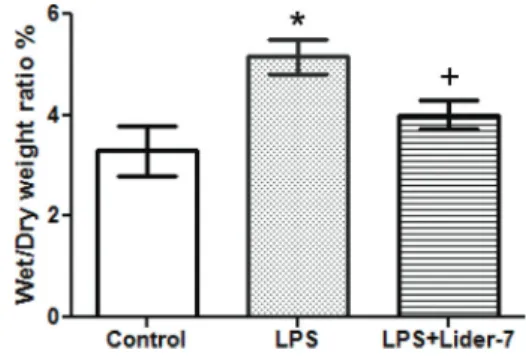
Effect of Lider-7 pre-treatment on right lung W/D ratio. The LPS group had a significantly higher W/D ratio than the healthy group, indicating the presence of pulmonary
edema (Po0.05). However, the W/D ratio in the LPS+
Lider-7 group was significantly decreased compared to the LPS group, indicating that Lider-7 attenuated the
degree of pulmonary edema induced by LPS (Po0.01;
Figure 2).
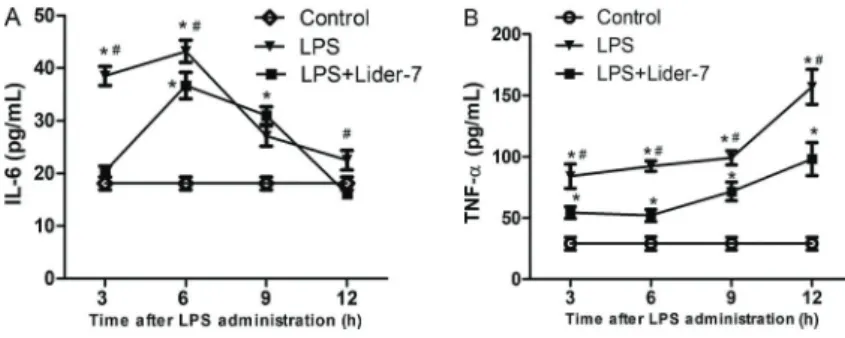
Effect of Lider-7 on the expression of pro-inflammatory cytokines of plasma. In the LPS group, the levels IL-6
significantly increased after LPS administration and reached
peak levels at 6 h. Thereafter, the levels decreased gradually to baseline at 12 h. However, the levels of the
late stage pro-inflammatory cytokine TNF-a increased
gradually and reached a peak at 12. In contrast, the rats
Figure 2.Comparison of the wet/dry ratio. The extent of pulmonary edema was assessed using the wet/dry ratio at 12 h after lipo-polysaccharide (LPS) infusion. Control group: n=5; LPS group: n=3; LPS+Lider-7 group: n=5. Data are reported as means±SD. *Po0.05, LPS group compared to control group; +Po0.05, LPS +Lider-7 group compared to LPS group (ANOVA).
Figure 1. Histopathological changes in lung tissue samples of the three groups. Hematoxylin and eosin (200 magnification).
A, Control group with normal lung structure.B, Lipopolysaccharide (LPS) group with increased alveolar wall thickness, edema, bleed-ing and infiltration of inflammatory cells. C, LPS+Lider-7 group showed less structure destruction and inflammatory infiltration.
pretreated with Lider-7 had significantly lower levels of
IL-6 (LPS+Lider-7 group vs LPS group: Po0.05 at
3, 6 and 12 h) and of TNF-a(Po0.05 at 3, 6, 9, and 12 h)
(Figure 3).
Discussion
The median lethal dose for Lider-7 tang was deter-mined as 8.9 g/kg on the result of acute toxicity studies carried out by the express method of Prozorovskii et al. (32). Therefore, we selected the dose of 90 mg/kg for this study. In the present study, a rat model of ALI was successfully established by the intravenous administration of LPS. We found that LPS exposure caused a dramatic increase in the W/D ratio, reflecting the pulmonary edema. Furthermore, histopathological analysis revealed a loss of epithelial integrity. Taken together, these manifestations confirmed the development of LPS-induced ALI. Interest-ingly, pretreatment with Lider-7 reduced the extent of his-topathological changes and secretion of pro-inflammatory cytokines in rat lung tissue.
Gram-negative sepsis is the most common risk factor of acute respiratory distress syndrome. LPS is the princi-pal component of the outer membrane of gram-negative bacteria and is a potent stimulator of rapid pro-inflammatory
cytokine production. The elevated expression of TNF-a
and IL-6 is an important step in the pathogenesis of ALI and acute respiratory distress syndrome (33). Many natural
substances such as sophoraflavanone G (34),
quinolizi-dine alkaloids (35–38), alantolactone (16–20) and
genipo-side (21) have shown the effect of decreasing LPS-induced inflammation via suppression of pro-inflammatory cytokine secretion.
Because S. alopecuroides is the main compound in
Lider-7-tang, we speculate that quinolizidine alkaloids had a major contribution to the effects observed. Moreover, sesquiterpene lactones have shown anti-inflammatory effects, so they might have assisted in the effects observed.
Consistently, our study showed that the levels of TNF-a
and IL-6 reached a peak at 6 h after LPS administration and then returned to baseline levels. The persistence of lung injury suggests that other late stage downstream pro-inflammatory cytokines may be involved in the progres-sion of ALI.
The current study demonstrated that Lider-7-tang 1) ameliorated histopathological changes that indicate lung injury, and 2) inhibited the release of pro-inflamma-tory cytokines in rats with ALI. Taken together, these results suggest that Lider-7-tang might be a potential candidate for the pre-treatment of LPS-induced ALI.
Acknowledgments
We thank the team of the Pharmacological Laboratory of the Institute of Traditional Medicine and Technology for their help during this study.
References
1. Martínez O, Nin N, Esteban A. Prone position for the treatment of acute respiratory distress syndrome: a review of current literature.Arch Bronconeumol2009; 45: 291–296, doi: 10.3892/mmr.2014.2226.
2. Wozniak K, Sleszycka J, Safianowska A, Wiechno W, Domagala-Kulawik J. Systemic inflammation in peripheral arterial disease with or without coexistent chronic obstruc-tive pulmonary disease: analysis of selected markers.Arch Med Sci2012; 8: 477–483, doi: 10.5114/aoms.2012.29525.
3. Matute-Bello G, Frevert CW, Martin TR. Animal models of acute lung injury. Am J Physiol Lung Cell Mol Physiol
2008;295:L379–L399, doi: 10.1152/ajplung.00010.2008. 4. Ware LB, Matthay MA. The acute respiratory distress
syndrome.N Engl J Med2000; 342: 1334–1349, doi: 10.1056/ NEJM200005043421806.
5. Beutler B, Rietschel ET. Innate immune sensing and its roots: the story of endotoxin.Nat Rev Immunol 2003; 3: 169–176, doi: 10.1038/nri1004.
Figure 3.Changes in the levels of pro-infl am-matory cytokines.A,Interleukin (IL-6);B,tumor necrosis factor (TNFa). Control group: n=5 for each time point; lipopolysaccharide (LPS) group: n=5 (3 and 6 h), n=4 (9 h) and n=3 (12 h); LPS +Lider-7 group: n=5 for each time point. Data are reported as means±SD. *Po0.05, LPS and LPS+Lider-7 groups compared to control group;
#
6. Piotrowski WJ, Majewski S, Marczak J, Kurmanowska Z, Górski P, Antczak A. Exhaled breath 8-isoprostane as a marker of asthma severity.Arch Med Sci2012; 8: 515–520, doi: 10.5114/aoms.2012.28639.
7. Jambalchoijidanzanperenlei. "Manag Rinchin Junai". Tradi-tional Medical Source Book. China: "Inner Mongolian medical treasurers" printing house; 1978.
8. Ligaa U, Davaasuren B, Ninjil N.Mongolian medicinal plants using in Western and Eastern Medicine. Ulaanbaatar: JKC printing; 2005.
9. Chang A, Cai Z, Wang Z, Sun S. Extraction and isolation of alkaloids of sophora alopecuroides and their anti-tumor effects in h22 tumor-bearing mice.Afr J Tradit Complement Altern Med2014; 11: 245–520248, doi: 10.4314/ajtcam.v11i2.3. 10. Küc¸ükboyaci N, Ozkan S, Adigüzel N, Tosun F. Character-isation and antimicrobial activity ofSophora alopecuroides
L. var.Alopecuroidesalkaloid extracts.Turk J Biol2011; 35: 379–385, doi: 10.3906/biy-0910-113.
11. Huang YX, Wang G, Zhu JS, Zhang R, Zhang J. Traditional uses, phytochemistry, and pharmacological properties of
Sophora alopecuroides L. Eur J Inflammation 2016; 14: 128–132, doi: 10.1177/1721727X16642779.
12. Han Y, Zhou Y, Liu Q. Antiendotoxic effects of sophora alopecuroides.Zhong Yao Cai2006; 29: 1066–1069. 13. Atta-Ur-Rahman Au, Choudhary MI, Parvez K, Ahmed A,
Akhtar F, Nur-E-Alam M, et al. Quinolizidine alkaloids from
Sophora alopecuroides. J Nat Prod 2000; 63: 190–192, doi: 10.1021/np990351v.
14. World Health Organization, Western Pacific Region. Medic-inal plants in Mongolia. Geneva: WHO; 2013. ISBN 987 92 9061 632 0.
15. Kwon J, Basnet S, Lee JW, Seo EK, Tsevegsuren N, Hwang BY, et al. Chemical constituents isolated from the Mongolian medicinal plantSophora alopecuroidesL. and their inhibitory effects on LPS-induced nitric oxide production in RAW 264.7 macrophages.Bioorg Med Chem Let2015; 25: 3314–3318, doi: 10.1016/j.bmcl.2015.05.062.
16. Stojanović-RadićZ, Comić Lj, Radulović N, BlagojevićP, Denić M, MiltojevićA, et al. Antistaphylococcal activity of
Inula heleniumL. root essential oil: eudesmane sesquiter-pene lactones induce cell membrane damage.Eur J Clin Microbiol Infect Dis 2012; 31: 1015–1025, doi: 10.1007/ s10096-011-1400-1.
17. Talib WH, Zarga MHA, Mahasneh AM. Antiproliferative, antimicrobial and apoptosis inducing effects of compounds isolated from Inula viscosa. Molecules 2012; 17: 3291– 3303, doi: 10.3390/molecules17033291.
18. Konishi T, Shimada Y, Nagao T, Okabe H, Konoshima T. Antiproliferative sesquiterpene lactones from the roots ofInula helenium. Biol Pharm Bull 2002; 25: 1370–1372, doi: 10.1248/bpb.25.1370.
19. Yan H, Haiming S, Cheng G, Xiaobo L. Chemical con-stituents of the roots ofInula helenium.Chem Nat Compd
2012; 48: 522–524, doi: 10.1007/s10600-012-0298-x. 20. Zhao YM, Zhang ML, Shi QW, Kiyota H. Chemical
constituents of plants from the genusInula(Review).Chem Biodivers2006: 3: 371–384, doi: 10.1002/cbdv.200690041. 21. Phatak RS. Phytochemistry, Pharmacological activities and intellectual property landscape of Gardenia jasminoides
Ellis: a review. Pharmacog J 2015; 7: 254–265, doi: 10.5530/pj.2015.5.1.
22. Jensen SR, Opitz SEW, Gotfredsen CH. Iridoids and phenylethanoids in Lagotis integrifolia and Wulfeniopsis amherstiana(Plantaginaceae).Biochem System Ecol2009; 37: 421–425, doi: 10.1016/j.bse.2009.04.013.
23. Kardan K, Gurav N, Solanki B, Patel P, Patel B. RP-HPLC method development and validation of gallic acid in poly-herbal tablet formulation.J Appl Pharm Sci2013; 3: 37–42. 24. StankovićMS. Total phenolic content,flavonoid concentra-tion and antioxidant activity of Marrubium peregrinum l.
extracts.Kragujevac J Sci2011; 33: 63–72.
25. Quettier DC, Gressier B, Vasseur J, Dine T, Brunet C, Luyckx MC, et al. Phenolic compounds and antioxidant activities of buckwheat (Fagopyrum esculentum Moench) hulls and flour. J Ethnopharmacol 2000; 72: 35–42, doi: 10.1016/S0378-8741(00)00196-3.
26. Singleton VL, Orthofer R, Lamuela-Raventos RM. Analysis of total phenols and other oxidation substrates and antioxidants by means of Folin-Ciocalteu reagent.Methods Enzymol1999; 299: 152–178, doi: 10.1016/S0076-6879(99)99017-1.
27. Shamsa F, Monsef H, Ghamooshi R, Verdianrizi V. Spectro-photometric determination of total alkaloids in some Iranian medicinal plants.Thai J Pharm Sci2008; 32: 17–20. 28. Trim A, Hill R. The preparation and properties of Aucubin.
Biochem J1952; 50: 310–319, doi: 10.1042/bj0500310. 29. Li G, Zhou CL. Galantamine protects against
lipopolysac-charide-induced acute lung injury in rats.Braz J Med Biol Res2016; 49: e5008.
30. Imanaka H, Shimaoka M, Matsuura N, Nishimura M, Ohta N, Kiyono H. Ventilator-induced lung injury is associated with neutrophil infiltration, macrophage activation, and TGF-beta 1 mRNA upregulation in rat lungs.Anesth Analg2001; 92: 428–436, doi: 10.1213/00000539-200102000-00029. 31. Chen F, Liu Z, Wu W, Rozo C, Bowdridge S, Millman A, et al.
An essential role for TH2-type responses in limiting acute tissue damage during experimental helminth infection.Nat Med2012; 18: 260–266, doi: 10.1038/nm.2628.
32. Prozorovskii VB, Prozorovskaya MP, Demchenko VM. Express method of determining the median effective dose and its error.Pharmacol Toxicol1978; 4: 497–500. 33. Giebelen IA, van Westerloo DJ, LaRosa GJ, de Vos AF, van der
Poll T. Local stimulation of alpha7 cholinergic receptors inhibits LPS-induced TNF-alpha release in the mouse lung. Shock
2007; 28: 700–703, doi: 10.1097/shk.0b013e318054dd89. 34. Guo C, Yang L, Luo J.SophoraflavanoneG fromSophora
alopecuroides inhibits lipopolysaccharide-induced infl am-mation in RAW264.7 cells by targeting PI3K/Akt, JAK/STAT and Nrf2/HO-1 pathways.Int Immunopharmacol2016; 38: 349–356, doi: 10.1016/j.intimp.2016.06.021.
35. Zhou Y, Wang N, Zhao J, Zhang YL, Wang DJ, Tong SJ, et al. Effects of sophoridine on the function of peritoneal macro-phages in mice.Liaoning J Tradition Chinese Med2000; 27: 84. 36. Liu T, Liu D, Wang J, Luo Ch. Effects of matrine, oxymatrine and sophordine on activity and TNF-asecretion of macro-phage RAW264.7. Chinese J Informat Trad Chinese Med
2010; 17: 31.
37. Zhang W, Zhang Y, Zhang T, DG Zhang, XM Liao. Studies on antibacterial and anti-inflammatory effect of alkaloid of