Controversies involving hypercapnic acidosis in
acute respiratory distress syndrome
Controvérsias acerca da acidose hipercápnica na síndrome do
desconforto respiratório agudo
INTRODUCTION
The benefit of ventilating a patient with acute lung injury (ALI)/acute respiratory distress syndrome (ARDS) using protective ventilatory strat-egy is a consensus in the literature(1,2), because the inappropriate
adjust-ment of ventilator may induce and/or exacerbate pulmonary and systemic inflammation(3,4) leading to multiple organ dysfunction(5,6) and
contrib-uting to a worse prognosis in ALI/ARDS patients. If on one hand, pro-tective ventilatory strategy with low tidal volume of 6 ml/kg and limited
plateau pressure (30-35 cmH2O) reduces the stress, on the other hand it
causes a condition referred to as permissive hypercapnia, with consequent respiratory acidosis.(1,2)
The effects of hypercapnic acidosis in ALI/ARDS are not completely understood. There are evidences that hypercapnic acidosis may, by itself,
Liliane Nardelli1, Patricia Rieken
MacedoRocco2, Cristiane Sousa
Nascimento Baez Garcia3
1. Master in Science, Laboratory of Pulmonary Investigation Researcher, Carlos Chagas Filho Biophysics Institute (IBCCF), Universidade Federal do Rio de Janeiro (UFRJ) – Rio de Janeiro (RJ), Brazil.
2. PhD, Associate Professor,
Universidade Federal do Rio de Janeiro (UFRJ) – Rio de Janeiro (RJ), Brazil. 3. Post-Doctor, Laboratory of Pulmonary Investigation Researcher, Carlos Chagas Filho Biophysics Institute (IBCCF), Universidade Federal do Rio de Janeiro (UFRJ) – Rio de Janeiro (RJ), Brazil.
ABSTRACT
Acute respiratory distress syn-drome is characterized by a diffuse inflammatory reaction of lung paren-chyma induced by a direct insult to the alveolar epithelium (pulmonary acute respiratory distress syndrome) or an indirect lesion through the vas-cular endothelium (extrapulmonary acute respiratory distress syndrome). The main therapeutic strategy for acute respiratory distress syndrome is the ventilatory support. However, mechanical ventilation can worsen lung injury. In this context, a protec-tive ventilatory strategy with low tidal volume has been proposed. The use of low tidal volume reduced the mortal-ity rate in acute respiratory distress syndrome patients, but result in hy-percapnic acidosis. The current arti-cle presents a literature review on the
effects of permissive hypercapnia in acute respiratory distress syndrome. To that end, we carried out a sys-tematic review of scientific literature based on established criteria for docu-mental analysis including clinical and experimental articles, using as data-bases MedLine, LILACS, SciELO, PubMed, and Cochrane. Hypercapnic acidosis has been considered by some authors as an inflammatory process modulator in acute respiratory dis-tress syndrome. However, clinical and experimental studies on hypercapnic acidosis effects have shown controver-sial results. Therefore it is important to better elucidate the role of hyper-capnic acidosis in acute respiratory distress syndrome.
Keywords: Acute respiratory distress syndrome; Permissive hypercania; Hip-ercapinic acidosis; Inlammation
Received fron laboratory of Pulmonary Investigation Carlos Chagas Filho Biophysics Institute (IBCCF), Universidade Federal do Rio de Janeiro (UFRJ) – Rio de Janeiro (RJ), Brazil.
Submitted on September 18, 2009 Accepted on November 19, 2009
Author for correspondence: Patricia Rieken Macedo Rocco Laboratory of Pulmonary Investigation Carlos Chagas Filho Biophysics Institute Universidade Federal do Rio de Janeiro Centro de Ciências da Saúde, Avenida Carlos Chagas Filho, s/n, Bloco G-014 - Cidade Universitária - Ilha do Fundão CEP: 21941-902 - Rio de Janeiro (RJ), Brazil.
Phone: +55 (21) 2562-6530/ Fax: +55 (21) 2280-8193
reduce lung injury by modulating the inflammatory
process.(7) However, experimental studies have shown
controversial results, with some studies showing no
im-provement(8) and others presenting worsening of lung
injury.(9) The controversies of the experimental studies
may be attributed to: (1) the hypercapnia induction method, either by changing ventilatory parameters(8)
or administration of a carbogenic gas mixture in the inspiratory extremity of the ventilator circuit;(7,9) and
(2) the ALI/ARDS model used. In addition, it is im-portant to emphasize that the human body tolerance to hypercapnic acidosis is so far unknown.
For years, hypercapnic acidosis was treated with sodium bicarbonate, being its use recommended by ADRSNet which describes the importance of buffer-ing acidosis and partial hypercapnia correction for a better ALI/ARDS patients’ survival.(2) However, the
impact of the buffering hypercapnic acidosis with so-dium bicarbonate in ALI/ADRS patients’ mortality remains to be clarified.
This paper discusses controversies on hypercapnic acidosis as well as the possible therapeutic effects of ALI/ARDS buffering.
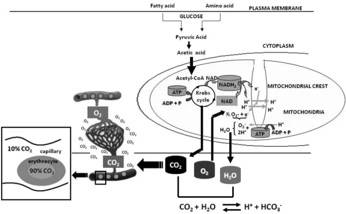
The CO2 chemistry
he main ventilation function is to eliminate carbon dioxide (CO2), the end-product of aerobic cell breath (Figure 1). CO2 molecules are eliminated by the cell while hydrogen (H+) molecules are captured by NAD
molecules, which become NADH2 (hydrogen ions
car-rier). In some reactions, ADP molecules are phospho-rilated generating ATP molecules, in which a relevant portion of cellular energy is stored. he NADH2 carrier delivers the H+ ions to a cytochrome chain at the
mito-chondrial crest, releasing energy. he H+ ions react with
oxygen molecules (O2) to form water (H2 O).
he CO2 released by the cell into the extracellular environment is transported by blood to the lungs dis-solved in the plasma with bicarbonate ions (HCO3-) and
bound to hemoglobin (carbamino-hemoglobin) and other
carbamin compounds. Only a minor portion of cells CO2
(10%) is transported dissolved in the plasma, while most of it (90%) enters the erythrocytes. Inside the erythro-cytes, CO2 combines with water to form carbonic acid which dissociates into H+ and HCO
3
-. Unlike plasma, this
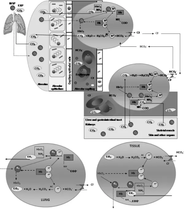
chemical reaction inside the erythrocyte is catalyzed by carbonic anhydrase (Figure 2).
Figure 1 – Carbon dioxide and cellular breathing.
Usually, alveolar ventilation is adjusted to keep arte- keep arte-rial carbon dioxide partial pressure (PaCO2) around
35-45 mmHg. PaCO2 changes are detected by
cen-tral and peripheral chemoreceptors. When the alveo-lar ventilation is increased (hyperventilation) or
de-creased (hypoventilation) in relation to CO2
produc-Figure 2 – Carbon dioxide transportation from tissues to lung.
Both in the periphery and in the lung, most of CO2 transportation occurs within the erythrocyte, where there is carbonic anhydrase. In the periphery of body, the CO2 difuses from the tissues to capillaries. In the lungs, the CO2 follows a reverse path spreading the pulmonary capillaries to the alveoli. In this context, the reactions are also contrary. In the periphery, the oxyhemoglobin (HbO2) dissociates into reduced hemoglobin (Hb) and O2. Hb bufers the H+ ions released from the CO
2 and H2O reaction as well as the H
+ ions released from the reaction of carbaminic compounds. O 2 goes to
the tissues and peripheral organs. In the pulmonary capillaries, the reduced hemoglobin (Hb) dissociates from the H+ ions and binds to O
2 forming
HbO2. he reoxygenation of Hb in the lungs is facilitated by intensive H+ ions release, which are required in reactions with bicarbonate ions (HCO 3
-)
and with carbaminic compounds. In periphery, about ¾ of HCO3- leaves the erythrocyte through a HCO 3
-/Cl- transportation (chloride deviation)
and the reverse mechanism takes place in lung capillary erythrocytes. HbO2 = oxyhemoglobin; Hb = reduced hemoglobin; AC = carbonic anhydrase; EXP = expiration; INSP = inspiration.
tion, there is a respiratory acid-base imbalance. When the CO2 elimination is insufficient in relation to its tissue production rate, PaCO2 increases, as well as the
H+ and HCO
3
- ions concentration, according to the
Hypercapnia in ALI/ARDS: from the bench to clini cal practice
Experimental studies
Experimental studies have reported controversial results regarding hypercapnia effects in ALI/ARDS.
Hypercapnia-induced respiratory acidosis may im-prove, mitigate, and eventually worsen lung injury (Chart 1). Since the results differ according to the model used to induce ALI/ARDS, the exposed data will be initially discussed according to this line.
Chart 1 - Experimental studies on the efects of hypercapnic acidosis in acute pulmonary injury and ventilator-induced lung injury
Animal Lung injury
model Hypercapnia Results
PaCO2
(mmHg) pH Time
Rabbits ex vivo (11)
1. ALI induced by purine and xhantine oxyda-se addition.
addition of FiCO2 = 5% or 25%
Attenuation of lung injury, possibly by endogenous xhantine-oxydase inhibi-tion. ~32 (5%) e ~120.2 (25%) ~7.39 (5%) e ~6.84 (25%) 25 min
2. ALI induced by ischemia-reperfusion
~31 (5%) e ~113 (25%) ~7.44 (5%) e ~6.89 (25%) 3 h Rabbits ex vivo (7)
ALI induced by ischemia-reperfusion
addition of FiCO2 = 25%
Pulmonary function improvement and reduced alveolar-capillary membrane permeability.
~110 ~7 40 min
Rabbits in vivo (12)
ALI induced by ischemia-reperfusion
addition of FiCO2 = 12%
Preservation of lung mechanics, attenu-ation of lung inlammattenu-ation, and reduc-tion of damage induced by free radicals.
~101 ~7.1 1 h e 45 min
Rabbits ex vivo (24)
VILI induced by barotrauma
addition of CO2 to achieve a PaCO2 = 70-100 mmHg
Reduction of VILI severity, attenuating the barotrauma-induced increment of NO production, weight gain, capillary iltration coeicient and BALF protein and hemoglobin.
~105 ~6.93 60 min
Rabbits in vivo (25)
VILI induced by volutrauma
addition of de FiCO2 = 12%
Reduction of VILI severity, attenua-ting the increment of lung wet-to-dry weight ratio, BALF protein concentra-tion, and lung injury.
80-100 ~7.1 4 h
Rats in vivo (13)
ALI induced by ischemia-reperfusion
addition of FiCO2 = 2,5%, 5%, 10%, and 20%
Lung mechanics preservation, attenu-ation of protein leakage and oxyge-nation improvement. Protection both prophylactic (15 min before) and the-rapeutic (15 min after reperfusion), which was dose-dependent with better protective efect with FiCO2 above 5%.
57.4 - 62.5 7.11 - 7.19 ?
Rats in vivo (15)
ALI induced by intratracheal instillation of E. coli
addition of CO2 = 12% before (prophylactic) or 30 min after (therapeutic) instillation
Hypercapnic acidosis attenuated en-dotoxin-induced ALI, with both pro-phylactic and therapeutic eicacy. Its beneicial actions were not mediated by the inhibition of peroxy-nitro induced proteins nitration.
70-80 ~ 7.1
4 h pro-phylactic
6 h thera-peutic
Chart 1 - Continuation
Animal Lung injury
model Hypercapnia Results
PaCO2
(mmHg) pH Time
Rats ex vivo (28)
VILI induced by volutrauma
addition of FiCO2 = 12%
Compared to normocapnia, hypercap-nic acidosis reduced the lung cells ca-pacity to repair cytoplasm membrane injury.
119 7.01 20 min
Rabbits in vivo(9)
ALI induced by intravenous administration of E. coli LPS
respiratory rate reduction
he increment of BALF protein and cells content, NO synthase expression, NO metabolites formation, and the lung wet-to-dry weight ratio, in addition to worse-ning of histological changes.
~ 60 ~7.17 6 h
Rabbits in vivo(26)
VILI induced by barotrauma
addition of CO2 to achieve a PaCO2 = 65-75 mmHg
VILI protection, with no change in oxy-genation, BALF cytokines contents, lung wet-to-dry weight ratio and histology.
65 - 75 ~ 7.25 6 h
Rats in vivo (8)
ALI induced by intratracheal instillation of E. coli
addition of FiCO2 = 5%
Hypercapnic acidosis did not change the ALI severity induced by intratrache-al instillation of E. coli either with our without antibiotics.
~64 ~7.17 6 h
Rats in vivo (16)
ALI induced by intratracheal instillation of E. coli
addition of FiCO2 = 5%
Worsening of ALI induced by bacterial infection, suggesting immunosuppressi-ve efect
~55.6 ~7.36 2 days
Rats in vivo (18)
ALI induced by intratracheal instillation of E. coli
addition of FiCO2 = 5%
Attenuation of airway pressure increase and the reduction of pulmonary com-pliance reduction and PaO2, being this action neutrophil-independent.
~61.5 ~7.13 6 h
Rabbits in vivo (19)
ALI induced by intratracheal instillation of E. coli respiratory rate reduction
Increment of nitrogen reactive species, BALF neutrophil and myeloperoxydase contents, and neutrophil adhesion via increased adhesion molecules expres-sion.
55–65 ? 4 h
Piglets in
vivo (39) Normal animals
addition of CO2 to achie-ve a PaCO2 = 35-45, 50-60, 60-70, 90-100 e 110-120 mmHg
A short term exposure to respiratory acidosis reduces the diaphragm contrac-tility proportional to the hypercapnia degree, and this change was partially reverted 60 minutes after exposure dis-continuation.
~40. ~54. ~68. ~95 e
~116. res-pectively ~7.46. ~7.34. ~7.27. ~7.03 e ~6.98. res-pectively
~15 min at each PaCO2
level
Rats in vivo (20)
ALI induced by early sepsis (6 h) or prolonged sepsis (96 h) resulted from CLP
addition of FiCO2 = 5%
In early sepsis, attenuation of hypoten-sion severity development, and reduc-tion of lactate accumulareduc-tion and central venous oxyhemoglobin levels, BALF neutrophil iniltration and lung wet-to-dry weight ratio.
~60 7.10 - 7.30 3 h
In prolonged sepsis, lung injury scores reduction.
Hypercapnia in ALI/ARDS induced by isch-emia-reperfusion
Some experimental studies on ALI induced by ischemia-reperfusion showed a protective effect of hypercapnic acidosis.(7,11) However, there are some
doubts related to the protective role of hypercapnic acidosis, i.e., if the beneficial effects were related to CO2 or to the acid pH, as hypercapnic acidosis buffering did not protect the lungs from
ischemia-reperfusion induced ALI(7) Furthermore, these
stud-ies were performed ex vivo, thus limiting the under-standing of the role of systemic hypercapnic acidosis.
To overcome such limitations, Laffey et al. done
in vivo studies using ALI models induced by
isch-emia-reperfusion.(12,13) They initially evaluated in
rabbits the effects of mechanical ventilation with a
N2 balanced 12% CO2 and 75% O2 gas mixture for
90 minutes in a experimental pulmonary ischemia-reperfusion.(12) They observed an improvement in
lung function and a reduction in alveolar-capillary permeability, suggesting a beneficial effect of hy-percapnic acidosis. They also evaluated the effects of therapeutic hypercapnia (increase in inhaled
CO2) in an ALI model induced by mesenteric
isch-emia-reperfusion and showed that pulmonary mi-crovascular permeability, compliance and oxygen-ation changes were mitigated, but the results were not significant.(13) Based on these results, we can
speculated that mechanical and gas exchange pa-rameters might not be ideal for assessing the effects
of CO2, especially if its action was more at the
cel-lular and molecular levels. Despite this, it should be highlighted that this experimental model is highly relevant for the clinical context since extrapulmo-nary ALI/ARDS presents high mortality rate.(14) It
is important to say that, in none of these in vivo ALI/ARDS models induced by ischemia-reperfu-sion, hypercapnic acidosis buffering was used in order to separate CO2 and pH effects, thus, de-termining which parameter is responsible for the protective effect.
Hypercapnia in ALI/ARDS induced by pneu-monia (pulmonary ALI/ARDS) and sepsis (extra-pulmonary ALI/ARDS)
In a model of ALI induced by intratracheal in-stillation of E. coli lipopolysaccharide (LPS),
hy-percapnia induced by inhalation of a high CO2
concentration-containing gas mixture yielded gas exchange improvement and lung inflammation re-duction associated with a decrease in nitric oxide (NO) end-products, nitrite and nitrosothiol in the bronchoalveolar lavage fluid (BALF) and lung tis-sue.(15) In contrast, in an E. coli LPS ALI model,
Lang et al. evidenced that hypercapnia induced by changes in respiratory frequency worsened lung in-jury, increasing BALF cells and proteins content,
and lung wet-to-dry weight ratio.(9) In addition, NO
synthase expression and NO metabolites formation were higher in LPS groups under hypercapnic con-ditions. Although both studies differ according to animal model and hypercapnia induction method, these data requires careful analysis.
Since 2005 Laffey’s group carried out some stud-ies to better understand the mechanisms of hyper-capnic acidosis induced by CO2 inhalation in an ALI model induced by E. coli intratracheal instil-lation(8,16-18), however their results were also
contro-versial.
Laffey’s group also evaluated the influence of an-tibiotic therapy (100 mg/kg of intravenous ceftri-axone). In contrast to his findings in experimental ALI induced by E. coli LPS(15), when ALI was
in-duced by intratracheal instillation of living bacte-ria, the severity of lung injury was not modulated by hypercapnic acidosis, neither in the presence nor absence of antibiotics.(8) These differences could be
attributed to the experimental model used. Further-more, it is important to highlight that the degree of lung injury in the study of O’Croinin et al. may have not been severe enough to allow hypercapnic acidosis effects.
In 2008, the same researchers analyzed the ef-fects of 5% CO2 inhalation during 48 hours in an ALI model induced by intratracheal instillation of E. coli.(16) They observed that hypercapnic acidosis
worsened lung injury and increased bacterial colo-nies count.(16) Although the interleukin (IL)-1β
as well as the infection extension and inflammatory mediators’ levels compared to those seen in normo-capnic animals. Therefore, prolonged hypernormo-capnic acidosis may be immunosuppressive and worsen the bacterial pneumonia if not treated. This brings up a dilemma for clinicians as, if on one hand the protective ventilatory strategies with tidal volume reduction and permissive hypercapnia are indicat-ed for ALI/ARDS, on the other hand, the benefits that it provides are uncertain. Attention should be paid to the fact that the studies O’Croinin et al.(8,16)
showed that hypercapnic acidosis may compromise the host response to bacterial invasion, allowing a greater bacterial growth and worsening lung injury. However, it is important to say that the duration of therapeutic hypercapnia also needs to be better investigated, in order to be safely used in clinical practice, since longer exposure periods (2 days)(16)
led to adverse effects, worsening lung injury and increasing bacterial growth.
Undoubtedly, the studies of Ni Chonghaile et al. have contributed to a better comprehension of the effects of the interaction between the antibiotic and hypercapnic acidosis in ALI/ARDS.(17,18) Using the
same ALI model induced by intratracheal instilla-tion of E. coli, they demonstrated that, without an-tibiotic therapy, hypercapnic acidosis reduced lung peak pressure and compliance. However, with anti-biotic therapy, which substantially reduces the con-tent of bacteria in the lung, hypercapnic acidosis significantly attenuated the extent of histological
injury induced pneumonia.(17) This study showed
the therapeutic potential of hypercapnic acidosis, since the effects of therapeutic hypercapnia were evaluated after pneumonia be installed (6 hours af-ter intratracheal instillation of E. coli). This is im-portant because in the clinical practice therapeutic intervention occurs only after the lung disease is established.
In fact, neutrophils are fundamental in ALI/ARDS pathogenesis. In this line, recently, Ni Chonghaile et al.(18) reported that in the absence of neutrophil
depletion, hypercapnic acidosis protected against pneumonia-induced ALI, attenuating the increase in airway pressure and the reduction in lung
com-pliance and arterial oxygen partial pressure (PaO2),
without changing the histological lesion. The lack
of some other parameters as mean airway pressure, inspiratory flow, and inspiratory:expiratory times rate has limited the understanding of the functional effects. Furthermore, the use of a 2 cmH2O PEEP does not appropriately reflects the clinical scenario, because these patients need higher PEEP levels.
The negative findings of Laffey et al. regarding therapeutic hypercapnia in ALI induced by intra-tracheal instillation of E. coli LPS(15) were contrary
to those of Liu et al.(19) who submitted rabbits,
in-stead of rats, to mechanical ventilation during 4 hours with moderate hypercapnia (~55-60 mmHg). They observed that lung injury worsens with an in-crease in reactive nitrogen species and neutrophil content in BALF. Furthermore, hypercapnia also increased in vitro and in vivo neutrophil adhesion associated with a raise in vascular cell adhesion molecule-1 (VCAM-1), intercellular adhesion mol-ecule-1 (ICAM-1), E-selectin and chemokines. It is important to highlight that these studies(15,19)
dif-fer according to the method to induce hypercapnia, since in the study of Laffey et al.(15) hypercapnia
was induced by increasing the CO2 inspired frac-tion, and in the Liu et al.(19)’s study by the
respira-tory rate reduction.
Finally, more recently, Costello et al. published a study investigating whether acute hypercapnic
acidosis induced by adding CO2 to the inspired air
would protect against the pulmonary and systemic organ lesions induced by severe sepsis induced by cecal ligation and perforation.(20) In early sepsis,
hy-percapnic acidosis, when compared to normocap-nia, attenuated the hypotension development and severity, and reduced the lactate accumulation, the central venous oxyhemoglobin levels, neutrophil infiltration and the lung wet-to-dry weight ratio. In prolonged sepsis, hypercapnic acidosis reduced the histological scores of lung injury. Despite the posi-tive findings, hypercapnic acidosis did not change the pulmonary and systemic bacterial load, both in early and late sepsis.
Hypercapnia in ventilator-induced lung injury
Ventilator-induced lung injury (VILI), reviewed by Nardelli et al.(21) is an important component of
ALI/ARDS pathogenesis, and may contribute to the high mortality rate.(22) The protective strategy with
low tidal volume and pressure limitation had a posi-tive impact on mortality rate reduction,(1,2) but it
may cause a CO2 increase leading to a hypercapnic
acidosis. Moreover, during mechanical ventilation, airway and alveoli epithelial cells and other lung parenchyma cells as fibroblasts and macrophages, undergo a variety of mechanical forces that activate several cell signaling cascades (revised by Garcia et al.),(23) some of which show pH or PCO
2
depen-dence. To better understand the role of hypercap-nic acidosis in VILI, Broccard et al. evaluated, in isolated rabbit lungs, the effects of two distinct gas
mixtures administration: the first with 5% CO2 and
the second with 25% CO2.(24) They observed a
re-duction in the severity of VILI associated with a de-crease in BALF protein concentration. In addition, high pressures mechanical ventilation increased the BALF nitric oxide (NOx) end-products contents, which were attenuated by hypercapnia. Although Broccard et al.(24)’s study is an ex vivo model, the
results showed that hypercapnic acidosis has a pro-tective effect in VILI. In vivo studies have corrobo-rated Broccard et al.(24)’s findings, showing that
hy-percapnic acidosis is protective in VILI.(25,26)
In these VILI models, it is also unknown if the hypercapnic acidosis protective effect (24-26) is
re-lated to either low pH or increased CO2. In this context, Caples et al. evaluated the effects of buff-ering hypercapnic acidosis with bicarbonate or tris-hydroxymethyl aminomethane (THAM) in isolated rat lungs under non-protective mechanical venti-lation.(27) The authors found worsening of repair
process under hypercapnia acidosis, confirming the previous findings by Doerr et al.(28) Hypercapnic
acidosis buffering, both with bicarbonate or TAM, protected against mechanical ventilation-associated pulmonary cell damage, suggesting a pH-dependent protective mechanism.
Hypercapnia effects in the microcirculation Hypercapnia may affect the local and systemic arterial blood flow distribution, as well as blood
oxygenation and tissues oxygen uptake, since respi-ratory acidosis: 1) right-shifts the hemoglobin dis-sociation curve, increasing the venous blood par-tial pressure of oxygen (PvO2) and, consequently, the O2 uptake in ischemic tissues, 2) reduces the intrapulmonary shunt by potentiating the hypoxic pulmonary vasoconstriction and directly acting on airways, and 3) increases the cardiac output, further
increasing the PvO2 and venous blood oxygen
con-tent (CvO2).(29)
Hypercapnia effects on microcirculation appear to be pH-dependent. In this context, Cardenas et al. showed that the changes in the cardiac output, organs blood flow and intracranial pressures during hypercapnia may be attenuated by acidosis correc-tion with sodium bicarbonate, with no adverse
he-modynamic effects.(30)
Additionally, the time of hypercapnia exposure also appears to modulate the effects on the micro-circulation, since Kiefer et al. demonstrated that acute PCO2 changes have no relevant effects on
splanchnic perfusion and metabolism.(31)
It is important to better understand the effects of hypercapnia on the microcirculation since micro-circulatory disorders may compromise tissues nutri-ents and oxygen supply, leading to organ failure.
Hypercapnia effects in the diaphragm
Patients with respiratory failure may have hyper-capnic acidosis in two main circumstances: short term (acute) or chronic. Hypercapnic acidosis that occurs in acute respiratory failure can be result from any injury to the lung parenchyma (pulmonary
ede-ma and ede-massive pulmonary embolism),(32) airway,(33)
pleura, chest wall, neuromuscular (spinal cord in-jury) or central nervous system (drug overdose).(34)
Chronic hypercapnic acidosis is seen in mechani-cally ventilated patients with limited plateau pres-sure and tidal volume (“protective strategy”).(35,36)
Although hypercapnia effects on pulmonary func-tion(35) and hemodynamics(30) have being evaluated,
few studies evaluated in vivo hypercapnia effects on diaphragm function.(37,38) It was observed that
evalu-tion, and [HCO3] by liver and kidneys, being thus called a “open buffer system”. CO2 is a conjugate acid which is membrane permeable and, by diffu-sion, overcomes the lipid layer; HCO3- is a base
which moves through membrane only helped by specific transporter proteins.
Some experimental protocol mentioned on this review did not include the hypercapnic acidosis buffering, thus leading to doubt regarding if the protective effects seen were due to the increased CO2 or the pH drop. There is one single ex vivo study conducted in rabbits, ventilated with 5%
CO2 and 95% O2, where the authors performed
a bicarbonate hypercapnic acidosis buffering. The authors concluded that hypercapnic acidosis en-tailed beneficial effect, and that buffering attenu-ated these effects, suggesting that the protective ef-fect is related to the acidosis and not to CO2.(7) This
finding is very interesting considering pulmonary vascular tonus, since hypercapnia, itself, is a potent vasodilator, while acidosis acts as a pulmonary vaso-constrictor. Hypercapnic acidosis results in pulmo-nary vasoconstriction, showing that the pulmopulmo-nary
vascular tonus is more sensitive to pH than to CO2.
This may suggest that direct pH and PCO2 effects are independent. Since it was an ex vivo study(7) it
neglects the hemodynamic changes from systemic circulation limiting the understanding of these data. Therefore, in vivo studies are required.
In a critical patient, acidosis promotes systemic changes, and the main organism defense is consti-tuted by bicarbonate/carbonic acid buffering system (Figure 3). Bicarbonate links to excessive hydrogen
producing CO2 which reaches the lungs to be
elimi-nated; this process would continue until pH is nor-malized, if not limited by progressive bicarbonate levels reduction. Cavaliere et al. studied the acid-base
balance changes and the CO2 elimination induced by
bicarbonate infusion in 10 patients, and found that during bicarbonate infusion the doses used increased
the total CO2 blood contents, while no tissue
reten-tion was found, since hemodynamics remained steady as well as the arterial-venous TCO2 difference.(42) In
the venous blood, bicarbonate infusion increased the
CO2 transportation capacity, probably by the pH
in-crement effect. CO2 elimination only increased after
a given bicarbonate amount was infused. ated the diaphragm contractility under
hypercap-nic conditions and after PaCO2 normalization.(39)
The authors showed that a short-term exposure to hypercapnic acidosis reduced diaphragm contractil-ity proportionally to the level of hypercapnia, being this change partially reverted 60 minutes after the exposure was discontinued. This is an interesting finding, because it may explain why asthmatic pa-tients or compensated obstructive pulmonary dis-ease did not recover immediately their diaphragm strength after a respiratory failure episode. How-ever, caution should be taken into account to ex-trapolate these results to a real setting, since the study was only performed on healthy diaphragms.
Clinical trials
Since 2000, the randomized multicenter trial comparing ventilated patients with 12 ml/kg versus 6 ml/kg tidal volume has been the mainstay for suc-cessive experimental studies regarding mechanical
ventilation in ALI/ARDS.(2) Tidal volume reduction
induces the increment of PaCO2 leading to
permis-sive hypercapnia. This hypercapnic acidosis, ac-cording to the ARDSNet protocol, was buffered by sodium bicarbonate, suggesting that this buffering was fundamental for these patients improved prog-nosis. In this context Kregenow et al. performed multiple logistic regression using the ARDSNet tri-al data, and found that hypercapnic acidosis was as-sociated to reduced mortality in the high tidal vol-ume patients group.(40) A meta analysis of six trials
involving 1297 adult ALI/ARDS patients compared low volume or low airway pressure ventilation ver-sus ventilation with tidal volumes between 10 and 15 ml/kg. The clinical heterogeneity and the dif-ferences between follow-up time and the degree of plateau pressures in the two trials make interpreting trial results more difficult. The long term effects on mortality are unknown, however the clinical benefit
of hypercapnic acidosis may not be ruled out.(41)
Hypercapnia buffering
Several buffers are responsible for maintain-ing body pH, among them the bicarbonate buffer which is highly relevant not only for buffering H+
ions, but also for the components concentration
respira-COMMENTS
Permissive hypercapnia has been tolerated and used in ALI/ARDS. herefore, it is essential to better understand the efects of hypercapnic acidosis, as well as CO2 and/or pH levels separately. here is still insuicient knowledge on hypercapnic acidosis beneits on respiratory system and distal organs to allow it to be consolidated in the thera-peutic arsenal of ALI/ARDS. Additionally, it is unclear whether hypercapnic acidosis should or not be bufered, and additional studies are necessary to clarify this issue.
ACKNOWLEDGEMENTS
Financial support by Centers of Excellence Program (PRONEX-FAPERJ), Brazilian Council for Scientiic and Technological Development (CNPq), Rio de Janeiro State
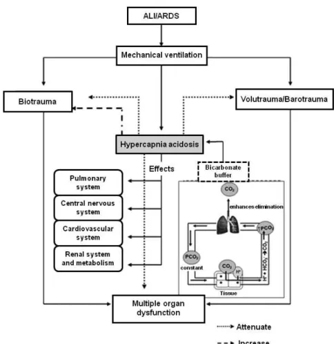
Figure 3 – Mechanical ventilation may contribute to acute lung injury (ALI) by causing direct structural damage (baro/volu-trauma) to the lung and activating the inlammatory response (bio(baro/volu-trauma) leading to multiple organ dysfunction.
Low tidal volume ventilation minimizes barotrauma, volutrauma and biotrauma, however promotes hypercapnic acidosis which acts on several or-gan systems. his hypercapnic acidosis may attenuate lung physical and inlammatory injuries; however, in some cases it increases infammation. In critically ill patients acidosis is usually bufered with bicarbonate (Figure modiied from Lafey et al., 2004(15)).
Research Supporting Foundation (FAPERJ), Coordina-tion for the Improvement of Higher EducaCoordina-tion Personnel (CAPES), and INCT-INOFAR.
RESUMO
hipercápnica. O presente artigo apresenta uma revisão da literatura acerca dos efeitos da acidose hipercápnica na síndrome do descon-forto respiratório agudo. Para tal, realizou-se uma revisão sistemática da literatura cientíica conforme critérios já estabelecidos para aná-lise documental incluindo artigos experimentais e clínicos sobre o tema, usando-se como bases de dados MedLine, LILACS, SciElo, PubMed, Cochrane. A acidose hipercápnica é defendida por alguns autores como moduladora do processo inlamatório da síndrome do
desconforto respiratório agudo. Entretanto, estudos clínicos e experi-mentais acerca dos efeitos da acidose hipercápnica têm demonstrado resultados controversos. Logo, é fundamental a realização de mais pesquisas para elucidar o papel da acidose hipercápnica na síndrome do desconforto respiratório agudo.
Descritores: Síndrome do desconforto respiratório agudo; Hipercapnia permissiva; Acidose hipercápnica; Inlamação
REFERENCES
1. Amato MB, Barbas CS, Medeiros DM, Magaldi RB, Schettino GP, Lorenzi-Filho G, et al. Efect of a protective-ventilation strategy on mortality in the acute respiratory distress syndrome. N Engl J Med. 1998;338(6):347-54. 2. Ventilation with lower tidal volumes as compared with
traditional tidal volumes for acute lung injury and the acute respiratory distress syndrome. he Acute Res-piratory Distress Syndrome Network. N Engl J Med. 2000;342(18):1301-8.
3. Tremblay L, Valenza F, Ribeiro SP, Li J, Slutsky AS. In-jurious ventilatory strategies increase cytokines and c-fos m-RNA expression in an isolated rat lung model. J Clin Invest. 1997;99(5):944-52.
4. Ranieri VM, Suter PM, Tortorella C, De Tullio R, Dayer JM, Brienza A, et al. Efect of mechanical ventilation on inlammatory mediators in patients with acute respiratory distress syndrome: a randomized controlled trial. JAMA. 1999;282(1):54-61.
5. Slutsky AS, Tremblay LN. Multiple system organ failure. Is mechanical ventilation a contributing factor? Am J Respir Crit Care Med. 1998;157(6 Pt 1):1721-5.
6. Ranieri VM, Giunta F, Suter PM, Slutsky AS. Mecha- Mecha-nical ventilation as a mediator of multisystem organ failure in acute respiratory distress syndrome. JAMA. 2000;284(1):43-4.
7. Lafey JG, Engelberts D, Kavanagh BP. Bufering hyper-capnic acidosis worsens acute lung injury. Am J Respir Crit Care Med. 2000;161(1):141-6.
8. O’Croinin DF, Hopkins NO, Moore MM, Boylan JF, McLoughlin P, Lafey JG. Hypercapnic acidosis does not modulate the severity of bacterial pneumonia-induced lung injury. Crit Care Med. 2005;33(11):2606-12. 9. Lang JD, Figueroa M, Sanders KD, Aslan M, Liu Y,
Chu-mley P, Freeman BA. Hypercapnia via reduced rate and ti-dal volume contributes to lipopolysaccharide-induced lung injury. Am J Respir Crit Care Med. 2005;171(2):147-57. 10. Kellum JA. Determinants of plasma acid-base balance.
Crit Care Clin. 2005;21(2):329-46.
11. Shibata K, Cregg N, Engelberts D, Takeuchi A, Fedorko L, Kavanagh BP. Hypercapnic acidosis may attenuate acute lung injury by inhibition of endogenous xanthine oxidase. Am J Respir Crit Care Med. 1998;158(5 Pt 1):1578-84.
12. Lafey JG, Tanaka M, Engelberts D, Luo X, Yuan S, Tanswell AK, et al. herapeutic hypercapnia reduces pul-monary and systemic injury following in vivo lung reper-fusion. Am J Respir Crit Care Med. 2000;162(6):2287-94.
13. Lafey JG, Jankov RP, Engelberts D, Tanswell AK, Post M, Lindsay T, et al. Efects of therapeutic hypercapnia on mesenteric ischemia-reperfusion injury. Am J Respir Crit Care Med. 2003;168(11):1383-90.
14. Montgomery AB, Stager MA, Carrico CJ, Hudson LD. Causes of mortality in patients with the adult respiratory distress syndrome. Am Rev Respir Dis. 1985;132(3):485-9.
15. Lafey JG, Honan D, Hopkins N, Hyvelin JM, Boylan JF, McLoughlin P. Hypercapnic acidosis attenuates endoto-xin-induced acute lung injury. Am J Respir Crit Care Med. 2004;169(1):46-56.
16. O’Croinin DF, Nichol AD, Hopkins N, Boylan J, O’Brien S, O’Connor C, et al. Sustained hypercapnic acidosis du-ring pulmonary infection increases bacterial load and wor-sens lung injury. Crit Care Med. 2008;36(7):2128-35. 17. Chonghaile MN, Higgins BD, Costello J, Lafey JG.
Hypercapnic acidosis attenuates lung injury induced by established bacterial pneumonia. Anesthesiology. 2008;109(5):837-48. Erratum in: Anesthesiology. 2009 Mar;110(3):689.
18. Ni Chonghaile M, Higgins BD, Costello JF, Lafey JG. Hypercapnic acidosis attenuates severe acute bacterial pneumonia-induced lung injury by a neutrophil-indepen-dent mechanism. Crit Care Med. 2008;36(12):3135-44. 19. Liu Y, Chacko BK, Ricksecker A, Shingarev R, Andrews
E, Patel RP, Lang JD Jr. Modulatory efects of hypercap-nia on in vitro and in vivo pulmonary endothelial-neu-trophil adhesive responses during inlammation. Cytokine. 2008;44(1):108-17.
20. Costello J, Higgins B, Contreras M, Chonghaile MN, Hassett P, O’Toole D, Lafey JG. Hypercapnic acidosis attenuates shock and lung injury in early and prolonged systemic sepsis. Crit Care Med. 2009;37(8):2412-20. 21. Nardelli LM, Garcia CSNB, Pássaro CP, Rocco PRM. En-
En-tendendo os mecanismos determinantes da lesão pulmonar induzida pela ventilação mecânica: [revisão]. Rev Bras Ter Intensiva. 2007;19(4):469-74.
role of protein-protein interaction in mechanosensation. Proc Am horac Soc. 2005;2(3):181-7.
23. Garcia CS, Prota LF, Morales MM, Romero PV, Zin WA, Rocco PR. Understanding the mechanisms of lung mecha-nical stress. Braz J Med Biol Res. 2006;39(6):697-706. 24. Broccard AF, Hotchkiss JR, Vannay C, Markert M, Sauty
A, Feihl F, Schaller MD. Protective efects of hypercapnic acidosis on ventilator-induced lung injury. Am J Respir Crit Care Med. 2001;164(5):802-6.
25. Sinclair SE, Kregenow DA, Lamm WJ, Starr IR, Chi EY, Hlastala MP. Hypercapnic acidosis is protective in an in vivo model of ventilator-induced lung injury. Am J Respir Crit Care Med. 2002;166(3):403-8.
26. Park CM, Lim SC, Kim YI, Kim KS, Oh IJ, Kim SO, Kim YC. Does hypercapnic acidosis, induced by adding CO2 to inspired gas, have protective efect in a ventilator-indu-ced lung injury? J Korean Med Sci. 2005;20(5):764-9. 27. Caples SM, Rasmussen DL, Lee WY, Wolfert MZ,
Hub-mayr RD. Impact of bufering hypercapnic acidosis on cell wounding in ventilator-injured rat lungs. Am J Physiol Lung Cell Mol Physiol. 2009;296(1):L140-4.
28. Doerr CH, Gajic O, Berrios JC, Caples S, Abdel M, Lymp JF, Hubmayr RD. Hypercapnic acidosis impairs plasma membrane wound resealing in ventilator-injured lungs. Am J Respir Crit Care Med. 2005;171(12):1371-7. 29. Hickling KG, Joyce C. Permissive hypercapnia in ARDS
and its efect on tissue oxygenation. Acta Anaesthesiol Scand Suppl. 1995;107:201-8.
30. Cardenas VJ Jr, Zwischenberger JB, Tao W, Nguyen PD, Schroeder T, Traber LD, et al. Correction of blood pH attenuates changes in hemodynamics and organ blood low during permissive hypercapnia. Crit Care Med. 1996;24(5):827-34.
31. Kiefer P, Nunes S, Kosonen P, Takala J. Efect of an acute increase in PCO2 on splanchnic perfusion and metabo-lism. Intensive Care Med. 2001;27(4):775-8.
32. Bouchama A, Curley W, Al-Dossary S, Elguindi A. Refrac-tory hypercapnia complicating massive pulmonary embo-lism. Am Rev Respir Dis. 1988;138(2):466-8.
33. Mutlu GM, Factor P, Schwartz DE, Sznajder JI. Severe sta-tus asthmaticus: management with permissive hypercapnia and inhalation anesthesia. Crit Care Med. 2002;30(2):477-80.
34. Gupta R, Haydock T. Severe hypercapnia caused by acute heroin overdose. Ann Emerg Med. 2004;43(5):665-6. 35. Hickling KG. Permissive hypercapnia. Respir Care Clin N
Am. 2002;8(2):155-69, v.
36. Hickling KG, Walsh J, Henderson S, Jackson R. Low mortality rate in adult respiratory distress syndrome using low-volume, pressure-limited ventilation with per-missive hypercapnia: a prospective study. Crit Care Med. 1994;22(10):1568-78.
37. Schnader JY, Juan G, Howell S, Fitzgerald R, Roussos C. Arterial CO2 partial pressure afects diaphragmatic func-tion. J Appl Physiol. 1985;58(3):823-9.
38. Yanos J, Wood LD, Davis K, Keamy M 3rd. he efect of respiratory and lactic acidosis on diaphragm function. Am Rev Respir Dis. 1993;147(3):616-9.
39. Jaber S, Jung B, Sebbane M, Ramonatxo M, Capdevila X, Mercier J, Eledjam JJ, Matecki S. Alteration of the piglet diaphragm contractility in vivo and its recovery after acute hypercapnia. Anesthesiology. 2008;108(4):651-8.
40. Kregenow DA, Rubenfeld GD, Hudson LD, Swenson ER. Hypercapnic acidosis and mortality in acute lung injury. Crit Care Med. 2006;34(1):1-7.
41. Petrucci N, Iacovelli W. Lung protective ventilation stra-tegy for the acute respiratory distress syndrome. Cochrane Database Syst Rev. 2007;(3):CD003844. Review.