Effect of melatonin and time of
administration on irradiation-induced
damage to rat testes
G. Take
1, D. Erdogan
1, F. Helvacioglu
1, G. Göktas
1, G. Ozbey
2, C. Uluoglu
2, B. Yücel
2,
Y. Guney
4, A. Hicsonmez
4and
S. Ozkan
31
Department of Histology and Embryology,
2Department of Pharmacology,
3Department of Public Healthy,
Medical School, Gazi University, Ankara, Turkey
4
Department of Radiation Oncology, Medical School, Ankara University, Ankara, Turkey
Correspondence to: G. Take, Department of Histology and Embriology, Medical School, Gazi University,
06500 Besevler, Ankara, Turkey
Fax: +90-312-212-4647. E-mail: [email protected]
The effect of ionizing irradiation on testes and the protective effects of melatonin were investigated by immunohistochemical and electron microscopic methods. Eighty-two adult male Wistar rats were divided into 10 groups. The rats in the irradiated groups were exposed to a sublethal irradiation dose of 8 Gy, either to the total body or abdominopelvic region using a 60Co source at a
focus of 80 cm away from the skin in the morning or evening together with vehicle (20% ethanol) or melatonin administered 24 h before (10 mg/kg), immediately before (20 mg/kg) and 24 h after irradiation (10 mg/kg), all ip. Caspace-3 immunoreactivity was increased in the irradiated group compared to control (P < 0.05). Melatonin-treated groups showed less apoptosis as indicated by a considerable decrease in caspace-3 immunoreactivity (P < 0.05). Electron microscopic examination showed that all spermatogenic cells, especially primary spermatocytes, displayed prominent degeneration in the groups submitted to total body and abdominopelvic irradiation. However, melatonin administration considerably inhibited these degenerative changes, especially in rats who received abdominopelvic irradiation. Total body and abdominopelvic irradiation induced identical apoptosis and testicular damage. Chronobiological assessment revealed that biologic rhythm does not alter the inductive effect of irradiation. These data indicate that melatonin protects against total body and abdominopelvic irradiation. Melatonin was more effective in the evening abdominopelvic irradiation and treated group than in the total body irradiation and melatonin-treated group.
Key words: Testes; Gamma-irradiation; Melatonin; Immunohistochemistry; Electron microscopy Presented at the 4th APICA (Asian-Pacific International Congress of Anatomists), 2005.
The present address of Y. Guney is Department of Radiation Oncology, Ankara Hospital,Ankara, Turkey
Received September 9, 2008. Accepted March 27, 2009
Introduction
Spermatogenesis is a long, complex and finely tuned process. During this process, the developing sperm cell is sensitive to endogenous or exogenous stresses. Expo-sure to reproductive cytotoxic agents may damage so-matic testicular cells or germ cells at different stages of differentiation, leading to a temporary or permanent im-pairment of fertility (1). Among reproductive toxic agents,
ionizing irradiation has been studied extensively, and is well known to affect testicular function, morphology and spermatogenesis (2,3). Irradiation of the testes can pro-duce reversible or permanent sterility in males (1).
Melatonin (N-acetyl-5-methoxytryptamine; MW = 232) is an endogenous compound secreted by the pineal gland. It is generally accepted that melatonin influences repro-duction via its action on the hypothalamus and the hypo-thesis has been raised that melatonin may modulate the secretion of gonadotropins (5,6). Once synthesized in the pineal gland, melatonin is quickly released into the blood-stream and then into other body fluids, such as bile, cere-brospinal fluid, saliva, ovarian follicular fluid, semen, and amniotic fluid. Small amounts of non-metabolized melato-nin are excreted into urine as well (7). Melatomelato-nin concen-trations in the body are typically lower during the day and reach maximal levels at night in the dark (8).
Several studies have established that melatonin is a highly efficient free radical scavenger and general antiox-idant. Melatonin was also found to scavenge the peroxyl radical, which is generated during peroxidation (9). This implied that melatonin, which is lipophilic and hydrophilic, has effects at the cell level but also within subcellular compartments (4,10-13).
In the present study, we investigated the effects of ionizing radiation applied either to the total body (TB) or only to the abdominopelvic (AP) region on the testes at two different times of day and the protective chronobiological effects of melatonin by immunohistochemical methods using anti-caspace-3, a marker for apoptosis, and electron microscopy to determine irradiation-induced testicular dam-age.
Material and Methods
Experimental protocol
Animals. The experimental protocol was approved by the local Ethics Committee for animal studies and con-ducted at Ankara University Faculty of Medicine. The experiments were performed on male Wistar rats weighing 250-300 g. The animals were fed a standard rat chow diet, had access to water ad libitum and were housed under controlled environmental conditions (light, temperature, feeding time, etc.). The animals were synchronized to a light-dark cycle (lights on from 8:00 am to 8:00 pm) starting at least 2 weeks before the beginning of the experiments. Eighty-two rats were randomly divided into 10 groups (N = 8 in each group). The treatment design of the groups is shown in Table 1. All experiments were performed during February-March to avoid the possibility of seasonal rhythms affecting the findings (Table 1).
Ionizing irradiation. The rats in the irradiated groups were exposed to 8 Gy applied to the TB or AP field following ketamine anesthesia (100 mg/kg) using a 60Co
source at two different times. Morning irradiation was performed 1 h following lights on (at 9:00 am) and evening irradiation was performed 13 h following lights on (at 9:00 pm).
Melatonin treatment. Melatonin or vehicle (20% etha-nol) was administered immediately before, immediately after and 24 h preceding TB or AP irradiation (melatonin dose: 10, 20, and 10 mg/kg, ip, respectively). Forty-eight hours after irradiation, all animals were sacrificed with ketamine and tissues were obtained.
Immunohistochemical procedure. Testis tissues from each group were fixed in neutral formalin for 72 h and processed for paraffin embedding. Sections of 4-5-μm thickness were processed for polylysine-covered micro-scope slides.
For immunohistochemical examination, slides were stored in a microwave oven in 10 mM Tris–HCl buffer, (LabVision, USA). Endogenous peroxidase activity was blocked with 3% hydrogen peroxide (LabVision). Epitopes were stabilized by application of serum blocking solution (LabVision) and slides were then incubated with caspase-3 (rabbit polyclonal antibody Ab-4, 1 mg/mL, NewMarker, USA) for 60 min at room temperature. Next, the biotinyl-ated secondary antibody (goat antirabbit, LabVision), was applied and streptavidin peroxidase (LabVision) was ap-plied to the slides. 3-Amino-9-ethylcarbazole (LabVision) was used as chromogen. Finally, the slides were counter-stained with hematoxylin and examined with a light micro-scope. A negative control was prepared during the stage of primary antibody application. Two independent observers
Table 1. Table 1. Table 1. Table 1.
Table 1. Treatment design for the various groups.
Group Time of TB AP Vehicle Melatonin irradiation irradiation irradiation treatment 1A Morning - - - -1B Evening - - - -2A Morning + - + -2B Evening + - + -3A Morning - + + -3B Evening - + + -4A Morning + - - + 4B Evening + - - + 5A Morning - + - + 5B Evening - + - + The experimental groups (N = 8 for each group) were as follows: group 1: control; group 2: total body (TB) irradiation and vehicle (20% ethanol); group 3: abdominopelvic (AP) irradiation and vehicle; group 4: melatonin with TB irradiation; group 5: melato-nin with AP irradiation. The rats in the irradiated groups were exposed to a sublethal irradiation dose of 8 Gy, either to the TB or AP region after ketamine anesthesia using a 60Co source at a
who were blind to the treatment regimen evaluated the immunolabeling scores separately.
Electron microscopic procedure
Tissues were fixed in phosphate buffer containing 2.5% glutaraldehyde (Sigma-Aldrich, USA) for 2-3 h, post-fixed in 1% osmium tetraoxide (Sigma-Aldrich) and dehydrated in a series of graded alcohols (50, 60, 70, 80, 90, 96 and 100% ethanol). After passing through propylene oxide (Sigma-Aldrich), the specimens were embedded in Araldite CY 212 (Ciba-Geigy, USA), (2-dodecen-1-yl)succinic an-hydride (Sigma-Aldrich), benzyldimethyl amine (Poly-Sciences Inc., USA) and dibutylphtalate (Sigma-Aldrich). The semi-thin sections were stained with toluidine blue (Sigma-Aldrich), and examined with a photomicroscope (Leica DM4000, Germany). After selection of appropriate specimens, thin sections were cut and stained with uranyl acetate (ProSciTech, Australia) and lead citrate (Sigma-Aldrich) and examined with an electron microscope (Carl Zeiss EM 900, Germany).
Statistical analysis
Labeling intensity was graded semiquantitatively and the HSCORE was calculated using the following equation: HSCORE = Pi (i + 1), where i is the intensity of labeling with a value of 1, 2, or 3 (weak, moderate or strong, respec-tively) and Pi is the percentage of labeled primary sperma-tocytes for each intensity, ranging from 0
to 100%. Statistical differences were cal-culated using the Mann–Whitney U–test. Data are reported as means ± SEM. A P value <0.05 was considered to be signifi-cant.
Results
Immunohistochemical results
Immunohistochemical examination of the testes of the control group (1A) re-vealed normal seminiferous tubule and interstitial tissue structures. No caspase-3 immunoreactivity was observed in some seminiferous tubules, while a weak immu-noreactivity in the spermatogonia and pri-mary spermatocytes was found in other seminiferous tubules in this group. Like-wise, no immunoreactivity was observed in the spermatids. The pattern of immu-noreactivity was exactly the same in the slides of morning and evening (1B) control groups (data not shown).
Figure 1. Figure 1.Figure 1. Figure 1.
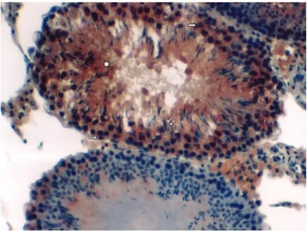
Figure 1. Total body irradiation group (morning). Asterisk: Degenerative seminifer-ous tubules; thin horizontal arrow: weak-to-moderate immunoreactivity in the sper-matogonia; thick horizontal arrows: primary spermatocytes displaying moderate-to-strong immunoreactivity; double oblique arrows: weak spermatid immunoreactivity (immunoperoxidase and hematoxylin staining, 200X).
In the TB-irradiated group (2A), while the integrity of interstitial tissue was conserved, the organized structure of cells forming the seminiferous tubules was completely disrupted and diffuse intercellular edema was observed in the morning group. In this group, spermatogenesis was absent in most seminiferous tubules. Caspase-3 immu-noreactivity was increased from weak to moderate in the spermatogonia, and from moderate to strong in primary spermatocytes. Weak immunoreactivity was observed in the spermatids (Figure 1). Moreover, some degenerative indications were found in the evening group slides (2B). Immunohistochemical evaluation revealed that caspase-3 immunoreactivity was significantly stronger than in the morning group specimens and weak to moderate immu-noreactivity was observed in spermatogonia. A strong reactivity was observed in primary spermatocytes although a weak reactivity was observed in the spermatids (data not shown).
ing group (3A). Caspase-3 immunoreactivity was similar to that observed in the morning specimens but stronger im-munoreactivity was detected in primary spermatocytes (data not shown).
In AP-irradiated and melatonin-treated morning tissue specimens (5A), prominent edema was observed both in the seminiferous tubules and the interstitial field. Caspase-3 immunoreactivity was prominent in some tubules but not in others. Spermatogonia and primary spermatocytes dis-played negative immunoreactivity. No immunoreactivity was present in the spermatids (data not shown). Although the AP-irradiated and melatonin-treated evening group (5B) showed more preserved structures than the morning group, caspase-3 immunoreactivity was prominent in some tubules but not in others. In the high-power-field examina-tions, very weak immunoreactivity was found in the sper-matogonia and primary spermatocytes, and negative reac-tivity in the spermatids. These findings were similar to those observed in controls (Figure 2).
Electron microscopy results
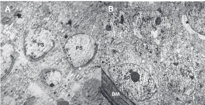
In the morning and evening control groups, the sper-matogenic cells that form seminiferous cords showed a normal ultrastructural appearance. The basement mem-branes of the seminiferous tubules were also normal (data not shown). In the TB-irradiated morning and evening groups, irregular basement membranes and disconnected junctional complexes were observed between the spermatoge-nic cells. Some primary spermatocytes looked like apoptotic bodies. A few acroso-mal cap formations were determined in these groups because of existing slower spermatogenesis (Figure 3, Panels A and B). In the TB-irradiated and melatonin-treated morning and evening groups, the appearance of the spermatogenic cell lines was almost normal, with primary sperma-tocytes identical to those of the control group. Abundant acrosomal cap formation was noted (data not shown).
On the other hand, different electron microscopy results were observed in the AP-irradiated morning and evening groups. In the morning groups (3A), all spermato-genic cell lines were separate from one another and their cytoplasm was filled with diffuse vacuoles (data not shown). In the tissue specimens obtained from the eve-ning groups (3B), the general structure was found to be similar to that observed in
Figure 2. Figure 2.Figure 2. Figure 2.
Figure 2. Abdominopelvic irradiation + melatonin (evening). Asterisk: Minimal degenerative changes in seminiferous tubules; thin horizontal arrow: negative-to-weak caspase-3 immunoreactivity in the spermatogonia; thick horizontal arrow: weak-to-moderate immunoreactivity in the primary spermatocytes; double oblique arrows: no immunoreactivity in spermatids (immunoperoxidase and hematoxylin staining, 200X).
evening group (4B), the tissue structure was similar to that observed in the morning group. Caspase-3 immunoreac-tivity was observed in a few spermatogonia and primary spermatocytes but was absent throughout the seminifer-ous tubules. At high magnification, immunoreactivity was weak in the spermatogonia, moderate in the primary sper-matocytes and negative in spermatids. Strongly immu-noreactive apoptotic bodies were observed within the lu-men of the tubules (data not shown).
morn-Figure 3. Figure 3.Figure 3. Figure 3.
Figure 3. Electron micrograph of the group submitted to total body irradiation. Panel A, Spermatogonium (Sp), lipid droplets (Lp), irregular basement membrane (BM), vacuolar formation of disconnected junctional complexes (black lozenge). Panel B, Primary spermatocytes (PS), a few acrosomal caps (asterisk) and apoptotic bodies (AB) (uranyl acetate and lead citrate staining, 3000X).
A
A
A
A
A
B
B
B
B
B
Figure 4. Figure 4.Figure 4. Figure 4.
Figure 4. Electron micrograph of the group submitted to abdominopelvic irradiation + melatonin administration. Panel A, In tissue samples obtained in the morning, primary spermatocytes show normal structure (PS), with some disconnecting areas between them (asterisk). Panel B, Tissue samples obtained in the evening are similar to control samples, with spermatogonia (Sp), primary spermatocytes (PS), acrosomal caps (asterisk), and basement membrane (BM) (uranyl acetate and lead citrate staining, 3000X).
A
A
A
the specimens taken in the morning, but wrinkling of base-ment membranes was observed (data not shown). In the AP-irradiated and melatonin-treated morning group (5A), separate junctional complexes were observed between the primary spermatocytes, but the remaining tissue seemed to be normal. A normal acrosomal cap formation was detected (Figure 4, Panel A). Finally, in the AP-irradiated and melatonin-treated evening group (5B) the spermatogenic cell line and tissue in general were similar to control (Figure 4, Panel B).
Statistical results
Compared to control, the number of immunoreactive primary spermatocytes was markedly increased in all ex-perimental groups, with significant differences between TB and control testes (P < 0.05). The number of apoptotic figures was decreased in the primary spermatocytes of the melatonin-treated TB group compared to cells from un-treated animals. The differences between the values of TB testes and AP testes were statistically significant (P < 0.05). Similarly, significant differences were observed be-tween TB morning and AP morning testes in terms of the reduction of apoptotic figures (P < 0.05). When the TB morning and melatonin-treated TB morning groups were compared to the TB evening and melatonin-treated TB evening groups, the number of apoptotic primary sperma-tocytes was found to be significantly increased in the evening groups (P < 0.05). But we noticed increased apoptotic figures in the AP morning and melatonin-treated AP morning group compared to the same group treated in the evening (P < 0.05). The statistical results are shown in Table 2.
Discussion
Radiation effects are particularly apparent in a rapidly proliferating tissue. The male germ cells in the
seminifer-ous epithelium of mammals are among the major cell systems at risk when animals are exposed to irradiation and provide an excellent model for studies in this area (1). Clear degeneration was caused by irradiation of testicular tissue in the present study.
Several researchers have reported the presence of the radiation-mediated degeneration especially in spermatogo-nia (1,3). Sapp et al. (3) determined that 72 h after radiation at slightly higher doses, the spermatogonial response begins to differ with type of irradiation. When exposed to X-rays, about 30% of the total spermatogonial population is lost, with 0.10-Gy exposure inducing a sharp decrease in the surviving fraction. However, with exposures from 0.1 to 0.8 Gy the decrease in survival fraction is much more gradual. Approximately an additional 40% of the sper-matogonial population is lost within this range of expo-sures. Thirty percent of the total population remain intact, even at 0.8 Gy. However, between 0.8 and 1 Gy there is another sharp decline in cells. In the present study, in contrast to the literature, apoptosis was lower in the sper-matogonia of samples taken from groups submitted to morning and evening TB and AP irradiation, a result prob-ably due to the short period of irradiation.
Different opinions exist in the literature about the pro-tective effects of melatonin against irradiation treatment (4,11). Badr et al.(4) suggested that the radioprotective effect of melatonin is not species-specific and acts in a similar way in different biological systems. Administration of melatonin after animal exposure to radiation had no significant effect. However, the effect of melatonin per se doubled the frequency of micronuclei in bone marrow polyerythrocytes compared to control. This suggests a cytotoxic effect of melatonin at the relatively high dose of 10 mg/kg body weight. The cytotoxic effect of exogenous aspirated melatonin noted in this study may be due to its abnormal concentration in the circulation.
In the present study, melatonin was found to have
Table 2. Table 2. Table 2. Table 2.
Table 2. Immunolabeling intensities of caspase-3 primary spermatocytes from the testes of all experimental groups.
Reaction Control Total body Total body-melatonin Abdominopelvic Abdominopelvic-melatonin
Morning Evening Morning Evening Morning Evening Morning Evening Morning Evening
Weak 20.4 ± 1.5 14.3 ± 0.1 5.6 ± 0.8a 3.0 ±1ab* 8.6 ± 0.5ac* 4.2 ± 0.7ad* 23.4± 1.1ab 27.8 ± 1.0 20.0 ± 2.3c 16.4 ± 1.1d
Moderate 43.9 ± 2.9 30.2 ± 2.0 74.7 ± 12.5* 26.2 ±3.9* 128.5 ± 2.34c* 73.61 ± 0.6d* 45.7± 1.9 43.0 ± 2.1 56.2 ± 4.0c 45.3 ± 1.4d
Strong 72.0 ± 2.7* 32.0 ± 3.06* 466.3 ± 19.02* 548.1 ±8.5b* 324.0 ± 3.06c* 436.7 ± 11.7d 240± 6.9bde 97.7 ± 2.9d* 103.1 ± 6.9ce* 41.03 ± 5.6d*
HSCORE values are reported as means ± SEM. aP < 0.05 control vs all experimental groups; *P < 0.05 morning vs all evening groups; bP < 0.05 total body morning vs all abdominopelvic morning groups; cP < 0.05 total body melatonin morning vs all abdominopelvic
melatonin morning groups; dP < 0.05 total body melatonin evening vs all abdominopelvic melatonin evening group; eP < 0.05
protective effects and to be more effective in the evening groups and in the endogenous form, a fact probably re-lated to the dose applied. Thus, in clinical therapy, the dose applied is thought to be a very important point in the progression of treatment.
Mornjakovic et al. (14) determined the volume density of the seminiferous epithelium, tubular lumen and testis interstitium in sham-pinealectomized adult Wistar rats af-ter melatonin treatment and whole-body irradiation with 8 Gy gamma rays. They found that melatonin is able not only to modify the quantitative characteristics of the semi-niferous tubules but also to reduce the effects originally produced by irradiation. Also, melatonin administration can significantly reduce the notorious effects of irradiation on Leydig cells (15). Taken together, several observations support the notion that melatonin has a radioprotective effect against X-ray irradiation. First, melatonin administration prior to irradiation prevented radiation damage to peripheral blood cells (16). Second, 6- and 8-Gy X-ray irradiation of rats was associated with increased malondialdehyde, myeloperoxi-dase and nitric oxide levels and decreased glutathione levels (17). All of these indices were reduced with melatonin pretreatment (18). Therefore, melatonin, by its free radical scavenging and antioxidant properties, ameliorates irradia-tion-induced cell damage (17).
In the present study, we investigated the protective effect of melatonin after both TB and AP applications. In TB-irradiated and morning melatonin-treated testis speci-mens (4A), the tissue structure was occasionally pre-served, whereas caspase-3 immunoreactivity was similar to that of the group undergoing radiation exposure without melatonin treatment. The 4B group showed results similar to control. In the group receiving both AP irradiation and melatonin treatment, no reaction was observed in the majority of the tubules in the tissue specimens taken in the morning. In light of these findings, we propose that AP irradiation has weaker effects on the spermatogonia than TB irradiation, and therefore the radioprotective effects of melatonin administration/treatment were more prominent in the group that underwent abdominal radiation.
In addition, our findings differed considerably from those in the literature, particularly in that we found promi-nent effects of radiation on primary spermatocytes, which were considerably improved following melatonin adminis-tration. We also found that the cells most sensitive to the difference between the morning and evening periods were primary spermatocytes. We attributed this sensitivity to the fact that the primary spermatocytes were in the phase of proliferation, and therefore displayed denser caspase-3 reactivity compared with the other cell groups when ex-posed to environmental factors such as irradiation.
The Sertoli and Leydig cells of seminiferous tubule and their basement membranes have been investigated in ultrastructural studies (19-21), but there are no studies in the literature dealing with the protective effects of melato-nin on the ultrastructure of radiated testes.
Hussein et al. (22) reported ultrastructural features of apoptosis (condensation of the nuclei, vacuolization of the cytoplasm, increased cytoplasmic density, and apoptotic bodies) in irradiated testes, which were absent when the irradiated animals were pretreated with melatonin. There was minimal depletion of Sertoli and Leydig cells following X-ray irradiation. Also, morphological features of apopto-sis were infrequent in these cells. The authors found that the protective effects included amelioration of germ-cell depletion and apoptotic changes.
References
1. Cordelli E, Fresegna AM, Leter G, Eleuteri P, Spano M, Villani P. Evaluation of DNA damage in different stages of mouse spermatogenesis after testicular X irradiation. Radiat Res 2003; 160: 443-451.
2. Sapp WJ, Philpott DE, Williams CS, Williams JW, Kato K, Miquel JM, et al. Comparative study of spermatogonial sur-vival after X-ray exposure, high LET (HZE) irradiation or spaceflight. Adv Space Res 1992; 12: 179-189.
3. Sapp WJ, Philpott DE, Williams CS, Kato K, Stevenson J, Vasquez M, et al. Effects of spaceflight on the spermatogo-nial population of rat seminiferous epithelium. FASEB J 1990; 4: 101-104.
4. Badr FM, El Habit OH, Harraz MM. Radioprotective effect of melatonin assessed by measuring chromosomal damage in mitotic and meiotic cells. Mutat Res 1999; 444: 367-372. 5. Kim JK, Lee CJ. Effect of exogenous melatonin on the
ovarian follicles in gamma-irradiated mouse. Mutat Res 2000; 449: 33-39.
6. Vera H, Tijmes M, Ronco AM, Valladares LE. Melatonin binding sites in interstitial cells from immature rat testes. Biol Res 1993; 26: 337-340.
7. Vijayalaxmi, Reiter RJ, Tan DX, Herman TS, Thomas CR Jr. Melatonin as a radioprotective agent: a review. Int J Radiat Oncol Biol Phys 2004; 59: 639-653.
8. Claustrat B, Geoffriau M, Brun J, Chazot G. [Melatonin in humans: a biochemical marker of the circadian clock and an endogenous synchronizer]. Neurophysiol Clin 1995; 25: 351-359.
9. Ianas O, Olinescu R, Badescu I. Melatonin involvement in oxidative processes. Endocrinologie 1991; 29: 147-153. 10. Pieri C, Marra M, Moroni F, Recchioni R, Marcheselli F.
Melatonin: a peroxyl radical scavenger more effective than vitamin E. Life Sci 1994; 55: L271-L276.
11. Reiter RJ. Interactions of the pineal hormone melatonin with oxygen-centered free radicals: a brief review. Braz J Med Biol Res 1993; 26: 1141-1155.
12. Reiter RJ. Functional pleiotropy of the neurohormone mela-tonin: antioxidant protection and neuroendocrine regulation.
Front Neuroendocrinol 1995; 16: 383-415.
13. Reiter R, Tang L, Garcia JJ, Munoz-Hoyos A. Pharmacolo-gical actions of melatonin in oxygen radical pathophysiol-ogy. Life Sci 1997; 60: 2255-2271.
14. Mornjakovic Z, Scepovic M, Kundurovic Z. [Morphometric aspects of seminiferous tubules in rats treated with melato-nin and whole body irradiation (stereologic analysis)]. Med Arh 1991; 45: 9-10.
15. Mornjakovic Z, Alicelebic S, Bilalovic N, Susko I. [Morpho-metric characteristics of Leydig cells after total irradiation of rats treated with melatonin]. Med Arh 1998; 52: 183-184. 16. Koc M, Buyukokuroglu ME, Taysi S. The effect of melatonin
on peripheral blood cells during total body irradiation in rats. Biol Pharm Bull 2002; 25: 656-657.
17. Sener G, Jahovic N, Tosun O, Atasoy BM, Yegen BC. Melatonin ameliorates ionizing radiation-induced oxidative organ damage in rats. Life Sci 2003; 74: 563-572.
18. Taysi S, Koc M, Buyukokuroglu ME, Altinkaynak K, Sahin YN. Melatonin reduces lipid peroxidation and nitric oxide during irradiation-induced oxidative injury in the rat liver. J Pineal Res 2003; 34: 173-177.
19. Pinon-Lataillade G, Velez de la Calle JF, Viguier-Martinez MC, Garnier DH, Folliot R, Maas J, et al. Influence of germ cells upon Sertoli cells during continuous low-dose rate gamma-irradiation of adult rats. Mol Cell Endocrinol 1988; 58: 51-63.
20. Dedov VI. [Ultrastructure of rat Sertoli and Leydig cells normally and under prolonged internal irradiation]. Tsitologiia 1980; 22: 1153-1156.
21. Sawada H, Esaki M. Electron microscopic observation of 137Cs-irradiated rat testis: production of basal laminae for germ cells, despite their absence. J Electron Microsc 2003; 52: 391-397.