J of Evolution of Med and Dent Sci/ eISSN- 2278-4802, pISSN- 2278-4748/ Vol. 3/ Issue 16/Apr 21, 2014 Page 4282
modification in procedure, and way of thinking in tackling the situation in present scenario with whatever things are there on trolley & in the armamentarium. We are presenting a case of intramedullary tibial implant – two V nails, put about 8 years ago having pain &sinus in upper third of leg. It was difficult to predict radiologically the nature of the intra- medullary implant & hence, difficulties during its removal. It is mandatory to discuss all the possible options with the patient and relatives prior to surgery. It is also necessary to explain at times, we might not be able to remove the implant and procedure have to be abandoned. The patient usually considers the removal of implant to be easier than the initial surgery and is usually unaware of the difficulties that can be encountered during removal and the fact that it may need a major procedure followed by a prolonged recovery time.
KEYWORDS: Impacted intramedullary nails, difficult extraction, vertical osteotomy.
INTRODUCTION: Removal of intramedullary nail is considered a routine procedure but may prove to be challenging. Bone ingrowth or overgrowth, damage to the proximal threads of the nail, cutting of the proximal part of nail including extracting eye, and broken nails or locking screws may complicate the removal of intramedullary nails.
It is important to assess the main reason for nail removal and the extent to which the surgeon and the patient are willing to go to do so. Infection, nonunion, migration of the implant, unremitting pain, deformity or re-fracture requiring fixation are indications for nail removal. It is difficult to assess radiologically the nature and number of implants to be removed as we have experienced in present case. Multiple techniques including universal extraction sets, guide wires with hooks, and multiple guide wires have been described for removal of implant.1-6
We present a case of couple of impacted V nails, one of which is proximally bend in transverse fashion (which we realized on exploration) with discharging sinus and a salvage procedure for nail extraction after failure of routine methods.
J of Evolution of Med and Dent Sci/ eISSN- 2278-4802, pISSN- 2278-4748/ Vol. 3/ Issue 16/Apr 21, 2014 Page 4283
The patient was pain free for 7 years and subsequently started complaining of leg pain with discharging sinus for which he did not pay much attention to, however, 6 months prior to evaluation the patient had increasing pain with limiting the activities of daily living. The patient reported at our institution where routine radiographs revealed intramedullary implant with K wire and screw in malleoli. Patient blood profile revealed Anemia (Hb 9 gm. %), raised ESR- 30 mm at the end of first hour, leukocytosis (TLC- 12900).
On physical examination, the patient had a well-healed surgical scar in the right leg in the region of the proximal tibia. There was a sinus below the tibial tuberosity with serous discharge. No rise in local temperature. The fracture was well healed clinically. The right knee was stable in varus and valgus with full range of motion. The ipsilateral ankle had fair range of motion. Neurovascular examination was normal. Overall alignment of the extremity was anatomic.
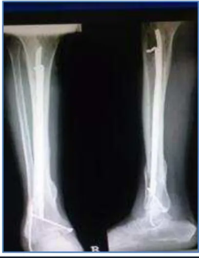
Preoperative radiographs showed a healed tibia and fibula fractures, an intramedullary implant, which was bend proximally with implants in malleoli.
The possible association of leg pain and retained hardware were discussed with the patient. The surgical risks were discussed extensively with the patient, as well as other risks, including continued pain after hardware removal and failure to remove the implant. Informed consent was taken. Surgery was performed under regional anesthesia. Malleolar implants – K wire and screw was removed without any difficulty.
Cortical window was made at the site of sinus between medial and posterior column of the tibia after confirmation of the proximal end of implant under C-arm IITV. There was no pus or granulation tissue at the site of sinus .We found the proximal portion of the implant- V nail in the metaphyseal area of the tibia. Outer V nail was bent acutely and transversely on the medial side.
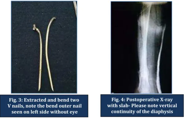
Proximal part of the nails including eye was cut by previous surgeon while insertion. Attempts to extract the nail through the window were futile. Nails were bent in U shape and hook of extractor was engaged in it.
J of Evolution of Med and Dent Sci/ eISSN- 2278-4802, pISSN- 2278-4748/ Vol. 3/ Issue 16/Apr 21, 2014 Page 4284
Still it was not possible to remove either of the nail. It was then decided to do a tibial osteotomy to remove the impacted nails. Accordingly, a linear and vertical osteotomy of the tibia was done distally inch by inch during attempted extraction without damaging the three Tibial columns. The shaft of Tibia was opened between the columns like a book and we could remove both V nails simultaneously successfully. The osteotomized tibia was kept in place and periosteum was sutured.
The patient was discharged after suture removal about 15 days with Above-knee cast. Follow-up of 1 month each, for first 3 months and then at the end of 6 months revealed uneventful recovery. Patient can walk with full weight bearing and healed sinus.
Fig. 2: Intraoperative photographs showing the attempted extraction of the nail
Fig. 3: Extracted and bend two V nails, note the bend outer nail
seen on left side without eye
Fig. 4: Postoperative X-ray with slab- Please note vertical
J of Evolution of Med and Dent Sci/ eISSN- 2278-4802, pISSN- 2278-4748/ Vol. 3/ Issue 16/Apr 21, 2014 Page 4285 DISCUSSION: Removal of orthopedic hardware is mostly easier said than done. Bony overgrowth, worn out screw threads and heads, stuck nails, broken implants and ingress of bone into all possible crevices of the implants. Absence of the eye makes extraction difficult and exasperating for the surgeon. Of these, intramedullary nail removal is considered a routine procedure but may prove to be challenging. Contrary to old belief that implants with infection are loose & easy to remove, it may not be the situation in all cases.
Incarcerated intramedullary nails have been described since Kuntscher’s time.1 Various reasons were listed for incarceration including ingrowth of bone into the inside of a clover leaf nail, bent nails, excessive callus formation closing the medullary canal, and bone in growth through the locking screw holes.
The material of the nail or the bone-metal-interface has not been mentioned as a factor in previous reports. We recommend that to extract an impacted double nail, an early decision should be made to do a vertical osteotomy without disturbing the three columns of the tibia to free the implant which heals with minimal morbidity.
Moral is reiteration of the dictum that removal of implant is not to be taken lightly where one might have to give up the procedure. It should be explained to the patient and relatives with informed consent is mandatory. All the same, one should not hesitate to do a vertical osteotomy and extract the intramedullary implants. Vertical osteotomy heals easily than the transverse one without any morbidity. There is hardly any difficulty in healing of osteotomy, weight bearing capacity and biomechanics following such procedure.
REFERENCES:
1. Kuntscher G, Maatz R. Hardware Removal of Intramedullary Nails: A surgical Technique. Leipzig, Georg Thieme Verlag 63-94, 1945.
2. Georgiadis GM, Heck BE, Ebraheim NA. Technique for removal of intramedullary nails when there is failure of the proximal extraction device: a report of three cases. J Orthop Trauma. 1997; 11(2):130-132.
J of Evolution of Med and Dent Sci/ eISSN- 2278-4802, pISSN- 2278-4748/ Vol. 3/ Issue 16/Apr 21, 2014 Page 4286
1. Varunjikar M. D. 2. S. C. Joshi 3. Bejoy E Jayan 4. A. M. Varunjikar 5. C. R. Joshi
PARTICULARS OF CONTRIBUTORS:
1. Associate Professor, Department of Orthopaedics, Vikhe Patil Memorial Hospital. 2. Consultant, Department of Anaesthesia, Vikhe
Patil Memorial Hospital.
3. Senior Resident, Department of Orthopaedics, Vikhe Patil Memorial Hospital.
4. Consultant, Department of Anaesthesia, Vikhe Patil Memorial Hospital.
Patil Memorial Hospital.
NAME ADDRESS EMAIL ID OF THE CORRESPONDING AUTHOR: Dr. M. D. Varunjikar,
Tathasthu, 54, Deshmukh Colony, Opposite Civil Hospital, Sadar Bazaar, Satara – 415001. E-mail: [email protected]