CLINICAL SCIENCE
I Department of Surgery, GATA – Ankara/Turkey II Department of Pathology, GATA – Ankara/Turkey III Department of Oncology, GATA – Ankara/Turkey Email: [email protected]
Tel.: 90 312.3045116
Received for publication on August 30, 2009 Accepted for publication on November 09, 2009
FASCIN EXPRESSION IN COLORECTAL
CARCINOMAS
Ismail Hakki Ozerhan,I Ersoz Nail,I Onguru Onder,III Mustafa Ozturk,II
Bulent Kurt,III Sadettin CetinerI
doi: 10.1590/S1807-59322010000200007
Ozerhan IH, Ersoz N, Onguru O, Ozturk M, Kurt B, Cetiner S. Fascin expression in colorectal carcinomas. Clinics. 2010;65(2):157-64.
PURPOSE: The purpose of this study was to investigate the signiicance of fascin expression in colorectal carcinoma.
METHODS: This is a retrospective study of 167 consecutive, well-documented cases of primary colorectal adenocarcinoma for which archival material of surgical specimens from primary tumor resections were available. We chose a representative tissue sample block and examined fascin expression by immunohistochemistry using a primary antibody against “fascin”. We calculated the “immunohistochemical score (IHS)” of fascin for each case, which was calculated from the multiplication of scores for the percentage of stained cells and the staining intensity.
RESULTS: Fascin immunoreactivity was observed in 59 (35.3%) of all cases with strong reactivity in 24 (14.4%), moderate reac-tivity in 25 (14.9%) and weak reacreac-tivity in 10 (6.0%) cases. Strong/moderate immunoreactivities were mostly observed in invasive fronts of the tumors or in both invasive and other areas. Fascin immunoreactivity scores were signiicantly higher in tumors with lymph node metastasis (p:0.002) and advanced stage presentation (p:0.007). There was no relation between fascin expression and
age, gender, depth of invasion, distant metastasis or histological grade (p>0.05). There was a higher and statistically signiicant
correlation between fascin immunoreactivity in the invasive borders of tumors and lymph node metastasis (r:0.747, p:0.005). In
stage III/IV tumors, two-year survival was 92.2% in tumors without fascin immunoreactivity, and only 60.0% in tumors with a fascin IHS>10 (p:0.003).
CONCLUSION: These indings suggest that fascin is heterogeneously expressed in approximately one third of colorectal carci-nomas with a signiicant association with lymph node metastasis, tumor stage and location. Moreover, these results indicate that fascin may have a role in the lymph node metastasis of colorectal carcinomas.
KEYWORDS: Colorectal carcinoma; Fascin; Prognosis; Tumor.
INTRODUCTION
Gaining motility and migratory capacity are important steps in the neoplastic transformation of a cell, enabling invasion and the metastatic potential of a tumor. Malignant cells have the ability to migrate, which requires rearrangements of the actin cytoskeleton facilitated by actin binding proteins. Fascin is a 55 kDa actin-binding globular
of the biliary duct, colon, ovary, pancreas and stomach are all negative for fascin.5 The expression of fascin in epithelial neoplasia has recently been described, with the inding that fascin expression is often up-regulated in several types of human neoplasms such as ovarian,6 breast,7 pancreatic,8 lung,9 skin,10 brain,11 stomach12 and esophageal tumors.13
In a cell culture model developed to examine the effect of fascin expression on the adhesive interactions, invasiveness and differentiation of colonic epithelial cells, Jawhari et al. reported marked effects on the organization of the actin cytoskeleton, cell-surface protrusions and focal adhesions in the absence of alterations in the protein levels of the major components of these structures. These effects correlate with alterations in cell movements on a two-dimensional matrix and an increased invasiveness in a three-dimensional matrix. The cells also showed increased proliferation and a decreased capacity for normal glandular differentiation in collagen gels.1 The forced expression of fascin in colorectal cancer cells increased their migration and invasion in cell culture and caused cell dissemination and metastasis in vivo. In contrast, the suppression of fascin expression by small interfering RNA reduced cell invasion. Although the expression of fascin in primary tumors correlated with the presence of metastases, fascin was not expressed in metastases themselves.14
Few clinicopathologic studies have been conducted on fascin expression in colorectal carcinomas,15-17 and some were performed on tissue microarrays.15,17 In one study of syndecan-1 immunoreactivity in colon adenocarcinomas, loss of syndecan-1 expression was correlated with strong fascin staining in the stroma.18 In a study on fascin transcription, fascin up-regulation appeared to have multiple effects, with CREB and AhR playing roles in those effects.19 Recently, it was demonstrated that Rac regulates the interaction of fascin with protein kinase C in cell migration in colon cancer cell lines.20 The aim of this study was to evaluate the immunohistochemical expression of fascin in conventional tissue sections of colorectal carcinomas and to analyze the relationship between fascin expression and clinicopathological features, TNM stage and patient survival.
MATERIALS AND METHODS
Patients
This was a retrospective study of 167 consecutive, well-documented cases of primary colorectal adenocarcinoma for which archival material of surgical specimens from primary tumor resections were available in the Department of Pathology of Gulhane Military Medical Academy. The patient sample consisted of 106 men and 61 women with a median age of 64 years (range 21–88 years). None of the patients had
received chemotherapy or radiation before surgery. All cases were reviewed for differentiation according to the World Health Organization classiication of colonic carcinomas. There were 6 (3.6%) grade 1, 131 (78.4%) grade 2 and 19 (11.4%) grade 3 carcinomas. Ten cases (6%) were mucinous carcinomas and one (0.6%) was a signet ring cell carcinoma. According to the TNM system of colorectal cancer staging, tumors were classiied as stage I (n =14), stage II [n =74, 21 stage IIA (T3N0M0) and 53 stage IIB (T4N0M0) cases], stage III [n =50, 3 stage IIIA (T1-T2N1M0), 27 stage IIIB (T3-T4N1M0) and 20 stage IIIC (anyTN2M0) cases] and stage IV (n =29, any T, any N M1 cases). The mean follow-up period for 151 patients was 28.29 months (range 1–67 months). The remaining 16 patients were lost to follow-up. By the time this study was undertaken, 36 patients died of their disease after a mean survival of 16.27 months (range 1–54 months).
Immunohistochemistry
We chose a representative block for each tumor for immunohistochemical analysis. A 4 μm thick section was cut from each formalin ixed and parafin-embedded tumor tissue block. The sections were deparaffinized, rehydrated, immersed in 3% H2O2 for 30 min and heated in a microwave at 750 watts in citrate buffer (pH 6.0) for 20 min. The tissue sections were then incubated with Ultra V block (Labvision, Freemont, CA, USA) for 5 min at room temperature before applying the primary antibody. Then, sections were automatically (autostainer-480-LV1, Labvision, USA) immunostained using a primary antibody against “fascin” (FCN01, 1:80, Mouse Monoclonal Ab, Neomarkers, Fremont, CA, USA) with the avidin-biotin peroxidase detection technique (UltraVision Large Volume Detection System, Anti-Polyvalent, horseradish peroxidase; LabVision, Fremont, CA, USA). The internal positive control was vascular endothelial cell fascin staining for the same slide. A negative control was obtained by applying the same protocol without the primary antibody.
Evaluation of Fascin Expression by Immunohistochemistry
1+ (weak) = weaker than endothelial cells, 2+ (moderate) = same intensity as endothelial cells and 3+ (strong) = stronger than endothelial cells.13 The intensity score for each case was also characterized on the basis of the predominant area of intensity (invasive fronts of the tumor, other areas, or both). The percentage of positively stained cells was assessed semi-quantitatively and grouped from 1 to 4 as follows: 1=0-10%, 2=11-25%, 3=26-50% and 4=51-100%. We calculated the fascin “immunohistochemical score (IHS)” (0-12) for each case by multiplying the semi-quantitative staining intensity score (0-3) and the percentage of cells that were fascin positive (0-4). According to the IHS, we categorized cases into 3 groups as follows: 0 (negative), IHS≤10 and IHS>10.
Statistical analysis
SPSS software v.13.0 was used for the statistical analysis. p<0.05 was considered to be statistically significant.
χ2 tests were used to measure the significance of the
relationships between the nominalized immunoreactive fascin intensity, distribution percentages and clinicopathological characteristics. Survival analysis was carried out by the Kaplan-Meier method.
RESULTS
Fascin immunoreactivity was detected in 59 (35.3%) of all cases. Fascin immunoreactivity was observed in the cytoplasm of neoplastic cells in the tumor. No fascin immunoreactivity was present in the adjacent non-neoplastic epithelium. Immunoreactivity was also observed in endothelial cells of vessels in the tumor stroma and was used as an internal control. Fascin immunoreactivity was strong in 10 (6.0%), moderate in 25 (14.9%) and weak in 24 (14.4%) cases. While strong immunoreactivity was generally (60.0%) widespread (>50%) in the tumors, weak immunoreactivity was often (66.7%) focal (<10%) (p:0.0001) (Table 1). Strong or moderate immunoreactivities were mostly observed (90.0% and 80.0%, respectively) in the invasive fronts of the tumors or in both the invasive fronts and other tumor areas. However, weak immunoreactivity was generally (58.3%) observed in other areas of the tumor (p:0.0001) (Table 1).
Both the intensity and distribution of fascin immunoreactivity were associated with lymph node metastasis and tumor stage (Table 2 and 3). The intensity of fascin immunoreactivity was also associated with tumor location. Tumors with fascin IHS>10 were larger than (4.9±3.3 vs. 4.5±1.7) those without fascin expression (p=0.005). The association between fascin expression and age, gender, depth of invasion, distant metastasis or histological grade was not significant (NS). Fascin immunoreactivity was present in 5 of 10 mucinous carcinomas, with weak positivity in 4 cases and moderate positivity in the remaining case. Moreover, one signet ring cell carcinoma was negative for fascin expression.
Table 1 - Comparison of fascin intensity with fascin distribution and location in colorectal carcinomas
Fascin Intensity
p
Weak (1+) (n=24)
(%)
Moderate (2+) (n=25)
(%)
Intense (3+) (n=10)
(%) Fascin distribution in the tumor
0-9% 16 (66.7) 8 (32.0) 1 (10.0)
0.0001
10-25% 4 (16.7) 10 (40.0) 1 (10.0)
26-50% 3 (12.5) 5 (20.0) 2 (20.0)
51-100% 1 (4.2) 2 (8.0) 6 (60.0)
Fascin location in the tumor
Invasive fronts or invasive fronts + other areas 10 (41.7) 20 (80.0) 9 (90.0)
0.0001
Only other areas 14 (58.3) 5 (20.0) 1 (10.0)
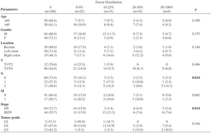
A fascin IHS >10 was statistically signiicantly more commonly observed in tumors with lymph node metastasis (p:0.005) and more often present in tumors located in the right colon (p:0.020) (Table 4). Similarly, higher stage (stage III/IV) tumors had signiicantly higher immunohistochemical scores (p:0.011). The percentage of cases with fascin IHS >10 was higher in the T3/T4 group (23.3%) compared to the T1/T2 group (5.9%). However, this difference was not statistically signiicant. Moreover, a mild to moderate correlation was observed between the number of positive lymph nodes and fascin immunoreactivity (r:0.373, p:0.001). There was a stronger correlation between fascin immunoreactivity in the invasive borders of tumors and lymph node metastasis (r:0.747, p:0.005).
In stage III/IV tumors, while two-year survival was 92.2% in tumors without fascin immunoreactivity, survival was 68.2% in tumors with fascin IHS≤10 and only 60.0% in tumors with fascin IHS>10 (log rank:11.836, p:0.003).
DISCUSSION
Fascin is expressed in many types of transformed epithelial cell lines and in several solid neoplasms. Fascin
expression has been correlated with tumor stage12, 13,21,22 and grade,9,12,22 pT class,12,13 lymph node involvement12,13,21,23 and recurrence.12 Furthermore, fascin expression is related to poor prognosis for some solid neoplasms because it facilitates invasion and metastasis.4,9,11-13,15,24,25
In the present study, no immunohistochemical staining was observed in the normal colonic epithelium adjacent to the tumor, in accordance with previous reports.1,15 Jawhari et al. demonstrated that fascin was not detectable in normal colonic epithelium, but was dramatically up-regulated in colorectal adenocarcinoma.1 Moreover, scattered fascin expression was demonstraed in control specimens of normal colonic glandular epithelia in the study by Tsai et al.17 Additionally, fascin was up-regulated in a proportion of adenomas, where its expression was often focal.15 Furthermore, only 16% of adenomas showed fascin expression in greater than 10% of the tumor cells. Finally, Tsai et al. found that higher fascin immunostaining scores were signiicantly associated with advanced dysplasia in colorectal adenomas.17
In this study, we observed fascin immunoreactivity in 35.3% of all cases and noted that fascin positivity was signiicantly higher in right colon adenocarcinomas. In a study Table 2 - Relationship between fascin intensity and clinicopathological characteristics in 167 colorectal carcinomas
Parameters Fascin Intensity p Absent (n=108) (%) Weak (1+) (n=24) (%) Moderate (2+) (n=25) (%) Intense (3+) (n=10) (%) Age ≤60 >60 50 (69.4) 58 (61.1) 7 (9.7) 17 (17.9) 9 (12.5) 16 (16.8) 6 (8.3)
4 (4.2) 0.258
Gender M F 64 (72.1) 44 (60.4) 16 (13.1) 8 (15.1) 19 (9.8) 6 (17.9) 7 (4.9)
3 (6.6) 0.422
Location Rectum Left colon Right colon 39 (69.6) 50 (71.4) 19 (46.3) 9 (16.1) 4 (5.7) 11 (26.8) 7 (12.5) 11 (15.7) 7 (17.1) 1 (1.8) 5 (7.1) 4 (9.8) 0.028 T T1/T2 T3/T4 12 (70.6) 96 (64.0) 5 (29.4) 19 (12.7) 0 25 (16.7) 0 10 (6.7) 0.070 N N0 N1 N2 68 (73.9) 23 (57.5) 17 (48.6) 16 (17.4) 2 (5.6) 6 (17.1) 4 (4.3) 14 (35.0) 7 (20.0) 4 (4.3) 1 (2.5)
5 (14.3) < 0.0001
M M0 M1 91 (66.4) 17 (56.7) 20 (14.6) 4 (13.3) 17 (12.4) 8 (26.7) 9 (6.6)
1 (3.3) 0.244
Stage I/II III/IV 64 (72.7) 44 (55.7) 14 (19.9) 10 (12.7) 6 (6.8) 19 (24.1) 4 (4.5)
6 (7.6) 0.011
by Hashimoto et al. using conventional tissue sections from 35 adenocarcinomas, 26% showed fascin expression in greater than 10% of tumor cells.15 When examining fascin expression in 228 advanced colonic adenocarcinoma patients, Puppa et al. found that fascin was detected in 71% of tumors.16 In both these studies, fascin immunoreactivity was more prevalent in tumors located in the proximal colon.15, 16
Interestingly, no association was identified between fascin expression and age or gender. This observation was in accordance with the clinically annotated tumors, where fascin immunoreactivity was not associated with age and gender.15 However, in a recent study, fascin correlated signiicantly with the female sex.16
Although immunohistochemical scores were higher for poorly differentiated tumors in the present study, this inding was not statistically signiicant. In a study by Tsai et al., fascin immunohistochemistry was performed in tissue microarrays of 91 surgical specimens. Tsai et al. found that higher fascin immunostaining scores were significantly associated with high-grade histopathological differentiation of colorectal adenocarcinomas.17 Similarly, another study found a signiicant correlation between fascin expression and tumor grade and mucinous differentiation.16 In the present study, no differences were observed in fascin expression
between mucinous and nonmucinous adenocarcinomas. In mucinous adenocarcinomas, fascin immunoreactivity was generally weak in half of the cases. However, more studies with a large number of cases are needed to analyze the role of fascin in mucinous colorectal adenocarcinomas.
We also noted an association between fascin expression and the N stage of colorectal carcinomas. Specifically, strong immunoreactivity in the invasive front of tumors was correlated with the number of metastatic lymph nodes. The association of fascin expression with lymph node involvement has also been documented for other gastrointestinal cancers such as oesophageal13 and gastric carcinomas,12 as well as pulmonary neoplasms.21, 26 Tsai et al. reported that higher fascin immunostaining scores were signiicantly associated with an advanced T stage of colorectal carcinomas.17 In their study, higher fascin scores were also related with more advanced M and N stages of colorectal carcinomas. However, in the study by Hashimoto et al. with clinically annotated colorectal carcinomas, fascin immunoreactivity was not associated with TNM stage.15 In a recent study, fascin expression was found to be signiicantly correlated with the number of metastatic lymph nodes, extranodal tumor extensions and the occurrence of distant metastases.16
Table 3 - Relationship between fascin distribution and clinicopathological characteristics in 167 colorectal carcinomas
In the present study, although the mean follow-up was not long; overall survival in stage III/IV tumors was shorter for cases with fascin expression compared to those without. In the study of Hashimoto et al., fascin expression was investigated using a tissue microarray containing cores of 158 colorectal adenocarcinomas with a mean of 38 months of clinical follow-up.15 Patients with stage III/ IV adenocarcinomas with strong fascin immunoreactivity had a worse prognosis than patients with low or no fascin immunoreactivity (3-year overall survival of 11% vs. 43% for fascin-negative patients; p = 0.023). Similarly, a study of fascin expression in 228 advanced colonic adenocarcinoma patients with a long follow-up period revealed that patients with fascin-expressing tumors had a shorter disease-free and overall survival compared to patients with fascin-negative tumors. Moreover, fascin immunoreactivity emerged as an independent prognostic factor in the multivariate analysis.16 Moreover, patients with the same tumor stages could be stratified in different risk categories for relapse and progression according to fascin expression.
The contribution of fascin to the migration of cancer cells and its relationship with beta-catenin were recently analyzed.14,27 Vignjevic et al. identiied fascin as a target of
beta-catenin-TCF signaling in colorectal cancer cells. Fascin mRNA and protein expression were increased in primary cancers in a stage-dependent manner. Fascin was exclusively localized at the invasive front of tumors that also displayed the nuclear localization of beta-catenin, which is a hallmark of activated Wnt signaling. Their studies show that fascin expression is tightly regulated during the development of colon cancer metastases and that it is a novel target of beta-catenin-TCF signaling. They proposed that transient up-regulation of fascin in colorectal cancer promotes the acquisition of migratory and invasive phenotypes that lead to metastasis. Moreover, they also found that the expression of fascin is down-regulated when tumor cells reach their metastatic destination, where migration ceases and proliferation is enhanced.14 To understand the functional and mechanistic contributions of fascin, Hashimoto et al. used inducible short hairpin RNA (shRNA) to knockdown fascin expression in human colon carcinoma cells derived from an aggressive primary tumor. Fascin depletion led to decreased numbers of ilopodia and an altered morphology of cell protrusions, decreased Rac-dependent migration on laminin, decreased turnover of focal adhesions, and decreased xenograft tumor development and metastasis in vivo. They further showed that both the actin bundling and active PKC-binding activities of fascin were required for the organization of ilopodial protrusions and tumor metastasis. Thus, these authors concluded that fascin contributes to carcinoma migration and metastasis through parallel pathways that have an impact on multiple subcellular structures that are needed for cell migration.27
Over the past 10 years, our knowledge of the genes and genetic pathways associated with cancer development and progression has been greatly expanded. From the perspective of colorectal carcinomas, one of the important carcinogenetic factors is epidermal growth factor receptor (EGFR), which contributes to the development and progression of several carcinomas and is overexpressed in 50-80% of colorectal cancers.28 Gain-of-function KRAS mutations lead to EGFR-independent activation of intracellular signaling pathways, resulting in tumor cell proliferation, protection against apoptosis, increased invasion and metastasis.29 For EGFR-targeted therapy, standardized testing for KRAS mutation status has been recommended.30 Another example is CD133, the expression of which correlates with liver metastasis and has a prognostic impact in colorectal carcinomas.31 The combination of CD133 and nuclear beta-catenin expression was shown to be a powerful tool to identify high-risk cases of stage IIA colon cancer.32
CONCLUSION
Fascin expression is present in approximately one third
Table 4 - Immunohistochemical scores of fascin expression
in colorectal carcinomas
Parameters
1. Jawhari AU, Buda A, Jenkins M, Shehzad K, Sarraf C, Noda M, et al. Fascin, an actin-bundling protein, modulates colonic epithelial cell invasiveness and differentiation in vitro. Am J Pathol. 2003;162:69-80. 2. Tao YS, Edwards RA, Tubb B, Wang S, Bryan J, McCrea PD.
beta-Catenin associates with the actin-bundling protein fascin in a noncadherin complex. J Cell Biol. 1996;134:1271-81.
3. Beachy PA, Karhadkar SS, Berman DM. Tissue repair and stem cell renewal in carcinogenesis. Nature. 2004;432:324-31.
4. Kureishy N, Sapountzi V, Prag S, Anilkumar N, Adams JC. Fascins, and their roles in cell structure and function. Bioessays. 2002;24:350-61. 5. Zhang FR, Tao LH, Shen ZY, Lv Z, Xu LY, Li EM. Fascin expression in
human embryonic, fetal, and normal adult tissue. J Histochem Cytochem. 2008;56:193-9.
6. Hu W, McCrea PD, Deavers M, Kavanagh JJ, Kudelka AP, Verschraegen CF. Increased expression of fascin, motility associated protein, in cell cultures derived from ovarian cancer and in borderline and carcinomatous ovarian tumors. Clin Exp Metastasis. 2000;18:83-8. 7. Grothey A, Hashizume R, Ji H, Tubb BE, Patrick CW Jr, Yu D, et
al. C-erbB-2/ HER-2 upregulates fascin, an actin-bundling protein associated with cell motility, in human breast cancer cell lines. Oncogene. 2000;19:4864-75.
8. Maitra A, Iacobuzio-Donahue C, Rahman A, Sohn TA, Argani P, Meyer R, et al. Immunohistochemical validation of a novel epithelial and a novel stromal marker of pancreatic ductal adenocarcinoma identiied by global expression microarrays: sea urchin fascin homolog and heat shock protein 47. Am J Clin Pathol. 2002;118:52-9.
9. Pelosi G, Pastorino U, Pasini F, Maissoneuve P, Fraggetta F, Iannucci A, et al. Independent prognostic value of fascin immunoreactivity in stage I nonsmall cell lung cancer. Br J Cancer. 2003;88:537-47. 10. Goncharuk VN, Ross JS, Carlson JA. Actin-binding protein fascin
expression in skin neoplasia. J Cutan Pathol. 2002;29:430-8. 11. Gunal A, Onguru O, Safali M, Beyzadeoglu M. Fascin expression
[corrected] in glial tumors and its prognostic significance in glioblastomas. Neuropathology. 2008;28:382-6.
12. Hashimoto Y, Shimada Y, Kawamura J, Yamasaki S, Imamura M. The prognostic relevance of fascin expression in human gastric carcinoma. Oncology. 2004;67:262-70.
13. Hashimoto Y, Ito T, Inoue H, Okumura T, Tanaka E, Tsunoda S, et al. Prognostic signiicance of fascin overexpression in human esophageal squamous cell carcinoma. Clin Cancer Res. 2005;11:2597-605.
14. Vignjevic D, Schoumacher M, Gavert N, Janssen KP, Jih G, Lae M, et al. Fascin, a novel target of beta-catenin-TCF signaling, is expressed at the invasive front of human colon cancer. Cancer Res. 2007;67:6844-53. 15. Hashimoto Y, Skacel M, Lavery IC, Mukherjee AL, Casey G, Adams JC. Prognostic signiicance of fascin expression in advanced colorectal cancer: an immunohistochemical study of colorectal adenomas and adenocarcinomas. BMC Cancer. 2006;6:241.
16. Puppa G, Maisonneuve P, Sonzogni A, Masullo M, Chiappa A, Valerio M, et al. Independent prognostic value of fascin immunoreactivity in stage III-IV colonic adenocarcinoma. Br J Cancer. 2007;96:1118-26. 17. Tsai WC, Chao YC, Sheu LF, Chang JL, Nieh S, Jin JS. Overexpression
of fascin-1 in advanced colorectal adenocarcinoma: tissue microarray analysis of immunostaining scores with clinicopathological parameters. Dis Markers. 2007;23:153-60.
18. Hashimoto Y, Skacel M, Adams JC. Association of loss of epithelial syndecan-1 with stage and local metastasis of colorectal adenocarcinomas: an immunohistochemical study of clinically annotated tumors. BMC Cancer. 2008;8:185.
19. Hashimoto Y, Loftis DW, Adams JC. Fascin-1 promoter activity is regulated by CREB and the aryl hydrocarbon receptor in human carcinoma cells. PLoS One 2009;4: e5130.
20. Parsons M, Adams JC. Rac regulates the interaction of fascin with protein kinase C in cell migration. J Cell Sci. 2008;121(Pt 17):2805-13. 21. Choi PJ, Yang DK, Son CH, Lee KE, Lee JI, Roh MS. Fascin
immunoreactivity for preoperatively predicting lymph node metastases in peripheral adenocarcinoma of the lung 3 cm or less in diameter. Eur J Cardiothorac Surg. 2006;30:538-42.
22. Yoder BJ, Tso E, Skacel M, Pettay J, Tarr S, Budd T, et al. The expression of fascin, an actin-bundling motility protein, correlates with hormone receptor-negative breast cancer and a more aggressive clinical course. Clin Cancer Res. 2005;11:186-92.
23. Zhang H, Xu L, Xiao D, Xie J, Zeng H, Cai W, et al. Fascin is a potential biomarker for early-stage oesophageal squamous cell carcinoma. J Clin Pathol. 2006;59:958-64.
24. Rodriguez-Pinilla SM, Sarrio D, Honrado E, Hardisson D, Calero F, Benitez J, et al. Prognostic signiicance of basal-like phenotype and fascin expression in node-negative invasive breast carcinomas. Clin Cancer Res. 2006;12:1533-9.
25. Roma AA, Prayson RA. Fascin expression in 90 patients with glioblastoma multiforme. Ann Diagn Pathol 2005;9:307-11.
REFERENCES
of colorectal adenocarcinomas and is more prevalent in right colon tumors. In this study, we noted an association between fascin expression and the N stage of colorectal carcinomas. Speciically, we observed strong immunoreactivity in the invasive front of tumors that was correlated with the number of metastatic lymph nodes. Moreover, we found that in
26. Pelosi G, Pasini F, Fraggetta F, Pastorino U, Iannucci A, Maisonneuve P, et al. Independent value of fascin immunoreactivity for predicting lymph node metastases in typical and atypical pulmonary carcinoids. Lung Cancer. 2003;42:203-13.
27. Hashimoto Y, Parsons M, Adams JC. Dual actin-bundling and protein kinase C-binding activities of fascin regulate carcinoma cell migration downstream of Rac and contribute to metastasis. Mol Biol Cell. 2007;18:4591-602.
28. Spano JP, Lagorce C, Atlan D, Milano G, Domont J, Benamouzig R, et al. Impact of EGFR expression on colorectal cancer patient prognosis and survival. Ann Oncol.2005;16: 102-8.
29. Ciardiello F, Tortora G. EGFR antagonists in cancer treatment. N Engl J. Med 2008;358:1160-74.
30. van Krieken JH, Jung A, Kirchner T, Carneiro F, Seruca R, Bosman FT, et al. KRAS mutation testing for predicting response to anti-EGFR therapy for colorectal carcinoma: proposal for an European quality assurance program. Virchows Arch. 2008;453:417-31.
31. Horst D, Scheel SK, Liebmann S, Neumann J, Maatz S, Kirchner T, et al. The cancer stem cell marker CD133 has high prognostic impact but unknown functional relevance for the metastasis of human colon cancer. J Pathol. 2009;219:427-34.