Evidence of progenitor cells in the adult human cochlea:
sphere formation and identification of ABCG2
Milene Massucci-Bissoli,* Karina Lezirovitz,* Jeanne Oiticica, Ricardo Ferreira Bento Departamento de Otorrinolaringologia, Faculdade de Medicina FMUSP, Universidade de Sao Paulo, Sao Paulo, SP, BR.
OBJECTIVES:The aim of this study was to search for evidence of stem or progenitor cells in the adult human cochlea by testing for sphere formation capacity and the presence of the stem cell marker ABCG2.
METHODS: Cochleas removed from patients undergoing vestibular schwannoma resection (n=2) and from brain-dead organ donors (n=4) were dissociated for either flow cytometry analysis for the stem cell marker ABCG2 or a sphere formation assay that is widely used to test the sphere-forming capacity of cells from mouse inner ear tissue.
RESULTS:Spheres were identified after 2-5 daysin vitro, and the stem cell marker ABCG2 was detected using flow cytometric analysis after cochlear dissociation.
CONCLUSIONS:Evidence suggests that there may be progenitor cells in the adult human cochlea, although further studies are required.
KEYWORDS: Progenitor Cells; Cochlea; ABCG2; Human.
Bissoli MM, Lezirovitz K, Oiticica J, Bento RF. Evidence of progenitor cells in the adult human cochlea: sphere formation and identification of ABCG2. Clinics. 2017;72(11):714-717
Received for publication onMarch 2, 2017;First review completed onMay 31, 2017;Accepted for publication onJuly 12, 2017 *Corresponding authors. E-mails: [email protected] / [email protected]
’ INTRODUCTION
Hearing loss is one of the most common sensorial deficiencies in humans. Although there exist several options for treat-ing heartreat-ing loss, none of these approaches can completely reestablish auditory physiology. Prosthetic devices require maintenance and have inherent daily limitations due to their electrical nature; many such devices are simply not designed to be used while sleeping. One possible strategy to reestablish auditory physiology is the replacement of lost sensorial cells. In theory, the discovery of adult stem cells in the cochlea would bring this possibility closer to clinical practice.
No prior studies have attempted to find evidence of stem cells in the adult human cochlea. The availability of human cochlear tissue is limited, and most authors have chosen to use cochleas from patients who are undergoing surgical procedures that suggest permanent hearing loss (1-4). The only references to human cochlear stem cells involve fetal specimens (5-6).
Sphere formation assays have been used to demonstrate the proliferative capacity of neonatal mammalian cochleas in different species (7-9). Spheres have also been identified in adult mice, although such spheres were not numerous, could
not differentiate in vitro and exhibited few developmental
markers (10). In neonatal mice, there also exists evidence of a side population of cochlear cells that express the stem cell marker ABCG2 and are capable ofin vitroproliferation,
self-renovation and differentiation (11-14).
The aim of this study was to identify evidence of progen-itor or stem cells in the adult human cochlea. To test our hypothesis, we used cochleas removed from patients under-going vestibular schwannoma (VS) resection and brain-dead organ donors; this article is the first report involving the use of cochlear tissue from organ donors.
’ MATERIALS AND METHODS
Patients
Cochlear samples were obtained from patients undergoing excision of a VS via a translabyrinthine approach (n=2) and brain-dead organ donors (n=4) at University of Sao Paulo Clinics Hospital. All patients or their legal representatives provided informed consent prior to tissue collection in accordance with requirements of local and national ethics committees.
Surgery in the VS group
A translabyrinthine approach was utilized by the same surgeon for both patients, and cochlear access was achieved as described by Browne and Fisch (15). After cochlear exposure, a wide cochleotomy was performed to allow for tissue removal. The membranous portion of the cochlea was collected into culture medium for immediate transport to the laboratory.
DOI:10.6061/clinics/2017(11)11
Copyright&2017CLINICS–This is an Open Access article distributed under the terms of the Creative Commons License (http://creativecommons.org/licenses/by/ 4.0/) which permits unrestricted use, distribution, and reproduction in any medium or format, provided the original work is properly cited.
No potential conflict of interest was reported.
714
Tissue collection in the organ donor group
To reach the cochlear promontory, three different approaches were tested. On one side, we used an endaural approach; on the remaining sides, retroauricular access was established to perform a mastoidectomy. On four sides, a posterior tympanotomy was conducted to gain access to the cochlear promontory. On one side, the posterior wall of the external auditory canal was drilled away to improve cochlear visualization. In all cases, after cochlear exposure, a wide cochleotomy was performed to allow for tissue removal. The membranous portion of the cochlea was collected into culture medium for immediate transport to the laboratory.
Sphere formation assay
To test the ability of removed cochlear tissue to form spheres, we used the protocol described by Oshima et al. (16). Briefly, tissue was inspected with an inverted microscope, dissociated with trypsin, mechanically passed through a 70 mm filter and cultured under non-adherent conditions
on defined media (DMEM-F12 with 2 B-27, 1 N2, 2 mM
glutamine, 2mL/mL ITS, 6 g/L glucose, 0.2mL/mL ampicillin,
20 ng/mL EGF, 10 ng/mL bFGF and 50 ng/mL IGF) for up to five days.
Flow cytometry
To verify the presence of the stem cell marker ABCG2, removed cochlear tissue was transferred to the laboratory in
Eagle’s minimum essential medium (EMEM; Vitrocell Embrio-life, Campinas, SP, Brazil). Due to limited equipment avail-ability, samples remained at 4o
C for up to 36 hours; they were then dissociated with trypsin and mechanically dissociated (16) for antibody incubation in accordance with the manufac-turer’s instructions.
We used an anti-ABCG2 antibody (Biolegend, 332020, San Diego, CA, USA) and its isotype (Biolegend, 401209), and flow cytometric analysis was performed on an Attunes
NxT Acoustic Focusing Cytometer (Thermo Fisher Scientific, Inc., Waltham, MA, USA).
’ RESULTS
Patients and tissue collection
The patients in the VS group (both female) were 61 and 63 years of age, and those in the organ donor group (2 males and 2 females) were 17-55 years of age. Both patients had moderate unilateral sensorineural hearing loss. In the organ donor group, six of the eight available sides were used. We have no auditory testing data from this group.
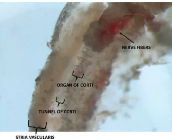
There was great variability in the quantities of tissue collected from patients and organ donors. In all cases, a por-tion of the organ of Corti and stria vascularis could be identified. In the organ donor group, the greatest quantity of tissue was collected when we used a retroauricular approach followed by mastoidectomy with drilling of the posterior wall of the external auditory canal. In the VS group, vari-ability in tissue quantities was mainly attributable to anatomical variations. Figure 1 depicts a section of the membranous portion of the cochlea shortly after its removal from a patient undergoing VS resection.
Identification of spheres
Three independent sphere formation assays were performed. Two of these assays used samples from the VS group (involv-ing one cochlea each), and the remain(involv-ing assay utilized a sample from the organ donor group (with two cochleas assessed in the same experiment). The evaluated cochleas were from 61- and 63-year-old female patients who were undergoing surgery and a 17-year-old male organ donor.
Spheres were identified in two of the three experiments (Figure 2).
Identification of ABCG2
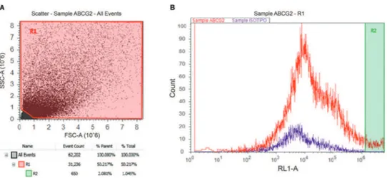
Four cochleas from three organ donors were used for flow cytometric identification of ABCG2. The donors included one 38-year-old male and 33- and 55-year-old females. There was a clear population of ABCG2-positive
Figure 1 -Membranous portion of the cochlea that was removed from a patient undergoing VS resection.
Figure 2 -Sphere identification for samples from 2A, a 61-year-old female undergoing VS resection at 5 DIV; 2B, a 17-year-old male organ donor at 3 DIV; and 2C, a 17-year-old male organ donor at 5 DIV.
715
CLINICS 2017;72(11):714-717 Evidence of progenitor cells in adult human cochlea
cells in the dissociated cochlea from the 33-year-old female (Figure 3).
Comparative table of results
’ DISCUSSION
This article is the first report describing sphere formation and the identification of ABCG2 in the adult human cochlea and the first investigation in which brain-dead organ donors were a source of cochleas used for research.
The accurate identification of stem cellsin vivoremains a
major obstacle to understanding in stem cell biology since there are no single and universal stem cell markers common to all adult stem cells (17).
To identify evidence of stem or progenitor cells in the adult human cochlea, we used a sphere-forming assay validated for neonatal mice (16) that has been used to test the sphere-forming capacity of cells from adult mice (10). We identified spheres in two out of three experiments; nevertheless, the number of spheres was minimal, and no further testing could be adequately performed for further characterization.
Prior research has demonstrated that neonatal mice have a cochlear side population that expresses the stem cell marker ABCG2 (11). Several authors have established that the direct identification of ABCG2 in this tissue is a marker of this side population (18-20). We have used flow cytometric analysis to reveal the presence of ABCG2 in adult human dissociated cochleas. We have been unable to sort and regrow ABCG2-positive populations due to the small number of cells obtained in our experiments, but testing to determine whether these cells behave similarly to mouse cochlear side population/ ABCG2-positive cells would be extremely valuable (12-14).
A greater number of cochlear cells could be obtained for the aforementioned experiments via the collection of more
cochleas from brain-dead organ donors, the standardization of the procedure for membranous cochlear resection, and the development of greater expertise with this procedure. Vari-ability and little reproducibility in the removal of membra-nous cochlear tissue have been reported previously (1). However, with more widespread use of this material, this issue can be solved.
We have demonstrated evidence of the presence of progen-itor cells in the adult human cochlea in the form of sphere formation and the detection of ABCG2. These findings do not conclusively establish the presence of stem cells in this tissue, but they do open a new field for stem cell research. We have reported the first use of brain-dead organ donors for cochlear resection for research purposes; this approach can be of inestimable value for hearing research.
’ ACKNOWLEDGMENTS
We thank Dr. Tharcísio Citrângulo Tortelli, Junior, for helping withflow cytometry analysis and doctors Fábio de Alencar Rodrigues, Junior, Francisco das Chagas Cabral, Junior, Paula Tardim Lopes and Ricardo Dourado Alves for performing the organ donor cochlear resections. Funding: This work was supported by the Brazilian National Council of Technological and Scientific Development (CNPq) in the form of a scholarship to thefirst author.
’ AUTHOR CONTRIBUTIONS
Bissoli MM was responsible for the manuscript preparation, cochlear prep-aration after its removal from patients and cell culture and staining for
flow cytometry. Lezirovitz K was responsible for co-mentoring, manuscript preparation andfigure selection. Oiticica J was responsible for the cochlear preparation after its removal from patients in selected cases and mentoring. Bento RF was responsible for the surgery, mentoring and manuscript preparation.
’ REFERENCES
1. Oghalai J, Holt JR, Nakagawa T, Jung TM, Coker NJ, Jenkins HA, et al. Harvesting human hair cells. Ann Otol Rhinol Laryngol. 2000;109(1):9-16, http://dx.doi.org/10.1177/000348940010900102.
2. Rask-Andersen H, Boström M, Gerdin B, Kinnefors A, Nyberg G, Engstrand T, et al. Regeneration of human auditory nerve. In vitro/ in video demonstration of neural progenitor cells in adult human and guinea pig spiral ganglion. Hear Res. 2005;203(1-2):180-91, http://dx.doi. org/10.1016/j.heares.2004.12.005.
3. Wei L, Rui G, Helge RA. Morphological Study of Surgically Obtained Human Cochlear Specimens – Technical Aspects. Journal of Otology.
2014;9(1):52-7, http://dx.doi.org/10.1016/S1672-2930(14)50010-4.
Figure 3 -Flow cytometric analysis of a dissociated cochlea from a 33-year-old female (3B). There exists a population of cells positive for ABCG2 (R2) that represents 1.04% of the total cell population (3A).
Age Gender Experiment Result
61 female sphere formation assay negative
63 female sphere formation assay positive
17 male sphere formation assay positive
38 male ABCG2 identification inconclusive
55 female ABCG2 identification inconclusive
33 female ABCG2 identification positive
716 Evidence of progenitor cells in adult human cochlea
4. Taylor RR, Jagger DJ, Saeed SR, Axon P, Donnelly N, Tysome J, et al. Characterizing human vestibular sensory epithelia for experimental studies: new hair bundles on old tissue and implications for therapeutic interventions in ageing. Neurobiol Aging. 2015;36(6):2068-84, http://dx. doi.org/10.1016/j.neurobiolaging.2015.02.013.
5. Chen W, Cacciabue-Rivolta DI, Moore HD, Rivolta MN. The human fetal cochlea can be a source for auditory progenitors/stem cells isolation. Hear Res. 2007;233(1-2):23-9, http://dx.doi.org/10.1016/j.heares.2007.06.006. 6. Chen W, Johnson SL, Marcotti W, Andrews PW, Moore HD, Rivolta MN.
Human fetal auditory stem cells can be expandedin vitroand differentiate into functional auditory neurons and hair cell-like cells. Stem Cells. 2009; 27(5):1196-204, http://dx.doi.org/10.1002/stem.62.
7. Malgrange B, Belachew S, Thiry M, Nguyen L, Rogister B, Alvarez ML, et al. Proliferative generation of mammalian auditory hair cells in culture. Mech Dev. 2002;112(1-2):79-88, http://dx.doi.org/10.1016/S0925-4773(01) 00642-6.
8. Li H, Liu H, Heller S. Pluripotent stem cells from the adult mouse inner ear. Nat Med. 2003;9(10):1293-9, http://dx.doi.org/10.1038/nm925. 9. Oiticica J, Barboza-Junior LC, Batissoco AC, Lezirovitz K,
Mingroni-Netto RC, Haddad LA, et al. Retention of progenitor cell phenotype in otospheres from guinea pig and mouse cochlea. J Transl Med. 2010;8:119, http://dx.doi.org/10.1186/1479-5876-8-119.
10. Lou X, Dong Y, Xie J, Wang X, Yang L, Tokuda M, et al. Comparing the cultivated cochlear cells derived from neonatal and adult mouse. J Transl Med. 2014;12:150, http://dx.doi.org/10.1186/1479-5876-12-150. 11. Savary E, Hugnot JP, Chassigneux Y, Travo C, Duperray C, Van De Water
T, et al. Distinct population of hair cell progenitors can be isolated from the postnatal mouse cochlea using side population analysis. Stem Cells. 2007;25(2):332-9, http://dx.doi.org/10.1634/stemcells.2006-0303. 12. Chen HC, Sytwu HK, Chang JL, Wang HW, Chen HK, Kang BH, et al.
Hypoxia enhances the stemness markers of cochlear stem/progenitor cells and expands sphere formation through activation of hypoxia-inducible
factor-1 alpha. Hear Res. 2011;275(1-2):43-52, http://dx.doi.org/10.1016/ j.heares.2010.12.004.
13. Chao TT, Wang CH, Chen HC, Shih CP, Sytwu HK, Huang KL, et al. Adherent culture conditions enrich the side population obtained from the cochlear modiolus-derived stem/progenitor cells. Int J Pediatr Otorhinolaryngol. 2013;77(5):779-84, http://dx.doi.org/10.1016/j.ijporl. 2013.02.010.
14. Chen HC, Lee JT, Shih CP, Chao TT, Sytwu HK, Li SL, et al. Hypoxia Induces a Metabolic Shift and Enhances the Stemness and Expansion of Cochlear Spiral Ganglion Stem/Progenitor Cells. Biomed Res Int. 2015; 2015:359537, http://dx.doi.org/10.1155/2015/359537.
15. Browne JD, Fisch U. Transotic approach to the cerebellopontine angle. 1992. Neurosurg Clin N Am. 2008;19(2):265-78, http://dx.doi.org/10.1016/ j.nec.2008.02.008.
16. Oshima K, Senn P, Heller S. Isolation of sphere-forming stem cells from the mouse inner ear. Methods Mol Biol. 2009;493:141-62, http://dx.doi. org/10.1007/978-1-59745-523-7.
17. Morrison SJ, Spradling AC. Stem cells and niches: mechanisms that pro-mote stem cell maintenance throughout life. Cell. 2008;132(4):598-611, http://dx.doi.org/10.1016/j.cell.2008.01.038.
18. Zhou S, Schuetz JD, Bunting KD, Colapietro AM, Sampath J, Morris JJ, et al. The ABC transporter Bcrp1/ABCG2 is expressed in a wide variety of stem cells and is a molecular determinant of the side-population pheno-type. Nat Med. 2001;7(9):1028-34, http://dx.doi.org/10.1038/nm0901-1028. 19. Watanabe K, Nishida K, Yamato M, Umemoto T, Sumide T, Yamamoto K, et al. Human limbal epithelium contains side population cells expressing the ATP-binding cassette transporter ABCG2. FEBS Lett. 2004;565(1-3): 6-10, http://dx.doi.org/10.1016/j.febslet.2004.03.064.
20. Pfister O, Oikonomopoulos A, Sereti KI, Sohn RL, Cullen D, Fine GC, et al. Role of the ATP-binding cassette transporter Abcg2 in the phenotype and function of cardiac side population cells. Circ Res. 2008;103(8):825-35, http://dx.doi.org/10.1161/CIRCRESAHA.108.174615.
717
CLINICS 2017;72(11):714-717 Evidence of progenitor cells in adult human cochlea