Prevalence of hearing impairment in
patients with mild cognitive impairment
Leonardo da Costa Lopes
1, Regina Miksian Magaldi
2, Mara Edwirges Rocha Gândara
3,
Ana Carolina de Barros Reis
4, Wilson Jacob-Filho
5Abstract – The correlation between hearing and cognition is well established in dementia, but not in mild cognitive impairment (MCI). Objective: The aim of the present study was to defi ne the prevalence of hearing impairment in elderly patients with MCI and in controls. Methods: Twenty-nine patients with MCI and 24 control subjects were analyzed. We evaluated memory and hearing impairments through clinical tests, including the Mini Mental Status Examination, Clinical Dementia Rating (CDR) and Hearing Handicap Inventory for the Elderly Screening (HHIE-S). Audiometries were performed in 22 patients with MCI and 19 subjects in a control group.
Results: MCI patients showed more hearing complaints (68.9%) compared to the control group (25%) (p=0.001). No differences in the intensity of hearing complaints, measured by the HHIE-S, were detected. Nonetheless, dif-ferences between mean hearing threshold (MCI group=23.4±11.3 dB and control group=16.0±10.1dB) (p=0.03) were identifi ed. Conclusions: There is a signifi cant association between MCI and hearing impairment. Hearing impairment in MCI patients may be a contributory factor to cognitive decline. This may however be related to the same neuropathological process, due to lesions of cortical areas related to hearing. The early diagnosis of hearing impairment in MCI patients may offer a more appropriate approach to this disease.
Key words: dementia, memory, hearing, audiometry, elderly, aged.
Prevalência de defi cit auditivo em pacientes com comprometimento cognitivo leve
Resumo – A relação entre audição e cognição está bem estabelecida em demências, porém não no comprome-timento cognitivo leve (CCL). Objetivo: Propomos um estudo para determinar a prevalência de defi cit auditivo em idosos portadores de CCL e controles. Métodos: Foram avaliados 29 pacientes com CCL e 24 controles. Ana-lisamos as perdas de memória e de audição através de testes, como o Mini Exame do Estado Mental, o Escore Clínico de Demência e o HHIE-S (Hearing Handicap Inventory for the Elderly Screening). Vinte e dois pacientes com CCL e 19 controles foram submetidos a audiometrias. Resultados: O grupo CCL apresentou mais queixas auditivas (68,9%) se comparado com o controle (25%) (p=0.001). Não foram encontradas diferenças na intensi-dade da queixa auditiva, medida pelo HHIE-S. Foram detectadas diferenças entre a média dos limiares auditivos de pacientes com CCL (23,4±11,3 dB) e de controles (16,0±10,1 dB) (p=0,03). Conclusões: Existe signifi cativa associação entre CCL e perdas auditivas. O defi cit auditivo em pacientes com CCL pode ser um fator contribuinte para o declínio cognitivo ou estar relacionado a um mesmo processo neuropatológico, devido à lesão de áreas corticais relacionadas à audição. O diagnóstico precoce de perdas auditivas em pacientes com CCL pode permitir uma abordagem mais adequada desta doença.
Palavras-chave: demência, memória, audição, audiometria, idosos.
1,2,3,5Geriatric Division of Hospital das Clínicas, Department of Internal Medicine, São Paulo University Medical School, São Paulo, SP, Brazil. 2,4 Otorhi-nolaryngology Division of Hospital das Clínicas, São Paulo University Medical School, São Paulo, SP, Brazil.
Leonardo da Costa Lopes – Rua Capote Valente, 668 / 164 - 05409-002 São Paulo SP - Brasil. E-mail: [email protected]
The relationship between hearing impairment and cog-nitive decline has previously been demonstrated,1 showing
that even mild or moderate hearing losses are correlated with poor performance in verbal memory.2. A link has
also been established among hearing loss, depression and functional decline,3 especially evident in individuals with
hearing threshold >35 dB.4
More than 90% of patients with Alzheimer disease (AD) have some kind of hearing loss.5 Although this
re-lationship between cognition and hearing has been well studied in patients with dementia, the association in those with mild cognitive impairment (MCI) has not yet been as-sessed. MCI is a clinical entity, fi rst described in the 1990s,6
cogni-tive aging and dementia.7 To date, no studies published
in the English language have evaluated the prevalence of hearing impairment in such patients compared to normal control groups from a cognitive standpoint.
This paper aimed to analyze the prevalence of hearing impairment and its quantitative characteristics in elderly patients with MCI compared with normal elderly subjects from a control group, according to the cognitive standpoint – through a comparative transversal study.
Methods
Twenty-nine patients with subjective memory com-plaints (MCI group) and 24 control-subjects without cog-nitive complaints (control group) were studied. Subjects were recruited from May to November of 2005. The MCI group was formed through assessment of subjects attend-ing a memory clinic at the Geriatrics Department of Hos-pital das Clínicas – São Paulo University Medical School (SG-HC/FMUSP). The control group was formed through assessment of subjects followed by a multidisciplinary care group from SG-HC/FMUSP in 2003 and 2004. The study was approved by the Ethics Commission of the Institution. The inclusion criteria were: (a) age of 60 years or over; (b) Clinical Dementia Rating (CDR) of 0.5 in patients with cognitive complaints and CDR of 0 in controls; (c) ac-ceptance of an informed written consent. The exclusion criteria were: (a) presence of memory complaints among controls; (b) presence of clinical signs of dementia, as well as scores below those expected for schooling level in the Mini Mental Status Examination (MMSE) as established in the following section; (c) presence of any central ner-vous system disease, psychiatric diseases, such as anxiety and depression, severe infl ammatory systemic diseases, as well as hypothyroidism, defi ciency of vitamin B12, syphi-lis, renal and liver insuffi ciency; (d) use of drugs that act on the central nervous system, mainly benzodiazepines, antidepressives and anticholinergic drugs; (e) current or previous use of acetylcholinesterase inhibitor; (f) current or previous use of hearing aids, or presence of visual and hearing losses severe enough to prevent clinical interview or proposed tests from being conducted.
All subjects were investigated for schooling level, co-morbidities, medications being used, and hearing and memory complaints. Moreover, the following medical questionnaires were used: the MMSE,8, CDR,9,
CAM-COG, a structured interview based on the cognitive sec-tion of Cambridge Examinasec-tion for Mental Disorders of the Elderly10 and validated for the Brazilian population,11
Rivermead Behavioral Memory Test, which is divided into Rivermead 1 for scores of standardized profi le and Riv-ermead 2 for scores of screening,12 Digit Span, which is
divided into Digit Span 1for direct-order numbers and Digit Span 2 for reverse-order numbers,13 Whispered Test14
and Hearing Handicap Inventory for the Elderly Screening – HHIE-S.15. Of the total subjects included in this study, 19
from the control group and 22 from the MCI group were submitted to an auditory examination, tonal audiometry and determination of speech reception threshold and per-centage rate of speech recognition tests.
Mini Mental State Examination
MMSE was assessed according to schooling level of the subject, using the criteria suggested for Brazilian population by Herrera et al.,16 considering a cut off point of <28 for
subjects with more than 8 years at school, <24 for subjects who studied from 4 to 7 years, <23 for subjects who stud-ied between 1 and 3 years, and <19 for illiterate subjects.
Hearing tests
Subjects were investigated for hearing complaints and analyzed according to the HHIE-S, a scale that puts questions regarding the auditory performance in habitual situations. It is self applicable and assesses the degree of auditory loss, al-though is not capable of quantifying the level of the losses.15
All subjects were submitted to the Whispered Test and auditory examination. Twenty-two subjects from the MCI group and 19 from the control group were randomly se-lected and submitted to tonal audiometry. All tests were executed by the same examiner.
Tonal audiometry was performed by the MIDIMATE 622 audiometer, employing AZ 7 and Zodiac 901 middle-ear analyzers, studying air and bone conduction, in an acousti-cally isolated environment. The hearing threshold for fre-quencies from 250 to 8000 Hz was determined, as well as for frequencies of 500, 1000 and 2000 Hz, where means of these values for each ear were calculated.17 The result of the best
ear was considered, in order to eliminate any confounding factor caused by unilateral hearing losses.18 Subjects who
had a cerumen plug during otoscopy underwent removal before audiometry. Considering levels of severity of hearing loss, the threshold up to 25 dB is usually defi ned as normal, between 25 and 40 dB indicative of mild loss, between 40 and 65 dB as moderate loss, and over 65 dB as severe loss.19
We also calculated mean hearing thresholds at high frequen-cies (4000, 6000 and 8000 Hz), as well as the speech recep-tion threshold and percentage rate of speech recognirecep-tion.
Statistical analysis
were averages of the hearing threshold obtained through audiometry. Continuous variables were compared using the Student t test while categorical variables were compared using the chi-square test, determining the p values. Pro-portions were compared using Fisher’s Exact Test. Binary logistic regressions were performed to assess risk ratios (RR). For calculations, the statistical program MINI TAB 14 was used.
Results
Of 135 subjects initially considered, 53 were included in this study –29 of whom were part of the MCI group and 24 of the control group. The main causes of exclusion are shown in Table 1.
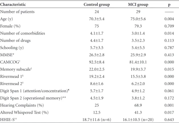
Table 2 shows clinical features of subjects and the result of tests applied.
The MCI group was older (75.0±5.6 years of age) than the control group (70.3±5.4 years of age), p<0.05. In ad-dition, a greater number of comorbidities was observed in the control group (4.1±1.7) compared to the MCI group (3.0±1.4), p<0.05. The most common diagnoses were similar for both groups: systemic arterial hypertension (54.1% and 58.6%), dyslipidemia (50% and 51.7%), and osteoarticular diseases (45.8% and 41.3%) respectively. The most common drugs used by both groups were also simi-lar: calcium carbonate (50% and 44.8%), statins (41.6% and 44.8%) and angiotensin-converting enzyme inhibitors (41.6% and 37.9%) respectively.
Regarding other clinical features, no statistical differ-ences between the groups were observed, nor in relation to the score on the MMSE and Digit Span.
However, scores of the two groups were different on the CAMCOG (92.5±8.4 and 81.4±10.1 for control and MCI group, respectively p<0.05) and in their subscale of memory (22.0±2.5 and 19.9±3.7, respectively, p<0.05), as were scores on the Rivermead Behavioral Test (Rivermead 1: 19.2±2.4 and 15.5±3.8; Rivermead 2: 8.6±1.6 and 6.2± 2.0, respectively, p<0.05).
The MCI group presented more hearing complaints (25% and 68.9%, control and MCI group, respectively, p<0.05). The risk ratio (RR) for patients with MCI pre-senting hearing complaints was 6.6. No signifi cant differ-ences in proportions of normal otoscopies were detected between the two groups. The Whispered Test was abnormal in 12.5% of control group subjects and 41.3% of the MCI group (p<0.05). The risk ratio (RR) of a patient with MCI being positive on the Whispered Test was 4.9. No differ-ences in the severity of hearing complaints were detected, according to the HHIE-S. The mean hearing threshold of the best ear assessed was statistically different between the two groups (16.0±10.1 dB and 23.4±11.3 dB, control and MCI group, respectively). Nevertheless, these thresholds lie within the range of audiometric normality. When we analyzed hearing means, classifying them individually as normal (up to 25 dB) or abnormal (>25dB), we observed that 10.5% of the control group subjects showed abnormal thresholds, while this was the case in 31.8% of the MCI group (p=0.09).
The analysis of hearing thresholds at high frequencies (4-8 KHz) revealed differences between control (33.2±15.4 dB) and MCI groups (46.2±20.1 dB), p<0.05. No signifi -cant differences between speech reception threshold and
Table 1. Causes of exclusion of the patients and control subjects.
Causes Patients (N=46) Causes Controls (N=36)
Depression 12 Depression 2
Dementia 8 Memory complaints 27
CDR=0 6 Language comprehension problems 2
Hypothyroidism 5 Hypothyroidism 1
B12 vitamin defi ciency 3 B12 vitamin defi ciency 1
Hearing aids 2 Hearing aids 2
Refusal 2 Refusal 1
Drugs with CNS effects 3
Metastatic cancer 1
Anxiety 1
Severe hearing impairment 1
Severe visual impairment 1
Severe CNS disease 1
percentage rates of speech recognition were detected. Table 3 shows the results of audiometric tests.
Discussion
Some papers have used a CDR score of 0.5 as a crite-rion for MCI, formally known as “mild dementia status”.6
Nonetheless, other authors have determined differences between subjects with CDR of 0.5 and with MCI, particu-larly if the criteria defi ned by Petersen are considered.20
However, these were reviewed recently by the European Alzheimer disease Consortium.21 Thus, diagnostic elements
for MCI were initially defi ned as the presence of memory complaints reported by the patient and/or relatives, with
decline of cognitive function in the last year, cognitive al-terations in a medical assessment, along with impairment of memory or other cognitive domains, without effect on daily life, and in the absence of dementia. According to these criteria, specifi c tests for assessment of cognitive losses were not devised, nor were performance defi cits that classify the disease. Given the absence of neuropsychologi-cal assessments in this paper, the criterion of CDR of 0.5 was chosen. All patients diagnosised as MCI cases were of the mnestic type.
The fact that the MCI group was more aged than the control group may be explained by the fact that these pa-tients at greater risk of developing dementia syndromes,
Table 2. Characteristics of groups.
Characteristic Control group MCI group p
Number of patients 24 29 ––––
Age (y) 70.3±5.4 75.0±5.6 0.004
Female (%) 75 79.3 0.709
Number of comorbidities 4.1±1.7 3.0±1.4 0.014
Number of drugs 4.4±1.7 3.5±2.3 0.113
Schooling (y) 5.7±3.5 5.4±5.5 0.787
MMSE* 26.5±2.8 25.9±2.9 0.413
CAMCOG† 92.5±8.4 81.4±10.1 0.000
Memory subscale‡ 22.0±2.5 19.9±3.7 0.015
Rivermead 1§ 19.2±2.4 15.5±3.8 0.000
Rivermead 2|| 8.6±1.6 6.2±2.0 0.000
Digit Span 1 (attention/concentration)¶ 5.7±1.7 4.9±1.2 0.061
Digit Span 2 (operational memory)** 4.5±1.9 3.8±1.2 0.172
Hearing Complaints (%) 25 68.9 0.001
Altered Whispered Test (%) 12.5 41.3 0.017
HHIE-S†† 18.7±11.6 (n=6) 16.1±10.5 (n=20) 0.643
*Mini Mental State Examination. Score: 0-30. Lower values represent higher defi cits; †Cambridge Examination for Mental Disorders. Score: 0-107. Lower values represent higher defi cits. Roth12 describes a cut-off of 79-80 for demented patients; ‡CAMCOG´s subscale related to memory. Score: 0-27. Lower values represent higher defi cits; §Rivermead’s Test (score of standardized profi le). Score: 0-24. Lower values represent higher defi cits. (greater than or equal to 22: normal; 17 to 21: mild defi cit; 10 to 16: moderate defi cit; lower than 10: severe defi cit); ||Rivermead’s Test (selection score). Score: 0-12. Lower values represent higher defi cits (greater than or equal to 1: normal; 7 to 9: mild defi cit; 3 to 6: moderate defi cit; lower than 3: severe defi cit); ¶Direct order. Score: 0-14. Lower values represent higher defi cits; **Reverse order. Score: 0-14. Lower values represent higher defi cits; ††Hearing Handicap Inventory for the Elderly Screening. Score: 0-40. Higher values represent higher defi cits; MCI, mild cognitive impairment.
Table 3. Audiometric tests.
Control group (n=19)
MCI group
(n=22) p
Hearing Threshold (dB, 0.5–4 KHz) 16.0±10.1 23.4±11.3 0.033
Hearing Threshold >25 dB (%) 10.5 31.8 0.092
Threshold – High Frequencies (in dB, 4–8 KHz) 33.2±15.4 46.2±20.1 0.024
where age represents a risk factor. As this study was not conducted following the case-control standard-paired ac-cording to age, we tried to detect markers in the sample of our population in order to ensure equality between the studied groups so as to facilitate comparison. Since the main objective of this study was related to hearing complaints, most likely intensifi ed by the aging process, we also investigated the frequency of hearing complaints in the medical literature. Among normal subjects, 33% of them aged between 64 and 74 years, and 45% of those aged between 75 and 84 years, tend to present hearing impair-ment,22 suggesting that the rate of 68.9% of hearing
com-plaints in patients with MCI found in this study, was not a consequence of age but associated with cognitive impair-ment. In the general elderly population, the prevalence of hearing impairment varies from 30-60% and may reach 79% in the demented.23
The only variable different between groups was the number of comorbidities. Despite their advanced age, patients with MCI presented a lower number of diseases compared to the control group. This data emphasizes the fact that hearing impairment seen in this group was not associated with worse general clinical condition.
In assessment with CAMCOG, we observed that even though none of the groups had scores that suggested de-mentia features, values differed from each other. Similarly, when Bottino24 analyzed 41 subjects, controls were detected
with normal memory and MMSE scores >28, CAMCOG mean of 91±2 whereas patients with MCI scored a mean of 82±4 (mean age of 73.05 years and schooling level of 5.61 years) – in line with that seen in our sample.
On the memory subscale of CAMCOG, scores also dif-fered, showing loss of memory in patients with MCI. In the Rivermead Behavioral Test, the MCI group obtained lower scores with a statistically signifi cant difference.
Alterations of memory observed were not a conse-quence of attention disorders, as suggested by the Digit Span 1 score (direct-order). The absence of differences in reverse-order of repetition of digits (Digit Span 2) sug-gested that operational memory (very short term) was similar in both groups. Furthermore, long-term memory is expected to be compromised in the MCI group, justify-ing the worse performance in cognitive tests.
In spite of the higher frequency of hearing complaints in patients with MCI, the severity of complaints was not different between both groups, demonstrated by scores on the HHIE-S. Vesterager25 demonstrated that the Handicap
does not correlate correctly with scores of self-perception of hearing losses. Nonetheless, there is the possibility that patients with MCI have a certain loss of critical ability in relation to their own hearing defi ciency.
There was difference between mean hearing thresh-olds obtained through audiometry between the frequencies of 500 and 2000 Hz, often used to defi ne the level of hy-poacusis. However, the thresholds observed in both groups remained within the range of normality. Speech reception threshold and percentage rate of speech recognition were also similar, suggesting that hearing complaints presented by the subjects cannot be accounted for by the differ-ences detected in frequencies from 500-2000 Hz. Among the elderly, neurosensory losses are common in 90% of cases,17 associated with presbyacusis. In presbyacusis there
is bilateral and gradual loss of hearing sensitivity for high-frequency sounds26, mainly in noisy environments. In the
analysis of high frequencies (4-8 KHz), we observed dif-ferences between the groups, with mild loss in the control group and moderate in the MCI group, suggesting that MCI patients have more severe presbyacusis, which may be caused by a cognitive worsening since this contributes to poor sound comprehension, but may also be a consequence of the neurodegenerative process related to memory losses. Another possibility is that this phenomenon is an indi-cator of severity of the process of cognitive loss and that hearing complaints of patients with MCI are related to al-terations in central auditory processing. Central auditory dysfunction was evident in studies on patients with mild AD, compared to controls of the same age. The peripheral auditory system however, seems to be similar in normal and demented patients.27
Presbyacusis may be accompanied by degeneration of central auditory structures and auditory cortex,28 formerly
called “central presbyacusis”.29 Hearing loss among patients
with dementia is not limited to peripheral alterations, but also involves reduction in speed of auditory processing.30
Thus, hearing losses may not be properly characterized by audiometric analysis, where assessment of the central audi-tory function is also required.28 Patients with AD show
evi-dence of degeneration of structures related to auditory pro-cessing, including the colliculus, medial temporal lobes, and auditory cortex, where neuritic plaques and neurofi brillar tangles have been detected in these areas.31 Furthermore,
here is histopathological evidence of impairment of the me-dial geniculate body and inferior colliculus in patients with AD.32 Moreover, prefrontal cortex is affected more
preco-ciously by amyloid-beta plaques in animal models,33 which
may reduce the response of auditory cortical neurons given the direct connections between these two areas.34 Prefrontal
cortex lesions are associated with an increase in amplitude of evoked potentials generated in the auditory cortex.35
hear-ing impairment compared to controls.17 Pekkonen36
inves-tigated how auditory processing is correlated with damage in superior cortical functions through the use magneto-encephalography techniques. Besides, he observed that inter-hemispheric auditory processing in AD is slower on the same side of the brain stimulated by sounds. In addi-tion, he observed neurodegenerative alterations in primary auditory cortex, as well as in thalamus and inferior collicu-lus.37 When Golob38 assessed motor reaction time after an
auditory stimulus, he detected alterations in modulation of auditory cortex of subjects with MCI resulting from neuro-pathological alterations in associative cortical areas.
However, in another study, subjects with CDR of 0.5 showed signs of central, and not peripheral, hearing defi -ciency.17 Studies employing positron emission tomography
(PET) have showed reduction of metabolism of glucose in the temporo-parietal region of patients with dementia, including the auditory cortex.39
The most important limitation of this study is the absence of grouping for age, which affects the conclusions. Future studies should evaluate larger groups, paired according to age, while addressing peripheral and central auditory losses. Early diagnosis of patients with MCI and the study of its epidemiological characteristics may allow an earlier clinical approach, particularly when risk factors are closely associated to the development of dementia, as is the case of hearing impairment.
Acknowledgments – CEREDIC (Center of Cognitive
Disorders) of Hospital das Clínicas - São Paulo University Medical School provided the venue for clinical assessments. A. Busse and S. Tamai and their team supplied data for the sample studied. C. de André assisted with the statistical analysis. L. Morillo and J. Curiati reviewed the manuscript.
References
1. Uhlmann RF, Larson EB, Rees TS, Koepsell TD, Duckert LG. Relationship of hearing impairment to dementia and cogni-tive dysfunction in older adults. JAMA 1989; 261:1916-1919. 2. van Boxtel MP, van Beijsterveldt CE, Houx P, et al. Mild hearing impairment can reduce verbal memory performance in a healthy adult population. J Clin Exp Neuropsychol 2000;22:147-154. 3. Kay DW, Roth M, Beamish P. Old age mental disorders in
Newcastle upon Tyne; II: a study of possible social and medi-cal causes. Br J Psychol 1964;110:668-682.
4. Gurland BJ, Kuriansky JB, Sharpe L, et al. The Comprehensive Assessment and Referral Evaluation (CARE): rationale, de-velopment, and reliability. Int J Aging Hum Dev 1977;8:9-42. 5. Gold M, Lightfoot LA, Hnath-Chisolm T. Hearing loss in a
memory disorders clinic: a specially vulnerable population. Arch Neurol 1996;53:922-928.
6. Luis CA, Barker WW, Loewenstein DA, et al. Conversion to Dementia among two groups with cognitive impairment. De-ment Geriatr Cogn Disord 2004;18:307-313.
7. Voisin T, Touchon J, Vellas B. Mild cognitive impairment: a no-sological entity? Curr Opin Neurol 2003;16(Suppl 2):S43-S45. 8. Folstein MF, Folstein SE, Mc Hugh PR. “Mini-mental state”.
A practical method for grading the cognitive state of patients for the clinician. J Psychiatr Res 1975;12:189-198.
9. Morris JC. The Clinical Dementia Rating (CDR): current ver-sion and scoring rules. Neurology 1993;43:2412-2414. 10. Roth M, Tym E, Mountjoy CQ, et al. CAMDEX : a
stan-dardised instrument for diagnosis of mental disorders in el-derly with special reference to the early detection of dementia. Br J Psychiatry 1986;149:698-709.
11. Bottino CMC, Stoppe Jr A, Scalco AZ, et al. Validade e con-fi abilidade da versão Brasileira do CAMDEX. Arq Neurop-siquiatr 2001;59(Suppl 3):20.
12. Wilson BA, Cockburn J, Baddeley A. The Rivermead Behav-ioral Memory Test. Reading: Thames Valley Test Co; 1991. 13. Wechsler D. WAIS-R Manual. New York: Psycological
Corpo-ration; 1981.
14. MacPhee GJ, Crowther JA, McAlpine CH. A simple screening test for hearing impairment in elderly patients. Age Ageing 1988;17:347-351.
15. Lichtenstein MJ, Bess FH, Logan AS. Validation of screening tools for identifying hearing-impaired elderly in primary care. JAMA 1988;259:2875-2878.
16. Herrera Jr E, Caramelli P, Nitrini R. Estudo epidemiológico populacional de demência na cidade de Catanduva: estudo de São Paulo, Brasil. Rev Psiq Clin 1998;25:70-73.
17. Yueh B, Shapiro N, MacLean CH, Shekelle PG. Screening and management of adult hearing loss in primary care. JAMA 2003;289:1976-1985.
18. Arlinger S. Audiometric profi le in presbycusis. Acta Otolar-yngol (Stockh) 1991;(Suppl 4/6):85-90.
19. Naramura H, Nakanishi N, Tatara K, et al. Physical and men-tal correlates of hearing impairment in the elderly in Japan. Audiology 1999;38:24-29.
20. Petersen RC, Smith GE, Waring SC, et al. Mild cognitive im-pairment. Clinical characterization and outcome. Arch Neu-rol 1999;56:303-308.
21. Portet F, Ousset P, Visser P, et al. Mild cognitive impairment in medical practice: critical review of the concept and new diagnostic procedure. Report of the MCI working group of the European Consortium on Alzheimer´s disease (EADC). J Neurol Neuosurg Psychiatry 2006;77:714-718.
22. Gold M, Lightfoot LA, Hnath-Chisolm T. Hearing loss in a memory disorders clinic. Arch Neurol 1996;53:922-928. 23. Herbst KG, Humphrey C. Hearing impairment and mental
Doença de Alzheimer, transtorno cognitivo leve e envelheci-mento normal: avaliação por medidas de ressonância mag-nética volumétricas. Rev Psiq Clin 1998;25:88-97.
25. Vesterager V, Salomon G. Psychosocial aspects of hear-ing impairment in the elderly. Acta Otolaryngol (Stockh) 1991;476(Suppl):215-220.
26. Working Group on Speech Understanding and aging (CHABA). Speech understanding and aging. J Acoust Soc Am 1988;83:859-893. 27. Gates GA, Karzon RK, Garcia P, et al. Auditory dysfunction in aging and senile dementia of the Alzheimer`s type. Arch Neurol 1995;52:626-634.
28. Strouse AL, Hall III JW, Burger MC. Central Auditory Pro-cessing in Alzheimer´s Disease. Ear Hear 1995;16:230-238. 29. Schuknecht HF, Woellner RC. Experimental and clinical study
of deafness for lesions of the cochlear nerve. J Laryngol Otol 1955;69:75-97.
30. Nebes R, Madden D. Different patterns of cognitive slowing produced by Alzheimer´s disease and normal ageing. Psychol Ageing 1988;3:102-104.
31. Jobst KA, Smith AD, Barker CS, et al. Association of atrophy of the medial temporal lobe with reduced blood fl ow in the posterior parieto-temporal cortex in patients with a clinical and pathological diagnosis of Alzheimer´s disease. J Neurol Neurosurg Psychiatry 1992;55:190-194.
32. Sinha UK, Hollen KM, Rodriguez R, Miller CA. Auditory
system degeneration in Alzheimer´s disease. Neurology 1993;43:779-785.
33. Haroutunian V, Perl DP, Purohit DP, et al. Regional distri-bution of neuritic plaques in the nondemented elderly and subjects with very mild Alzheimer disease. Arch Neurol 1998;55:1185-1191.
34. Alexander GE, Newman JD, Symmes D. Convergence of prefrontal and acoustic inputs upon neurons in the supe-rior temporal gyrus of the awake squirrel monkey. Brain Res 1976;116:334-338.
35. Alho K, Woods DL, Algazi A, Knight RT, Naatanen R. Lesions of frontal cortex diminish the auditory mismatch negativity. Electroenceph Clin Neurophysiol 1994; 91:353-362. 36. Pekkonen E, Huotilanen M, Virtanen J, et al. Age-related
func-tional differences between auditory cortices: A whole-head magnetic study. NeuroReport 1995;6:1803-1806.
37. Pekkonen E, Huotilanen M, Virtanen J, et al. Alzheimer´s dis-ease affects parallel processing between the auditory cortices. NeuroReport 1996;7:1365-1368.
38. Golob EJ, Johnson JK, Starr A. Auditory event-related poten-tials during target detection are abnormal in mild cognitive impairment. Clin Neurophysiol. 2002 Jan;113(1):151-161. 39. Benson DF, Kuhl DE, Hawkins RA, Phelps ME, Cummings