Efficacy of bacterial cellulose membrane for the treatment
of lower limbs chronic varicose ulcers: a randomized and
controlled trial
Eficácia da membrana de celulose bacteriana no tratamento de úlceras venosas
de membros inferiores: estudo randomizado e controlado
Luciana Marins cavaLcanti1; FLávia cristina Morone Pinto2; GLícia Mariade oLiveira3; saLvador viLar correia LiMa2; José LaMartine de andrade aGuiar2; esdras Marques Lins1.
INTRODUCTION
C
hronic venous disease (CVD) of the lower limbs (LL) is common and occurs due to abnormal venous system function caused by valvular insufficiency, which may be associated with obstruction of blood flow1,2. Its incidence is higher in women and only 30% of men are affected, which represents 3% to 5% of the population over 65 years old1,3-6. It causes significant social impact, is prone to complications, such as infection and hemorrhage, limits quality of life and causes psychological changes.The severity of LL CVD can be determined based on an international classification that considers clinical manifestation, etiological factors, anatomical distribution of the disease and pathophysiological findings (CEAP). By means of a score, the lesions can be divided into seven classes (0 to 6), the most severe clinical manifestation being the open ulcer (CEAP 6)7.
Currently, numerous materials are used as dressings for the treatment of chronic venous ulcers
(CVU), most of them expensive and, therefore, not available in the Brazilian Unified Health System (SUS)8,9. The bacterial cellulose (BC) membrane, a biopolymer made from sugarcane molasses, has been developed at the Experimental Station of Sugar Cane in Carpina (EECC), Federal University of Pernambuco, Brazil (UFRPE)10. Several studies, Including experimental analyzes and clinical trials have shown that BC is non-toxic, biocompatible and effective for tissue remodeling11-14.
The aim of this study was to evaluate the efficacy of BC membrane dressing in the treatment of chronic varicose ulcers of the lower limbs.
METHODS
It is a prospective, randomized, controlled inter-vention study in which we evaluated 25 patients with CVD ulcers (CEAP 6)7 located in the lower limbs. These patients were treated at the Angiology and Vascular Surgery Service of the Clinics Hospital of the Federal
1 - Department of Angiology and Vascular Surgery, Clinics Hospital, Federal University of Pernambuco, Recife, Pernambuco State, Brazil. 2 - Post-gra-duation Program in Surgery, Department of Surgery, Federal University of Pernambuco, Recife, Pernambuco State, Brazil. 3 - Department of Nursing, Federal University of Pernambuco, Recife, Pernambuco State, Brazil.
A B S T R A C T
Objective: to evaluate the efficacy of Bacterial Cellulose (BC) membrane dressings in the treatment of lower limb venous ulcers. Methods:
we carried out a prospective, randomized, controlled study of 25 patients with chronic venous ulcer disease in the lower limbs from the An-giology and Vascular Surgery Service of the Federal University of Pernambuco Hospital and from the Salgado Polyclinic of the County Health Department, Caruaru, Pernambuco. We randomly assigned patients to two groups: control group, receiving dressings with triglyceride oil (11 patients) and experimental group, treated with BC membrane (14 patients). We followed the patients for a period of 120 days. Results:
There was a reduction in the wound area in both groups. There were no infections or reactions to the product in any of the groups. Patients in the BC group showed decreased pain and earlier discontinuation of analgesic use. Conclusion: BC membrane can be used as a dressing for the treatment of varicose ulcers of the lower limbs.
University of Pernambuco (HC/UFPE) and at the Salgado Polyclinic, of the County Health Department in Caruaru, Pernambuco (PS / Caruaru / PE).
We randomly divided the participants in two groups: an experimental group that used BC membrane dressings (14 patients) and a control group that received conventional treatment with triglyceride oil (11 patients), a reference for the treatment of varicose ulcers in HC/ UFPE and PS/Caruaru/PE. We performed the random selection with the software Randomizer (Urbaniak, G. C., & Plous, S. 2013, Version 4.0).
The sample was calculated based on the expected frequency of active or cured ulcers (3.6%) in the population with CVD5, considering the acceptable margin of error (5%), the confidence level (95%) and the level of heterogeneity (50%). The calculation was based on a normal distribution.
Research participants were submitted to anamnesis, including questions about previous treatments and clinical examinations. The study included adults, regardless of age and gender, with diagnosis of varicose ulcers of the lower limbs, CEAP 6, infected or not. The presence of peripheral pulses was the decisive criterion for inclusion.
We excluded children and adolescents, as well as patients with the following conditions: neuropathy, arterial, lymphatic or malignant ulcers, and anemia.
The BC membrane
The BC membrane is an exopolysaride obtained from sugarcane molasses, composed of stable polymerized sugars. Its size ranged from 2x2 to 6x60 cm and thickness, from 0.01 to 0.02 mm. It was perforated and packaged separately in surgical grade-type envelopes13. BC dressings were previously sterilized with 25kGy gamma irradiation and were donated by POLISA® Biopolymers for Health, and incubated at the EECC/UFRPE.
Dressing technique
We performed the clinical evaluation of the lesion based on the MEASURE15 methodology and
applied the dressing according to the standard operating procedures (SOP) of the HC/UFPE and PS/Caruaru/PE. We carried out the following steps: debridement; cleaning with saline solution (0.9%); wound swab for evaluation of contaminating bacteria; application of BC membrane or oils with essential fatty acids (EFA) or containing medium chain triglycerides (MCT).
After applying the coverage, we placed a secondary dressing (gauze) and elastic cotton bandage on the wounds of all patients. We performed weekly follow-up visits and instructed the patients to remain for 48 hours without changing the dressing. They were also instructed to remove the secondary bandage (gauze and bandages) prior to initiating personal hygiene and, during the bath, moisten the BC dressing by washing the area normally without removing it. BC membrane change was done weekly under medical supervision.
Evaluated outcomes
We considered the healing process within 120 days as the primary outcome. We collected sociodemographic information, medical history, primary diagnosis and comorbidities, as well as drug use, at the first clinical visit. We based the lesions clinical evaluation on the MEASURE15 acronym, evaluating the following parameters: M (measure, E (exudate), A (appearance), S (suffering), U (undermining), R (reevaluate) and E (edge).
We assessed the wound healing process by direct measurement of the lesion with a millimeter ruler and through the analysis of the images captured with a digital camera in all dressings exchanges during clinical consultation. We captured the lesion areas from the photographs with the software Image-Pro®, version 6.0 for WindowsTM.
We classified treatment efficacy according to: the degree of healing and size of the wound area; the characteristics of the tissue during the healing process; and the number of wounds completely healed.
interfere directly in the healing process. In addition, the presence of chronic and recurrent lesions may indicate resistance to other treatment protocols.
We performed a descriptive analysis for the sociodemographic data and statistical inference for the clinical data (MEASURE). We evaluated all data with the software GraphPad Prism®, version 3.0. We expressed the frequencies as percentages, using the Fisher exact test or the chi-square test. We presented continuous data as mean and standard deviation, studied by means of difference tests. We chosen the nominal level of 0.05 to reject the null hypothesis.
The study followed the ethical recommendations of the National Health Council, the Helsinki Declaration and the Nuremberg Code for studies with human beings and was approved by the Institutional Ethics in Research Committee (No 1.117.265, CEP/CCS/UFPE). We formally informed participants about the study and invited them to attend. All patients enrolled in the study signed an informed consent form (ICF).
RESULTS
Male participants constituted 54.5% of the control group and 50.0% of the experimental group, the majority with low level of schooling and retirees. The mean age of participants was 60 ± 17 years in the control group, compared with 61 ± 14 years in the experimental one. On the degree of functional independence, that is, the ability to walk without help, in the control group this corresponded to 72.7%, and in the experimental group, to 78.6%.
Regarding previous disease history, 18.2% had diabetes mellitus (DM), 18.2% had systemic arterial hypertension (SAH) and 9.1% had malignant neoplasms. In the BC group there were 35.7% with SAH, 28.6% with DM and 7.14% with malignant neoplasia. The mean body mass index (BMI) was 29.0 ± 8.0 kg/m2 in the control group and 32.0 ± 8.0kg/m2 in the experimental group.
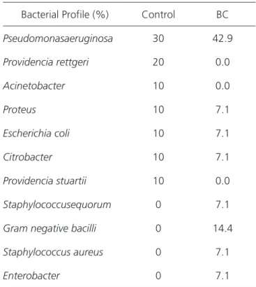
The swab culture was positive in 90.1% of the cases in the control group and in 100% in the experimental group (Table 1). The mean hemoglobin count was 13.0g/100ml in both groups.
Most ulcers were located in the right lower limb, seven (63.6%) in the control group and eight (57.1%) in the BC group. The most frequently affected site was the medial malleolus, five (45.4%) in the control and seven (50.0%) in the BC one. The wound area, measured during the initial clinical evaluation, was 50.0 ± 59.0cm2 in the control group and 54.0 ± 57.0cm2 in the BC. After 30 days (first reevaluation), the area was 31.0 ± 26.0cm2 in the control group and 55.0 ± 54.0cm2 in the BC group. After 120 days (second reevaluation), the wound area in the control group was 36.0 ± 27.0cm2 and 54.0 ± 49.0cm2 in the BC. There was no statistically significant difference between groups (p=0.5748). There was also no significant difference between the groups (p=0.7120) when the wounds were grouped by the mean area size at any of the evaluation times (initial, 1st or 2nd reevaluations).
The number of clinically healed wounds was similar in both groups, three (27.27%) in the control group and two (14.28%) in the BC membrane one. The analysis of the healing process by the Image-Pro Plus software revealed a statistically significant difference between the groups in the initial evaluation (p=0.0096), as well as in the first (p=0.0096) and in the second
Table 1. Bacteria found in secretion cultures (initial assessment).
Bacterial Profile (%) Control BC
Pseudomonasaeruginosa 30 42.9
Providencia rettgeri 20 0.0
Acinetobacter 10 0.0
Proteus 10 7.1
Escherichia coli 10 7.1
Citrobacter 10 7.1
Providencia stuartii 10 0.0
Staphylococcusequorum 0 7.1
Gram negative bacilli 0 14.4
Staphylococcus aureus 0 7.1
(p=0, 0156) reevaluations. The amount (p=0.9928) and the quality (p=0.9921) of exudates was not significant between the groups, although in the BC group the absence of exudates was more evident.
Pain intensity, measured by the analogue scale for pain, was lower in the BC group compared with controls (p=0.0357) in the second reevaluation, with earlier interruption of analgesic use by these patients. There was no difference in the other follow-up times (initial and first reevaluation).
In the control group, 63.6% of the patients had loss of subcutaneous tissue (deeper) in the initial evaluation. After 120 days, loss of epidermis (superficial wound) was more frequent (62.5%). In the BC group at baseline, participants with subcutaneous loss were also more common (57.14%) and, after 120 days, loss of epidermis was present in 83.33% of the cases. There was a significant difference between groups (p<0.0001). After 120 days of evaluation, patients with granulation tissue were 25% in the control group and 41.7% in the BC group. Epithelial tissue was present in 37.5% of the control group and 25.0% in the BC group. We also observed wounds with both types of tissue (granulation + epithelial) in the second reassessment in the control group, in 12.5% and in the BC group, in 25%, without statistical significance (p=0.6946).
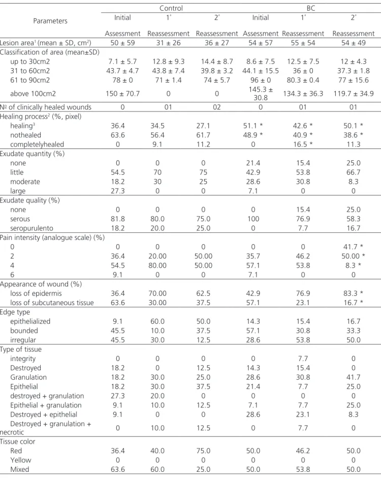
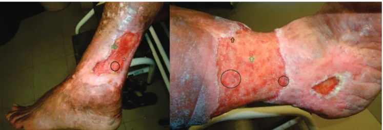
All baseline and reevaluation measurement parameters (30 or 120 days after initial assessment) can be seen in Table 2 and are shown in Figures 1 and 2.
DISCUSSION
The assessment of the sociodemographic profile showed that the majority of patients with varicose ulcers of the lower limbs were, on average, 60 years old. They had a low level of education, evidenced by a high illiteracy rate, observed both in the control and in the experimental group (36% and 50%, respectively). These results are similar to those described in the literature16,17. CVD of the lower limbs is insidious and progressive, and is usually aggravated by the difficulty presented by patients in taking proper care of their health, which determines the evolution to the disease’s most severe forms16,18,19. Low educational level is also related to the lack of access to medical care, since most of these patients depend exclusively on the Unified Health System (SUS) and therefore are often evaluated only when they present ulcers in advanced stages (CEAP 6).
In this study, most patients were male, contrary to the literature16,20. Most were married, which may have contributed to adherence to treatment in both groups16,19. A high percentage of patients, more than 60% in both groups, was unemployed and this data has also been widely described in the literature2,5,16,19.
On the functional level, most participants were classified as independent, but almost all presented some degree of difficulty in walking, similar to what was found in the literature16,21,22. Advanced CVD (CEAP 6) usually evolves with venous claudication (pain in walking), which,
Table 2. Evaluation of ulcers by MEASURE, initial assessment, after 30 and 120 days.
Parameters
Control BC
Initial
Assessment
1ª
Reassessment
2ª
Reassessment
Initial
Assessment
1ª
Reassessment
2ª
Reassessment
Lesion area1 (mean ± SD, cm2) 50 ± 59 31 ± 26 36 ± 27 54 ± 57 55 ± 54 54 ± 49
Classification of area (mean±SD)
up to 30cm2 7.1 ± 5.7 12.8 ± 9.3 14.4 ± 8.7 8.6 ± 7.5 12.5 ± 7.5 12 ± 4.3
31 to 60cm2 43.7 ± 4.7 43.8 ± 7.4 39.8 ± 3.2 44.1 ± 15.5 36 ± 0 37.3 ± 1.8
61 to 90cm2 78 ± 0 71 ± 1.4 74 ± 5.7 96 ± 0 80.3 ± 0.4 77 ± 15.6
above 100cm2 150 ± 70.7 0 0 145.3 ±
30.8 134.3 ± 36.3 119.7 ± 34.9
No of clinically healed wounds 0 01 02 0 01 01
Healing process2 (%, pixel)
healing3 36.4 34.5 27.1 51.1 * 42.6 * 50.1 *
nothealed 63.6 56.4 61.7 48.9 * 40.9 * 38.6 *
completelyhealed 0 9.1 11.2 0 16.5 * 11.3
Exudate quantity (%)
none 0 0 0 21.4 15.4 25.0
little 54.5 70 75 42.9 53.8 66.7
moderate 18.2 30 25 28.6 30.8 8.3
large 27.3 0 0 7.1 0 0
Exudate quality (%)
none 0 0 0 0 15.4 25.0
serous 81.8 80.0 75.0 100 76.9 58.3
seropurulento 18.2 20.0 25.0 0 7.7 16.7
Pain intensity (analogue scale) (%)
0 0 0 0 0 0 41.7 *
2 36.4 20.00 50.00 35.7 46.2 50.00 *
4 54.5 80.00 50.00 57.1 53.8 8.3 *
6 9.1 0 0 7.1 0 0
Appearance of wound (%)
loss of epidermis 36.4 70.00 62.5 42.9 76.9 83.3 *
loss of subcutaneous tissue 63.6 30.00 37.5 57.1 23.1 16.7 *
Edge type
epithelialized 9.1 60.0 50.0 14.3 15.4 16.7
bounded 45.5 10.0 37.5 57.1 30.8 33.3
irregular 45.5 30.0 12.5 28.6 53.8 50.0
Type of tissue
integrity 0 0 0 0 7.7 0
Destroyed 18.2 0 12.5 14.3 15.4 0
Granulation 18.2 30.0 25.0 28.6 30.8 41.7
Epithelial 18.2 30.0 37.5 21.4 7.7 25.0
destroyed + granulation 27.3 20.0 0 0 0 0
Epithelial + granulation 9.1 10.0 12.5 7.1 7.7 25.0
Destroyed + epithelial 9.1 0 0 28.6 23.1 8.3
Destroyed + granulation +
necrotic 0 10.0 12.5 0 7.7 0
Tissue color
Red 36.4 40.0 75.0 50.0 46.2 50.0
Yellow 0 0 0 0 0 0
Mixed 63.6 60.0 25.0 50.0 53.8 50.0
The values are percentages (%) or average followed by the standard deviation (mean ± SD).* Statistically significant if p<0.05. 1 Manual evaluation
of wound area, conducted by clinical evaluation. 2Evaluation of the healing process by Image-Pro Plus. 3Evaluation on the amount of granulation
although not preventing ambulation, makes it difficult and slow.
As for comorbidities, the main ones were diabetes mellitus and arterial hypertension, common in the predominant age group of the studied patients20. Although these diseases are not the cause of CVD, when present, they can aggravate the patient’s condition and interfere with the ulcer healing process. Diseases that may interfere with wound healing have also been observed, such as rheumatoid arthritis and uterine cancer, in which therapy requires the use of steroidal anti-inflammatory agents and antineoplastic drugs23,24.
Another factor that interferes with the healing process is obesity. In the control group, most participants had pre-obesity, whereas in the BC group, most were classified as grade-1 BMI25. Several studies have shown that high BMI contributes to the prolongation of wound healing time26. In a study that evaluated 50 patients with lower limb venous ulcers, the authors identified obesity in 46% of the patients27.
The majority of the participants, in both groups (control: 90%, BC: 100%), had positive cultures for the wounds’ secretion swabs. The microorganism most frequently found in both groups was Pseudomonas aeruginosa. This finding is expected and confirms the high rate of lower limbs CVU contamination27,28. The longer the ulcer is active or the more frequent the relapses, the greater the chances of contamination, increasing the risk of infection, which in turn slows down the healing
process. The knowledge of the CVU’s bacteriological profile can guide early empirical antibiotic therapy in cases where there is a clinical diagnosis of wound infection29.
In the control group, the wounds were treated with substances based on essential fatty acids (EFA) or containing medium chain triglycerides (MCT), and then covered with gauze and bandages (foot and leg). Although this type of dressing may not be the gold standard for CVU treatment, it has been routinely used in other clinics in the SUS due to its low cost. Its use and advantages have been described in the literature5,8,18,22.
The use of BC for the treatment of LL CVD is promising, since it is non-toxic11, biocompatible12 and promotes tissue remodeling. In addition, the BC membrane is made from a renewable source, whose raw material is the sugar cane, of low cost10, allowing its use in the SUS. There were no cases of cutaneous hypersensitivity reactions and BC-induced dermatitis. There have been reports of dermatitis and pain due to the use of MCT30. There was no evidence of MCT dermatitis in this study.
Follow-up of with the MEASURE15 methodology occurred for 120 days. The healing process of this type of injury occurs slowly and, in many cases, complete wound healing is only obtained after a long period of time, sometimes more than 12 months5,27, especially in cases of large ulcers6, similar to those presented by the majority of the participants of this study.
The 120 day period was chosen as sufficient to evaluate the CVU response to BC dressings. At the end of
the observation period, there was a reduction in the wound area in both groups. This difference was not statistically significant because several patients had extensive lesions and of different sizes in the first evaluation, which may justify the high standard deviation in relation to the mean area, which, therefore, compromises the results of the evaluation. To correct this potential error, wounds were classified according to their mean area or percentage (30cm2, 31-60 cm2, 61-90 cm2 and above 100cm2) in each group, and then the comparison was made by group classification.
In the BC group, ulcers were more superficial at the end of the observation period in more than 80% of the patients (versus 60% in the control group). This may indicate that BC dressings acted as an inducer of tissue remodeling, stimulating the granulation process14. This is important because the ulcer healing depends not only on the epidermal proliferation at the margins of the lesion but also on the growth of the granulation tissue from the central area. Other studies have evaluated the application of BC membrane in animals and humans. The authors also observed an increase in granulation tissue, infection control and reduction of healing time12,13,31-35.
All participants in the two groups reported decreased pain and discontinuation of analgesics. The participants who used BC dressing reported feeling more comfort and less pain than those in the control group. The BC membrane promoted self-care, including the possibility
of bathing, without having to worry about the dressing. There was no restriction on wetting the dressing, though were recorded some reports of dressing loss. The BC membrane is a wet dressing that favors hygiene, adheres well to the wound bed and spontaneously detaches once the wound is healed13. BC studies demonstrate that this is an innovative, effective, safe and low cost material13,31.
This study is pioneer in the use of the BC membrane in the treatment of wounds resulting from peripheral vascular diseases in humans. It establishes the scientific basis for further research in the field of vascular surgery, including the use of BC for the treatment of other types of wounds caused by vascular diseases, such as ulcers, or even as dressings for other skin wounds, such as neuropathic ulcers and those caused by burns.
In addition, various structural modifications can be made to the BC membrane to optimize its properties as a dressing, increasing its water absorption capacity and antimicrobial activity or associating it with a controlled release system of antibiotics or other active principles.
Our study allowed us to observe that the Bacterial Cellulose membrane has the ideal properties as a dressing, for maintaining the humidity in the bed of the wound, absorbing excess exudates, limiting infectious processes and protecting the lesion against mechanical trauma. It is an effective alternative to the dressings used for the treatment of chronic varicose ulcers of the lower limbs.
Objetivo: avaliar a eficácia de curativos com membrana de Celulose Bacteriana (CB) no tratamento de úlceras venosas de membros inferiores. Métodos: estudo prospectivo, randomizado e controlado de 25 pacientes com úlceras decorrentes de doença venosa crô-nica nos membros inferiores provenientes do Serviço de Angiologia e Cirurgia Vascular do Hospital de Clícrô-nicas da Universidade Federal de Pernambuco e da Policlínica do Salgado da Secretaria Municipal de Saúde, Caruaru, Pernambuco. Os pacientes foram distribuídos aleatoriamente em dois grupos: grupo controle, que recebeu curativos com óleo de triglicerídeos (11 pacientes) e grupo experimental, tratado com membrana de CB (14 pacientes). Os pacientes foram acompanhados por um período de 120 dias. Resultados: houve uma redução na área de ferida em ambos os grupos. Não houve infecção ou reações ao produto em nenhum dos grupos. Pacientes do grupo CB mostraram diminuição da dor e interrupção mais precoce do uso de analgésicos. Conclusão: a membrana de CB pode ser usada como curativo para o tratamento de úlceras varicosas dos membros inferiores.
Descritores: Úlcera Varicosa. Celulose. Saccharum. Cicatrização. R E S U M O
REFERENCES
1. Souza EM, Yoshida WB, Melo VA, Aragão JA, de Oli-veira LA. Ulcer due to chronic venous disease: a socio-demographic study in northeastern Brazil. Ann Vasc Surg. 2013;27(5): 571-6.
2. Eberhardt RT, Raffetto JD. Chronic venous insufficien-cy. Circulation. 2005;111(18):2398-409.
4. Graham ID, Harrison MB, Nelson EA, Lorimer K, Fisher A. Prevalence of lower-limb ulceration: a systematic review of prevalence studies. Adv Skin Wound Care. 2003;16(6):305-16.
5. Abbade LP, Lastória S. Venous ulcer: epidemiology, physiopathology, diagnosis and treatment. Int J Der-matol. 2005;44(6):449-56.
6. Robertson L, Evans C, Fowkes FG. Epidemiology of chronic venous disease. Phlebology. 2008;23(3):103-11. 7. Eklöf B, Rutherford RB, Bergan JJ, Carpentier PH,
Gloviczki P, Kistner RL, et al. Revision of the CEAP classification for chronic venous disorders: consensus statement. J Vasc Surg. 2004;40(6):1248-52.
8. Borges EL, Caliri MHL, Haas VJ. Systematic review of topic treatment for venous ulcers. Rev Latino-Am En-fermagem. 2007;15(6):1163-70.
9. Fan K, Tang J, Escandon J, Kirsner RS. State of the art in topical wound-healing products. Plast Reconstr Surg. 2011;127 Suppl 1: 44S-59S.
10. Paterson-Beedle M, Kennedy JF, Melo FAD, Lloyd LL, Medeiros V. A cellulosic exopolysaccharide pro-duced from sugarcane molasses by a Zoogloea sp. Carbohydr Polym. 2000;42(4):375-83.
11. Pinto FC, De-Oliveira AC, De-Carvalho RR, Gomes--Carneiro MR, Coelho DR, Lima SV, et al. Acute toxicity, cytotoxicity, genotoxicity and antigeno-toxic effects of a cellulosic exopolysaccharide ob-tained from sugarcane molasses. Carbohydr Polym. 2016;137:556-60.
12. de Lucena MT, de Melo Junior MR, de Melo Lira MM, de Castro CM, Cavalcanti LA, de Menezes MA, et al. Biocompatibility and cutaneous reactivity of cel-lulosic polysaccharide film in induced skin wounds in rats. J Mater Sci Mater Med. 2015;26(2):82.
13. Martins AGS, Lima SVC, Araújo LAP, Vilar FO, Caval-cante NTP. A wet dressing for hypospadias surgery. Int braz j urol. 2013;39(3):408-13.
14. Fragoso AS, Silva MB, de Melo CP, Aguiar JL, Rodri-gues CG, Medeiros PL, et al. Dielectric study of the adhesion of mesenchymal stem cells from human umbilical cord on a sugarcane biopolymer. J Mater Sci Mater Med. 2014;25(1):229-37.
15. Keast DH, Bowering CK, Evans AW, Mackean GL, Burrows C, D’Souza L. MEASURE: a proposed assessment framework for developing best practice recommendations for wound assessment. Wound Repair Regen. 2004;12(3 Suppl):S1-17.
16. Salome GM, Blanes L, Ferreira LM. Evaluation of de-pressive symptoms in patients with venous ulcers. Rev Bras Cir Plast. 2012;27(1):124-9.
17. Aguiar JLA, Lins EM, Marques SRB, Coelho ARB, Rossiter RO, Melo RJV. Surgarcane biopolymer patch in femoral artery angioplasty on dogs. Acta Cir Bras. 2007;22(Suppl 1):77-81.
18. Brito CKD, Nottingham IC, Victor JF, Feitoza SMS, Silva MG, Amaral HEG. Venous ulcer: clinical as-sessment, guidelines and dressing care. Rev Rene. 2013;14(3):470-80.
19. De Vasconcelos Torres G, Fernandes Costa IK, da Silva Medeiros RK, Almeida de Oliveira AK, Gomes de Sou-za AJ, Parreira Mendes FR. CaracteriSou-zação das pes-soas com úlcera venosa no Brasil e Portugal: estudo comparativo. Enfermería Global. 2013;32:75-87. 20. Oliveira BGRB, Nogueira GA, Abreu AM, Carvalho
MR. Caracteriação dos pacientes com úlcera venosa acompanhados no ambulatório de reparos de feri-das. Rev eletrônica enferm. 2012;14(1):156-63. 21. Santos RFFN, Porfírio GJM, Pitta GBB. A diferença na
qualidade de vida de pacientes com doença venosa crônica leve e grave. J vasc bras. 2009;8(2):143-7. 22. Sant´Ana SMSC, Bachion MM, Santos QR, Nunes
CAB, Malaquias SG, Oliveira BGRB. Úlceras veno-sas: caracterização clínica e tratamento em usuários atendidos em rede ambulatorial. Rev bras enferm. 2012;65(4):637-44.
23. Drugs that delay wound healing. Prescrire Int. 2013;22(137):94-8.
24. Zitelli J. Wound healing for the clinician. Adv Derma-tol. 1987;2:243-67.
26. Pierpont YN, Dinh TP, Salas RE, Johnson EL, Wright TG, Robson MC, et al. Obesity and surgi-cal wound healing: a current review. ISRN Obes. 2014;2014:638936.
27. Afonso A, Barroso P, Marques G, Gonçalves A, Gon-zalez A, Duarte N, et al. Úlcera crônica do membro inferior: experiência com cinqüenta doentes. Angiol Cir Vasc. 2013;9(4):148-53.
28. Vicentim AL, Carvalho RCO, Weckwerth PH, Gatti MAN. Etiologia da microbiota presente em úlceras venosas de usuários de bota de unna. Salusvita. 2009;28(1):65-72.
29. Lipsky BA. Empirical therapy for diabetic foot in-fections: are there clinical clues to guide antibiotic selection? Clin Microbiol Infect. 2007;13(4):351-3. 30. Ferreira AM, Souza BMV, Rigotti MA, Loureiro MRD.
Utilização dos ácidos graxos no tratamento de feri-das: uma revisão integrativa da literatura nacional. Rev esc enferm USP. 2012;46(3):752-60.
31. Silveira FCA, Pinto FCM, Caldas Neto SS, Leal MC, Cesário J, Aguiar JLA. Treatment of tympanic mem-brane perforation using bacterial cellulose: a ran-domized controlled trial. Braz j otorhinolaryngol. 2016;82(2):203-8.
32. Sulaeva I, Henniges U, Rosenau T, Potthast A. Bac-terial cellulose as a maBac-terial for wound treatment:
properties and modifications. A review. Biotechnol Adv. 2015;33(8):1547-71.
33. Abreu TC, Lima RP, Souza VSB, Campos Júnior O, Albuquerque AV, Aguiar JLA, et al. The biopolymer sugarcane as filling material of critical defects in rats. Acta Cir Bras. 2016;31(1):53-8.
34. Teixeira FMF, Pereira MF, Ferreira NLG, Miranda GM, Aguiar JLA. Spongy film of cellulosic polysaccharide as a dressing for aphthous stomatitis treatment in rabbits. Acta Cir Bras. 2014;29(4):231-6.
35. Coelho MCOC, Carrazoni PG, Monteiro VLC, Melo FAD, Mota RA, Tenório Filho F. Biopolímero produzi-do a partir da cana-de-acúcar para cicatrização cutâ-nea. Acta Cir Bras. 2002;17(Suppl 1):11-3.
Received in: 18/10/2016
Accepted for publication: 18/12/2016 Conflict of interest: none.
Source of funding: Brazil’s Science, Technology, and Innovation Ministry (MCTI); Financier of Studies and Projects (FINEP); National Counsel for Technological and Scientific Development (CNPq).
Mailing address:
Flávia Cristina Morone Pinto