* Study conducted at the University of São Paulo School of Medicine – FMUSP – São Paulo (SP) Brazil.
1. Masters in Rehabilitation Sciences. University of São Paulo, School of Medicine – FMUSP – São Paulo (SP) Brazil. 2. PhD in Sciences. University of São Paulo, School of Medicine – FMUSP – São Paulo (SP) Brazil.
3. Radiologist at the Hospital das Clínicas Radiology Institute, University of São Paulo School of Medicine – FMUSP – São Paulo (SP) Brazil. 4. PhD in Radiology. University of São Paulo, School of Medicine – FMUSP – São Paulo (SP) Brazil.
5. Professor of Physical Therapy. University of São Paulo, School of Medicine – FMUSP – São Paulo (SP) Brazil.
Correspondence to: Celso Ricardo Fernandes de Carvalho. Av. Dr. Arnaldo, 455, sala 1216, CEP: 05408-040, São Paulo, SP, Brasil. Tel 55 11 3066-7317. Fax 55 11 3091-7462. E-mail: [email protected] or [email protected]
Submitted: 12 August 2006. Accepted, after review: 14 November 2006.
Ultrasound evaluation of diaphragmatic mobility
in different postures in healthy subjects*
Wellington Pereira dos Santos Yamaguti1, Elaine Paulin2, Simone Shibao3, Sérgio Kodaira3,
Maria Cristina Chammas4, Celso Ricardo Fernandes Carvalho5
Abstract
Objective: To assess, using ultrasound, the effects that changes in body position have on diaphragmatic mobility in healthy subjects during spontaneous breathing. Methods: The study involved seven healthy female volunteers, all of whom were nonsmokers, within normal weight range, and free of any cardiopulmonary disease. They were submitted to pulmonary function testing and ultrasound evaluation of the mobility of the right diaphragm by the craniocaudal displacement of the left branch of the portal vein using an ultrasound device in mode B. The mobility of the right diaphragm was evaluated in right decubitus and in left decubitus. The order of evaluation was previously determined in a random drawing. Results: The average mobility of the right diaphragm in right decubitus (51.30 ± 9.69 mm) was significantly higher (p = 0.03) than that observed in left decubitus (45.93 ± 10.37 mm). Conclusion: The results suggest that, during spontaneous ventilation, the dependent portion of the diaphragm presents greater mobility than the nondependent portion, and that the technique used was sufficiently sensitive to detect variations in diaphragmatic mobility related to changes in posture.
Methods
Subjects and case study
This was a cross-sectional observational study. Data were collected at the Hospital das Clínicas Radiology Institute, University of Sao Paulo School of Medicine. The study included seven healthy, female nonsmoking volunteers who were within the normal range for weight (body mass index between 18.5 and 25 kg/m2) and had no prior medical history
of cardiopulmonary diseases. The study participants were submitted to pulmonary function tests and ultrasound evaluations on the same day. The study design was approved by the Ethics Committee of the Hospital (protocol nº 914/04), and all partici-pants gave written informed consent.
Parameters analyzed
Pulmonary function assessment: spirometry (Microquark; Cosmed, Rome, Italy) was performed as per the standards recommended by the American Thoracic Society and the I Consenso Brasileiro de Espirometria (First Brazilian Consensus on Spirometry).(12,13) The individuals were instructed to
rest for five to ten minutes before the test, and all procedures were described thoroughly, emphasizing the need to avoid leakage around the mouthpiece and to perform maximum inspiration, followed by maximum and sustained expiration, until the evalu-ator instructed them to stop. The room where the tests were performed was quiet, and all tests were conducted in the morning to avoid circadian influ-ences. The individuals were instructed to remain seated and to use a nose clip during the tests. The volume-time and flow-volume curves were inter-preted in accordance with the acceptability criteria recommended by the American Thoracic Society,(12)
and the best of three reproducible curves was selected (variance < 5%). Based on this curve, forced vital capacity (FVC) and forced expiratory volume in one second (FEV1) were calculated and analyzed using the values predicted by other authors.(14)
Ultrasound analysis
Right diaphragmatic mobility was evaluated by determining the craniocaudal displacement of the left branch of the portal vein using a B-mode ultra-sound device (Logic 500, Pro Series®; General Electric
Introduction
Body positioning (kinetic therapy) is a nonin-vasive therapeutic intervention, with significant effects on and benefits for pulmonary function and oxygenation. Kinetic therapy influences ventilation distribution, perfusion, alveolar opening pressure and diaphragmatic mechanics.(1)
Differences in the diaphragmatic move-ment pattern have been observed in healthy individuals during positive pressure mechanical ventilation and/or spontaneous respiration in different postures.(2,3) Some authors report that,
during mechanical ventilation, diaphragmatic mobility is significantly greater in the nonde-pendent zones, and that, during spontaneous respiration, diaphragmatic mobility is greater in the dependent zones, where the oppositional force to diaphragmatic displacement (abdominal hydrostatic pressure) tends to be greater.(2)
Diaphragmatic mobility evaluations have traditionally been performed using fluor-oscopy.(4) Although this method is considered the
gold standard, it presents some limitations, such as diaphragm visualization with a single angle of incidence, requirement to perform correc-tive calculations and patient exposure to ionizing radiation.(5) Over the past few years, ultrasound has
also been used to evaluate diaphragmatic mobility, since it offers some advantages over fluoroscopy: portability; no exposure to ionizing radiation; and direct quantification of diaphragmatic move-ment.(6-10)
Despite the advantages of ultrasound scan-ning, direct visualization of the diaphragm presents methodological difficulties that depend on trans-ducer positioning.(7) A recently validated ultrasound
method directly evaluates diaphragmatic mobility by means of placing the transducer perpendicular to the craniocaudal axis, using the subcostal abdom-inal window to visualize the displacement of the left branch of the hepatic portal vein as an indi-rect measurement for the displacement of the right diaphragm.(11)
(ii) diaphragmatic mobility variance of 45% for the different positions(2); (iii) standard deviation of
25% for intersubject diaphragmatic mobility;(2) and
(iv) discriminatory power of 80%, which established a sample of four individuals. The level of statistical significance was set at 5% (p < 0.05) (Statistical Package Sigma Stat 3.1).
Results
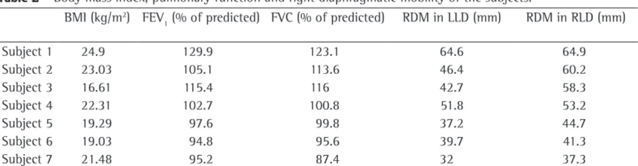
The anthropometric characteristics and pulmo-nary function of the individuals are shown in Table 1, which confirms that pulmonary function was within normal limits (FEV1 = 110.00 ± 11.02% of predicted and FVC = 107.84 ± 16.03% of predicted). Table 2 shows individual data in reference to body mass index, pulmonary function parameters and right diaphragmatic mobility measured in LLD and RLD.
All of the individuals studied presented greater mobility in the right diaphragm when evaluated in RLD (dependent decubitus) (Figure 2). The greatest diaphragmatic mobility variation between the positions was 26.76% (subject 3) and the lowest variation was 0.46% (subject 1).
Figure 3 presents the mean values for right diaphragmatic mobility in RLD (51.30 ± 9.69 mm) and LLD (45.93 ± 10.37 mm). Mean right diaphragm excursion was significantly greater when the indi-viduals were positioned in RLD (p = 0.03). Analysis of the mean variation of diaphragmatic mobility in the different positions, revealed a variation of 10.47%.
Discussion
In the present study, right diaphragmatic mobility was measured in RLD (dependent) and LLD Medical Systems, Milwaukee, WI, USA).(11) The
ultra-sound technician used a 3.5 MHz convex transducer positioned in the right subcostal region, with the incidence angle perpendicular to the craniocaudal axis, in the direction of the inferior vena cava. Next, an intraparenchymal portal branch was identified in the field of vision and its position was traced with the curser during the forced inspiration and expira-tion. The craniocaudal displacement of these points was considered to be the amount of right diaphrag-matic mobility (Figure 1). Right diaphragdiaphrag-matic mobility was evaluated in the right lateral decubitus (RLD) and left lateral decubitus (LLD) positions, and the posture evaluation order was previously defined by a random drawing. All tests were performed by the same radiologist, who had no knowledge of the pulmonary function results or the study objective. Three measurements were taken in each position, and the highest value was selected for statistical analysis.
Statistical analysis
Data distribution was evaluated using the Kolmogorov-Smirnov test, and the sample size of seven individuals was calculated in accord-ance with the following assumptions: (i) analysis using the paired t-test for repeated measurements;
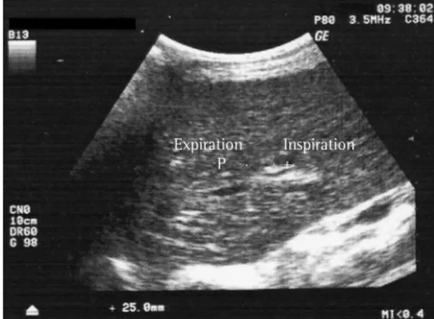
Expiration Inspiration
P +
Figure 1 - Ultrasound evaluation of craniocaudal displacement of the left branch of the hepatic portal vein. The vessel position was marked with the caliper during forced inspiration and expiration. Craniocaudal displacement of these points was recorded in millimeters and recorded as the degree of right diaphragmatic mobility.
Table 1 - Anthropometric characteristics and pulmonary function values of the subjects.
Variable Individuals (n = 7)
Age (years) 24.25 ± 2.65
BMI (kg/m2) 20.95 ± 2.81
FEV1 (predicted %) 110.00 ± 11.02 FVC (predicted %) 107.84 ± 16.03
FEV1/FVC (%) 106.14 ± 5.97
The greater diaphragmatic movement generated in the zones with higher hydrostatic pressure during spontaneous respiration can be attributed to certain mechanisms. In the dependent portion, the weight of the abdominal viscera displace the diaphragm in the cephalic direction reducing the diaphragm’s curvature radius.(15) Based on the Laplace law, the
transdiaphragmatic pressure in this region tends to be greater than that of the diaphragm muscle, for the same tension.(16) In addition, the fibers of
the hemidiaphragm in the dependent portion are stretched by the cephalic displacement, which can generate greater pressure and consequently greater movement due to the more favorable tension-length ratio.(17) Another factor to be considered is that, in
the lateral decubitus position, the mobility of the (nondependent) using a new ultrasound evaluation
technique. The results show that the right diaphragm presents greater mobility when the individuals are positioned over the dependent decubitus.
One group of authors evaluated diaphrag-matic mobility using fluoroscopy in three healthy individuals positioned in supine and lateral posi-tions and confirmed that the dependent porposi-tions of the diaphragm present greater craniocaudal displacement than do the nondependent portions,(2)
coinciding with the results of this study. The authors report that the greater displacement of the dependent portion of the diaphragm could be a result of greater oppositional force to muscle displacement (abdominal hydrostatic pressure) in this region.
Table 2 - Body mass index, pulmonary function and right diaphragmatic mobility of the subjects. BMI (kg/m2) FEV
1 (% of predicted) FVC (% of predicted) RDM in LLD (mm) RDM in RLD (mm)
Subject 1 24.9 129.9 123.1 64.6 64.9
Subject 2 23.03 105.1 113.6 46.4 60.2
Subject 3 16.61 115.4 116 42.7 58.3
Subject 4 22.31 102.7 100.8 51.8 53.2
Subject 5 19.29 97.6 99.8 37.2 44.7
Subject 6 19.03 94.8 95.6 39.7 41.3
Subject 7 21.48 95.2 87.4 32 37.3
BMI: body mass index; FEV1: forced expiratory volume in one second; FVC: forced vital capacity; RDM: right diaphragmatic mobility; LLD: left lateral decubitus; and RLD: right lateral decubitus.
65
60
55
50 45
40
35
30
Right diaphragmatic
mobility (mm)
Subjects
1 2 3 4 5 6 7
RLD LLD
Figure 2 - Evaluation of right diaphragmatic mobility in seven healthy individuals positioned in right lateral decubitus (RLD, dependent decubitus) and left lateral decubitus (LLD, nondependent decubitus). Note that all individuals present greater right diaphragmatic mobility when positioned over the dependent decubitus.
55
50
45
40
35
Right diaphragmatic
mobility (mm)
RLD LLD
Position *
the subcostal abdominal window to obtain a direct image of the hemidiaphragm,(27) that requires that
the transducer be angled cranially to visualize the posterior portion of the diaphragm,(28) and the
craniocaudal muscle displacement is oblique to the transducer incidence angle, which jeopardizes the accuracy of the mobility measurement.
The ultrasound method used in the present study enables evaluation of right diaphragmatic mobility by using a transducer incidence angle perpendic-ular to the craniocaudal axis, through the subcostal abdominal window, throughout the respiratory cycle, even during deep inspiration. Although this method evaluates the displacement of the left branch of the portal vein as the indirect measurement of the right diaphragm displacement, this evaluation is made without the methodological difficulties of the other ultrasound evaluation methods described above.
One limitation reported by some authors who used the subcostal abdominal window to evaluate diaphragmatic mobility is the possible interfer-ence of the transducer on abdominal movements during respiration.(20,24) These authors argue that the
application of the transducer over the abdominal wall could modify individual respiratory patterns, which could compromise the measurement during basal respiration evaluations. We believe that such an influence would have been minimal in the present study, since the individuals were encour-aged to voluntarily make one maximum inspiration and expiration, to measure the displacement of the structures identified by the evaluator.
Calculation of the sample size in the present study was established based on the data obtained by other authors,(2) who reported a 45% variation
in diaphragmatic mobility for the different posi-tions and standard deviation of 25% among the individuals.(2) However, the results obtained in this
study reveal a diaphragmatic mobility variation for the different positions of 10.47% and a standard deviation of 6.56% among the individuals. One possible explanation for this discrepancy could be the fact that, in that study,(2) diaphragmatic mobility
was evaluated during positive pressure ventilation. Despite the differences reported between the varia-tion obtained and that expected, the sample size of seven individuals was sufficient to detect a signifi-cant difference in diaphragmatic mobility depending on body position.
dependent portion of the chest cavity is restricted, which can favor craniocaudal displacement of the diaphragm in this region, since the ribcage assumes the function of a fixed point for thoracic movement during the respiratory cycle.(1)
Various imaging methods have been used to evaluate diaphragmatic mobility, including fluor-oscopy, computed tomography, nuclear magnetic resonance imaging and ultrasound. Fluoroscopy has been considered the most reliable method of quantitatively evaluating the degree of craniocaudal diaphragmatic movement during spontaneous respiration.(18) During fluoroscopy evaluation,
diaphragmatic mobility is measured by the displace-ment of the highest point of the hemidiaphragms. Although it is a relatively simple method and regional pulmonary ventilation analysis can also be performed, this technique presents some disad-vantages that should be considered: (i) radiation exposure; (ii) image amplitude due to the divergence of the X-rays, which requires corrective calculations; and (iii) a lack of three-dimensional information regarding diaphragmatic movement.(5)
One group of authors recently used computed tomography and nuclear magnetic resonance imaging to conduct a detailed anatomical study of different portions of the diaphragm and chest cavity.(19,20) Other authors described the
diaphrag-matic mobility in normal individuals using sequential magnetic resonance images during basal respira-tion.(21) These authors report some positive aspects
of using this method in diaphragmatic movement examinations, such as the possibility of digitalizing the images, which offers a more accurate analysis of the movement, and the fact that this technique does not depend on the evaluator, minimizing inter- and intra-observer measurement variations. However, equipment size and cost limit the feasibility and availability of these methods.
Ultrasound scanning has proven to be a prom-ising tool for evaluating diaphragmatic function, since it is a simple, reproducible and portable method that eliminates the risk of exposure to ionizing radiation and directly quantifies diaphrag-matic movement.(22-24) The hemidiaphragm can be
directly visualized by positioning the transducer on the midaxillary line between the intercostals spaces.(7,9,10,25) However, visualization of the muscle
cholecystectomy: a sonographic study. Anesth Analg. 2001;92(3):755-61.
9. Houston JG, Fleet M, Cowan MD, McMillan NC. Comparison of ultrasound with fluoroscopy in the assessment of suspected hemidiaphragmatic movement abnormality. Clin Radiol. 1995;50(2):95-8.
10. Houston JG, Angus RM, Cowan MD, McMillan NC, Thomson NC. Ultrasound assessment of normal hemidiaphragmatic movement: relation to inspiratory volume. Thorax. 1994;49(5):500-3.
11. Toledo NS, Kodaira SK, Massarollo PC, Pereira OI, Mies S. Right hemidiaphragmatic mobility: assessment with US measurement of craniocaudal displacement of left branches of portal vein. Radiology. 2003;228(2):389-94.
12. Standardization of spirometry, 1994 Update. American Thoracic Society. Am J Respir Crit Care Med. 1995;152(3):1107-36.
13. Sociedade Brasileira de Pneumologia e Tisiologia Con -senso Brasileiro sobre Espirometria. J Pneumol. 1996;22(3):105-64.
14. Knudson RJ, Lebowitz MD, Holberg CJ, Burrows B. Changes in the normal maximal expiratory flow-volume curve with growth and aging. Am Rev Resp Dis. 1983;127(6):725-34. 15. Ibanez J, Raurich JM. Normal values of functional residual
capacity in the sitting and supine positions. Intensive Care Med. 1982;8(4):173-7.
16. Whitelaw WA, Hajdo LE, Wallace JA. Relationships among pressure, tension and shape of the diaphragm. J Appl Physiol. 1983;55(6):1899-905.
17. Farkas GA, Roussos C. Diaphragm in emphysematous hamsters: sarcomere adaptability. J Appl Physiol. 1983;54(6):1635-40. 18. Verschakelen JA, Deschepper K, Jiang TX, Demedts M.
Diaphragmatic displacement measured by fluoroscopy and derived by respitrace. J Appl Physiol. 1989;67(2):694-8. 19. Iwasawa T, Kagei S, Gotoh T, Yoshiike Y, Matsushita K,
Kurihara H, et al. Magnetic resonance analysis of abnormal diaphragmatic motion in patients with emphysema. Eur Respir J. 2002;19(2):225-31.
20. Takazakura R, Takahashi M, Nitta N, Murata K. Diaphragmatic motion in the sitting and supine positions: healthy subject study using a vertically open magnetic resonance system. J Magn Reson Imaging. 2004;19(5):605-9.
21. Gierada DS, Curtin JJ, Erickson SJ, Prost RW, Strandt JA, Goodman LR. Diaphragmatic motion: fast gradient-recalled-echo MR imaging in healthy subjects. Radiology. 1995;194(3):879-84.
22. Gottesman E, McCool FD. Ultrasound evaluation of the paralyzed diaphragm. Am J Respir Crit Care Med. 1997;155(5):1570-4.
23. Gerscovich EO, Cronan M, McGahan JP, Jain K, Jones CD, McDonald C. Ultrasonographic evaluation of diaphragmatic motion. J Ultrasound Med. 2001;20(6):597-604.
24. Jiang JR, Tsai TH, Jerng JS, Yu CJ, Wu HD, Yang PC. Ultrasonographic evaluation of liver/spleen movements and extubation outcome. Chest. 2004;126(1):179-85.
25. Wait JL, Nahormek PA, Yost WT, Rochester DP. Diaphragmatic thickness-lung volume relationship in vivo. J Appl Physiol. 1989;67(4):1560-8.
26. Gibson GJ. Diaphragmatic paresis: pathophysiology, clinical features and investigation. Thorax. 1989;44(11):960-70. 27. Diament MJ, Boechat MI, Kangarloo H. Real-time
sector ultrasound in the evaluation of suspected
As stated earlier, some studies have evaluated the influence of body posture changes on diaphragmatic mobility in individuals during spontaneous respira-tion or controlled mechanical ventilarespira-tion.(2,3,29) Due
to the difficulties involved in fluoroscopy evalua-tions, the number of patients evaluated by these authors (three to four patients) is always a limiting factor. One significant advantage of this new method is that diaphragmatic mobility can be eval-uated quickly, with no risk to the patient, and can even be performed at the bedside.(24) This method
could have some clinical applications such as moni-toring the impact of respiratory and neuromuscular diseases on diaphragm mechanics, quantifying the benefits of pulmonary rehabilitation programs directed at diaphragmatic training(30) and evaluating
diaphragmatic mobility in patients with diaphrag-matic paralysis.
Our results suggest that, during spontaneous ventilation, the mobility of the dependent portion of the diaphragm is greater than is that of the nonde-pendent portion, and that the technique used was sensitive enough to detect diaphragmatic mobility variations for the different body positions. These results substantiate the use of ultrasound scanning to evaluate diaphragmatic displacement in clinical practice.
References
1. Dean E, Ross J. Oxygen transport - The basis for contemporary cardiopulmonary physical therapy and its optimization with body position and mobilization. Physical Therapy Practice. 1992;1(4):34-44.
2. Froese AB, Bryan AC. Effects of anesthesia and paralysis on diaphragmatic mechanics in man. Anesthesiology. 1974;41(3):242-55.
3. Krayer S, Rehder K, Vettermann J, Didier EP, Ritman EL. Position and motion of the human diaphragm during anesthesia-paralysis. Anesthesiology. 1989;70(6):891-8 4. George RB, Weil H. Fluorodensimetry. A method for
analyzing regional ventilation and diaphragm function. JAMA. 1971;217(2):171-6.
5. Gierada DS, Slone RM, Fleishman MJ. Imaging evaluation of the diaphragm. Chest Surg Clin N Am. 1998;8(2):237-80. 6. Harris RS, Giovannetti M, Kim BK. Normal ventilatory
movement of the right hemidiaphragm studied by ultrasonography and pneumotachography. Radiology. 1983;146(1):141-4.
7. Houston JG, Morris AD, Howie CA, Reid JL, McMillan N. Technical report: quantitative assessment of diaphragmatic movement - a reproducible method using ultrasound. Clin Radiol. 1992;46(6):405-7.
diaphragmatic motion in mechanically ventilated patients. Eur J Ultrasound. 2000;11(3):205-11.
30. Paulin E, Brunetto AF, Carvalho CRF. Efeitos de programa de exercícios físicos direcionado ao aumento da mobilidade torácica em pacientes portadores de doença pulmonar obstrutiva crônica. J Pneumol. 2003;29(5):287-94. abnormalities of diaphragmatic motion. J Clin Ultrasound.
1985;13(8):539-43.
28. Haber K, Asher M, Freimanis AK. Echographic evaluation of diaphragmatic motion in intra-abdominal diseases. Radiology. 1975;114(1):141-4.