* Study carried out at the Rio Grande do Sul Department of Health Hospital Sanatório Partenon, Porto Alegre (RS) Brazil.
1. PhD in Pulmonology. Universidade Federal do Rio Grande do Sul – UFRS, Federal University of Rio Grande do Sul – Porto Alegre (RS) Brazil. 2. PhD in Clinical Medicine. Universidade Federal do Rio Grande do Sul – UFRS, Federal University of Rio Grande do Sul – Porto Alegre (RS) Brazil. 3. Radiologist at the Rio Grande do Sul Department of Health Hospital Sanatório Partenon, Porto Alegre (RS) Brazil.
4. Director Emeritus of the Pavilhão Pereira Filho da Santa Casa de Misericórdia, Porto Alegre (RS) Brazil.
5. Specialist in Pulmonology. Universidade Federal do Rio Grande do Sul – UFRS, Federal University of Rio Grande do Sul – Porto Alegre (RS) Brazil. 6. Physician at the Rio Grande do Sul Department of Health Hospital Sanatório Partenon, Porto Alegre (RS) Brazil.
7. Masters in Pulmonology. Universidade Federal do Rio Grande do Sul – UFRS, Federal University of Rio Grande do Sul – Porto Alegre (RS) Brazil. Correspondence to: Dr. Pedro Dornelles Picon. Rua Filipinas, 295, CEP 91050-020, Porto Alegre, RS, Brasil.
Tel/Fax 55 51 3340-0660. E-mail: [email protected]
Submitted: 13 September 2006. Accepted, after review: 20 November 2006.
Differences in the clinical and radiological presentation of intrathoracic
tuberculosis in the presence or absence of HIV infection*
Pedro Dornelles Picon1, Maria Luiza Avancini Caramori2, Sérgio Luiz Bassanesi2, Sandra Jungblut3,
Marcelo Folgierini3, Nelson da Silva Porto4, Carlos Fernando Carvalho Rizzon5,
Roberto Luiz Targa Ferreira6, Tânia Mariza de Freitas6, Carla Adriane Jarczewski7
Abstract
Objective: To describe the differences in the clinical and radiological presentation of tuberculosis in the presence or absence of HIV infection. Methods: A sample of 231 consecutive adults with active pulmonary tuberculosis admitted to a tuberculosis hospital were studied, assessing HIV infection, AIDS, and associated factors, as well as re-evaluating chest X-rays. Results: There were 113 HIV-positive patients (49%) Comparing the 113 HIV-positive patients (49%) to the 118 HIV-negative patients (51%), the former presented a higher frequency of atypical pulmonary tuberculosis (pulmonary lesions accompanied by intrathoracic lymph node enlargement), hematogenous tuberculosis, and pulmonary tuberculosis accompanied by superficial lymph node enlargement, as well as presenting less pulmonary cavitation. The same was found when HIV-positive patients with AIDS were compared to those without AIDS. There were no differences between the HIV-positive patients without AIDS and the HIV-negative patients. Median CD4 counts were lower in HIV-positive patients with intrathoracic lymph node enlargement and pulmonary lesions than in the HIV-positive patients with pulmonary lesions only (47 vs. 266 cells/mm3; p < 0.0001), in
HIV-positive patients with AIDS than in those without AIDS (136 vs. 398 cells/mm3; p < 0.0001) and in patients with atypical pulmonary
tuberculosis than in those with other forms of tuberculosis (31 vs. 258 cells/mm3; p < 0.01). Conclusion: Atypical forms and disseminated
disease predominate among patients with advanced immunosuppression. In regions where TB prevalence is high, the presence of atypical pulmonary tuberculosis or pulmonary tuberculosis accompanied by superficial lymph node enlargement should be considered an AIDS-defining condition.
tive sputum smear microscopy; having undergone HIV testing; and having undergone simple antero-posterior and lateral chest X-ray.
The data were obtained through the re-eval-uation of chest X-rays and the review of medical charts. The variables analyzed were as follows: age; gender; alcoholism; use of illicit drugs; corticosteroid use; duration of symptoms; presence of multifocal disease, diabetes mellitus, neoplasias, and oppor-tunistic diseases; results of HIV testing; number of CD4 T lymphocytes; and radiological findings.
Sputum smear microscopy was performed using the Ziehl-Neelsen technique. The HIV testing was performed using the ELISA method, and posi-tive results were confirmed by Western blot. The CD4 counts were obtained using flow cytometry. Alcoholism and drug addiction were considered present when they had been registered on the medical chart by the treatment team. Multifocal disease was defined by the presence of superficial lymph node enlargement in patients with pulmo-nary TB.
The chest X-rays were independently evalu-ated by two radiologists, who had no knowledge of the results of HIV testing, in order to identify the types of TB; the location of the lesions in the lung segments; and the presence of cavitary lesions, hilar or mediastinal lymph node enlargement, and pleural effusion. In case of inconsistency between the eval-uations, the chest X-rays were jointly examined and, in the event of the inconsistency persisting, a third radiologist was consulted so that a consensus could be reached. The types of TB were characterized using the following classification:
1) Classic pulmonary TB: pulmonary lesions unaccompanied by hilar or mediastinal lymph node enlargement;
2) Tuberculous pneumonia: homogeneous consolidation with air bronchograms, with or without intrathoracic lymph node enlargement;
3) Hematogenous TB: diffuse pulmonary infil-trate with or without intrathoracic lymph node enlargement;
4) Tuberculoma: nodular lesion without intrathoracic lymph node enlargement; 5) Mediastinal lymph node TB: intrathoracic
lymph node enlargement as a single lesion; 6) Atypical pulmonary TB: non-cavitated foci of
consolidation located in the perihilar region
Introduction
The epidemic of acquired immunodeficiency syndrome (AIDS) has caused changes in the clin-ical and radiologclin-ical presentation of tuberculosis (TB) in adults, as described at the beginning of the 1980s.(1) In HIV-infected (HIV-positive) patients,
atypical intrathoracic forms (non-cavitated perihilar pulmonary infiltrates or pulmonary infiltrates in the middle and lower thirds of the lungs accompanied by hilar or mediastinal lymph node enlargement, resembling the lesions seen in primary TB) appeared, and cases of hematogenous TB, TB at multiple sites, and pulmonary TB with normal chest X-rays became common.(1-9)
The differences found in the presentation of TB in HIV-positive patients are due to cellular immunodeficiency secondary to the progressive destruction of CD4 lymphocytes by HIV,(4,6,9,10) and
the tuberculous lesions can result from the reactiva-tion of old foci or from re-infecreactiva-tion.(4,6,8) Therefore,
CD4 counts in peripheral blood are used to eval-uate the immunological status of patients infected with the virus, and the counts thus obtained can be correlated with the clinical and radiological presentation of TB.(3,6-12) When TB manifests in
HIV-positive patients with relatively preserved immunity(CD4 > 350 cells/mm3), the clinical and
radiological presentation is similar to that seen in individuals who are not infected with HIV (HIV-negative individuals).(6) In patients with low
CD4 counts, especially lower than 200 cell/mm3, TB
can have an atypical presentation.(3,6-11)
The objective of the present study was to describe the differences in the clinical and radiological pres-entation of intrathoracic TB in HIV-positive and HIV-negative patients, as well as to determine the correlation between CD4 lymphocyte counts and the presence of atypical forms of TB in HIV-positive patients living in an area of high prevalence of TB/ HIV co-infection.
Methods
posi-by the hospital facility based on the safeguards ensuring data confidentiality and patient privacy.
Results
Of the 231 patients included in the study, 69.7% were male, 60.6% were Caucasian, 59.7% were alco-holics, and 36.2% were illicit drug users. The mean age was 37.7 ± 12.9 years. The delay between the onset of symptoms and the diagnosis ranged from 10 to 540 days (median, 60 days) and was longer than 90 days in 48% of the patients. Diabetes mellitus was identified in 4.3% of the cases and multifocal disease in 13%. None of the patients used corticosteroids or had neoplasia. A total of 113 patients (48.9%) tested positive for HIV.
There were no differences between the HIV-positive and the HIV-negative patients in terms of gender, race, alcoholism, or presence of diabetes mellitus (Table 1), nor were any differences observed in terms of duration of symptoms: 60 days (range, 10-360 days) and 90 days (range, 14-540 days), respectively (p = 0.284). However, the HIV-positive patients were younger (34.3 ± 9.3 vs. 41.1 ± 15 years; p < 0.0001), used illicit drugs more frequently, and more often presented multifocal disease in compar-ison with the HIV-negative patients (Table 1). or in the anterior segment of the upper lobe
and in the basal segments of the lower and middle lobes or of the lingula, accompanied by intrathoracic lymph node enlargement; 7) Pleural TB: pleural effusion without
pulmo-nary or mediastinal lesions; and 8) Pulmonary TB with normal chest X-ray. For the purpose of this study, the patients infected with HIV who presented associated oppor-tunistic diseases, such as pneumocystosis, cerebral toxoplasmosis, esophageal candidiasis, cryptococ-cosis, histoplasmosis, cytomegalovirus infection, or Kaposi’s sarcoma, were considered to have AIDS.
The results are presented as mean and standard deviation or percentage of patients with a given characteristic. Non-normally distributed data (CD4 and duration of symptoms) were normalized by logarithmic transformation, for the purpose of analysis, and are presented as median and minimum and maximum values. The analysis was carried out using the chi-square test, Fisher’s exact test, and the Student’s t-test. Values of p < 0.05 were consid-ered significant.
Since the study used secondary data obtained by the review of medical charts, there was no interfer-ence with the patients or with the treatment routine. The permission to use the information was granted
Table 1 - Distribution of demographic, clinical, and radiological variables by HIV test results.
Variable Total HIV+ HIV− p
n % n %
Gender Male Female 161 70 80 33 70.8 29.2 81 37 68.6 31.4 0.722 Race Caucasian Non-Caucasian 140 91 68 45 60.2 39.8 72 46 61.0 39.0 0.896
Alcoholisma Yes
No 138 79 66 36 64.7 35.3 72 43 62.6 37.4 0.749
Use of illicit drugsb Yes
No 79 139 65 41 61.3 38.7 14 98 12.5 87.5 <0.0001
Multifocal disease Yes
No 30 201 27 86 23.9 76.1 3 115 2.5 97.5 <0.0001
Diabetes mellitus Yes
No 10 221 2 111 1.8 98.2 8 110 6.8 93.2 0.103
Intrathoracic lymph node enlargement Yes No 24 207 23 90 20.4 79.6 1 117 0.8 99.2 <0.0001
Pulmonary cavitation Yes No 188 43 80 33 70.8 29.2 108 10 91.5 8.5 <0.0001
Pleural effusion Yes
No 38 193 21 92 18.6 81.4 17 101 14.4 85.6 0.392
those with pulmonary lesions only - 266 cells/mm3
(range, 7-1288 cells/mm3) (p < 0.0001). The CD4
counts were also lower in the patients with AIDS - 136 cells/mm3 (range, 3-1288 cells/mm3) - than in
the HIV-positive patients without AIDS - 398 cells/ mm3 (range, 272-689 cells/mm3) (p < 0.0001).
Median CD4 counts by type of TB, which were lower in the patients with atypical pulmonary TB - 31 cells/mm3 (range, 3-71 cells/mm3) - than in those
with any of the other types (p < 0.01), are shown in Figure 3. A total of 30 patients had CD4 counts 200 cells/mm3, and 27 had counts > 200 cells/mm3.
The group with CD4 counts ≤ 200 differed from the group with CD4 counts > 200 by presenting a higher frequency of intrathoracic lymph node enlargement (50 vs. 3.7%; p = 0.0001) and less pulmonary cavi-tation (53.3 vs. 85.2%; p = 0.010).
The comparison between the HIV-positive and the HIV-negative patients revealed that the former presented a significantly higher frequency of pulmonary lesions accompanied by intrathoracic lymph node enlargement, as well as presenting less pulmonary cavitation, and that there was no differ-ence in terms of pleural effusion accompanied by pulmonary lesions (Table 1). Of the 23 HIV-positive patients with intrathoracic lymph node enlargement, 16 were classified as having atypical pulmonary TB, 5 were classified as having hematogenous TB, and 2 were classified as having tuberculous pneumonia. The only HIV-negative patient with lymph node enlargement had hematogenous TB.
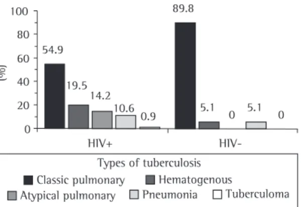
In terms of the types of TB, the distribution was different in the HIV-positive and in the HIV-negative patients (p < 0.001) (Figure 1). Atypical pulmonary TB occurred only in the HIV-positive patients, who also presented higher frequencies of hematogenous TB and tuberculous pneumonia and, consequently, a lower frequency of classic pulmonary TB. There were no cases of mediastinal lymph node TB, pleural TB, or TB with normal chest X-ray.
Of the 113 HIV-positive patients, 79 (99.9%) were classified as having AIDS. Of those, only 16 (20.3%) used antiretroviral drugs, all of them using those drugs irregularly and for no more than 90 days. The patients with AIDS differed from the HIV-positive patients without AIDS by presenting a higher frequency of multifocal disease and of intrathoracic lymph node enlargement accompanied by pulmonary lesions, as well as by presenting less pulmonary cavi-tation (Table 2). These patients also differed in terms of the distribution of the types of TB (p < 0.0001) (Figure 2). Of the 16 cases of atypical pulmonary TB, 15 occurred among the patients with AIDS.
The HIV-positive patients without AIDS differed from the HIV-negative patients only by being younger (36.4 ± 10.5 vs. 41.1 ± 15 years; p = 0.045) and by using illicit drugs more frequently (60.6 vs. 12.5%; p < 0.0001). There were no differ-ences between these two groups in terms of the other demographic, clinical, or radiological charac-teristics (Tables 1 and 2; Figures 1 and 2).
In the present study, CD4 counts were measured in 57 HIV-positive patients and ranged from 3 to 1288 cells/mm3 (median, 195 cells/mm3). The counts
were lower in the patients with mediastinal lymph node enlargement accompanied by pulmonary lesions - 47 cells/mm3 (range, 3-268 cells/mm3) - than in
(%)
HIV+
HIV-100
80
60
40
20
0
0 5.1 0 5.1 89.8
54.9
19.5 14.2
10.6 0.9
Types of tuberculosis
Classic pulmonary Hematogenous
Atypical pulmonary Pneumonia Tuberculoma
Figure 1 - Frequency of the types of tuberculosis in adults with positive sputum smear microscopy by results of HIV testing.
100
80
60
40
20
0
(%)
AIDS Não AIDS
Types of tuberculosis
Atypical pulmonary
Classic pulmonary Hematogenous
Pneumonia Tuberculoma
2.9 2.9 8.8 85.3
0 0
11.4 19 27.8 41.8
Discussion
Some studies in the literature report higher frequencies of intrathoracic lymph node enlargement accompanied by pulmonary lesions in HIV-positive patients (mean of 23.3%) than in HIV-negative patients (mean of 8.9%).(2-5,11,13-16) In studies in which the
differences between the two groups were not signifi-cant, this may have occurred due to the lower level of immunosuppression of the HIV-patients included or to the lower prevalence of co-infection in the popula-tion studied.(11,16) The high frequencies of lymph node
enlargement in HIV-negative patients found in some studies, and which are higher than those observed in the present study,(3-5,15) can be explained by the
occur-rence of primary TB in countries of low TB prevalence or by racial factors, since lymph node enlargement is more common in Blacks, which could explain the higher rates found in some studies conducted in Africa and in Haiti.(3-5,15) In the present study, the HIV-patients
with lymph node enlargement and pulmonary lesions had lower CD4 counts than did those with pulmonary lesions only -47 cells/mm3 (range, 3-268 cells/mm3)
vs. 266 cells/mm3 (range, 7-1288 cells/mm3) - similarly
to what was observed in another study -45 cells/ Multifocal disease occurred in 30 patients,
27 of whom (23.9%) were HIV-positive, and 3 of whom (2.5%) were HIV-negative (p < 0.0001). All of the HIV-patients with multifocal disease also presented other opportunistic diseases. In 63.2% of the 17 patients with multifocal disease whose CD4 counts were measured, the counts were
≤ 200 cells/mm3.
Table 2 - Distribution of demographic, clinical, and radiological variables by the presence or absence of AIDS in HIV-positive patients.
Variable Total With AIDS Without AIDS p
n % n %
Gender Male Female 80 33 54 25 68.4 31.7 26 8 76.5 23.5 0.384 Race Caucasian Non-Caucasian 68 45 49 30 62.0 38.0 19 15 55.9 44.1 0.541
Alcoholisma Yes
No 66 36 42 27 60.9 39.1 24 9 72.7 27.3 0.241
Use of illicit drugsb Yes
No 65 41 45 28 61.6 38.4 20 13 60.6 39.4 0.919
Multifocal disease Yes
No 27 86 27 52 34.2 65.8 0 34 0.0 100.0 <0.0001
Diabetes mellitus Yes
No 2 111 0 79 0.0 100.0 2 32 5.9 94.1 0.089
Intrathoracic lymph node enlargement Yes No 23 90 22 57 27.8 72.2 1 33 2.9 97.1 0.003
Pulmonary cavitation Yes No 80 33 50 29 63.3 36.7 30 4 88.2 11.8 0.007
Pleural effusion Yes
No 21 92 16 63 20.3 79.7 5 29 14.7 85.3 0.487
aNo information available for 11 patients (10 with AIDS; 1 without AIDS); and bno information available for 7 patients (6 with AIDS;
1 without AIDS).
300 250 200 150 100 50 0 Cell s/mm 3 266 171 136 31
Classic pulmonary Hematogenous
Pneumonia Atypical pulmonary
in the literature as a separate entity. Some authors classify the TB cases that manifest in the form of homogeneous consolidation in a lobe or segment in adults as being primary TB, whereas others consider such cases to be post-primary TB. However, such cases could represent the lung involvement after the aspiration of caseating material from a ganglion of progressive primary TB or from a ganglion in which there was a reactivation of old foci (post-primary TB).(23) Since it has been observed that the
frequency of intrathoracic lymph node involvement caused by TB is higher in HIV-positive patients than in HIV-negative patients, it is expected that the number of cases of this type of TB will increase.
In the present study, the lower frequency of classic pulmonary TB observed in the HIV-positive patients (as compared to the HIV-negative patients - 54.9 vs. 89.8%), in the patients with AIDS (as compared to the HIV-positive patients without AIDS - 41.8 vs. 85.3%), and in the patients with CD4 ≤ 200 cells/mm3 (as compared to those with
higher CD4 counts - 26.7 vs. 70.4%) was due to the high number of cases of hematogenous TB and of atypical pulmonary TB in these patients.
The lower frequency of pulmonary cavitation found in the HIV-positive patients than in the HIV-negative patients, associated with the level of immunosuppression, is in accordance with what has been reported in the literature.(2-5,13,15,16,19,24)
This frequency was lower in the patients with AIDS than in the HIV-positive patients without AIDS (63.3 vs. 88.2%) and in the patients with CD4 counts ≤ 200 cells/mm3 than in those with CD4
counts > 200 cells/mm3 (53.3 vs. 85.2%). A study
conducted in Africa found cavitation in 56.2% of the patients with CD4 counts <200 cells/mm3
and in 79.1% of the patients with CD4 counts
≥ 200 cells/mm3.(24) Other studies have found lower
values: 28.9% and 15.4% in patients with CD4 counts < 200 cells/mm3; and 53% and 66.7% in
those with CD4 counts > 200 cells/mm3.(8,11) The high
rate of cavitation found in the patients evaluated in the present study, both in the HIV-positive (70.8%) and in the HIV-negative (91.5%) patients, is partly due to the delay in diagnosing TB, since they were patients with a long duration of symptoms prior to hospitalization. A study that evaluated patients with a prolonged duration of symptoms (an average of 127 days in the HIV-positive patients and of 143 days in the HIV-negative patients) showed high rates mm3 (range, 18-245 cells/mm3) vs. 299 cells/
mm3 (range, 34-644 cells/mm3).(3) Intrathoracic
lymph node enlargement was more common in the patients with AIDS than in the HIV-positive patients without AIDS (27.2 vs. 2.9%), as well as being more common in the HIV-positive patients with CD4 counts
≤ 200 cells/mm3 than in those with CD4 counts
> 200 cells/mm3 (50 vs. 3.7%). Higher prevalences
of lymph node enlargement in HIV-positive patients with lower CD4 counts have also been found in other studies.(6,8,9,11) Therefore, intrathoracic lymph node
enlargement in adults with pulmonary TB strongly suggests the presence of severe immunosuppres-sion. This has also been observed in studies recently conducted in Brazil.(17,18)
The cases of atypical pulmonary TB in the present study occurred only in HIV-positive adults with low CD4 counts - 31 cells/mm3 (range, 3-71 cells/
mm3) - similar to what has been observed in the
literature.(3,6,8,9) In such patients, the HIV-induced
impairment of cellular immunity allows the development of pulmonary infiltrates in any region of the lung, typically without cavitations but accom-panied by intrathoracic lymph node enlargement. This finding is in accordance with those of studies that have found atypical forms in immunosuppressed patients.(1-4,6-9,11-13,18-20) This form of presentation can
be considered a true case of primary TB in areas of low TB prevalence, where the primary infection can occur later in adults.(21,22) However, in Brazil, it is
more likely that it is a case of post-primary TB by endogenous reactivation or by recent re-infection in a patient with immunodeficiency.
Hematogenous TB was also more prevalent in the HIV-positive patients, especially in those with a higher level of immunosuppression, than in the HIV-negative patients. High frequencies of this severe form of TB in HIV-positive patients have been iden-tified in several studies.(1,3,8,13,15,16,20) However, among
those that compared HIV-positive and HIV-negative patients, the difference in the frequency of hema-togenous TB was significant only in the series with the greatest number of cases studied,(15) which is in
accordance with what has been observed in a study conducted in Brazil.(16)
cavitation, frequency of multifocal disease, or distri-bution of the types of TB. These findings differ from those obtained in a study in which the HIV-positive patients without AIDS presented a higher frequency of intrathoracic lymph node enlargement and of disease in more than one organ, as well as presenting less pulmonary cavitation, than did the HIV-negative patients, although the mean CD4 counts in the HIV-positive patients - 133 cells/mm3 (range,
11-677 cells/mm3) - were similar to those obtained
in the present study - 136 cells/mm3 (range,
3-1288 cells/mm3).(3) The explanation for this apparent
contradiction is that HIV-positive patients who live in areas of high TB prevalence, as is the case of the area where the present study was conducted, acquire TB earlier, when their immunity is still preserved, and develop typical forms of the disease.(4) In view
of this, in such areas, only HIV-positive patients with atypical pulmonary TB (atypical radiological profiles being predictors of CD4 counts < 200 cells/mm3)(7) or
with multifocal disease should be considered to have AIDS. This is in accordance with a study conducted in Africa,(24) in which it was concluded that the value
of a simple diagnosis of pulmonary TB for predicting the level of immunity in HIV-positive patients is limited since the disease develops in a broad spec-trum of CD4 counts, and is in accordance with the criteria for definition of AIDS cases used in Brazil.(27)
The introduction of the highly active antiret-roviral therapy (HAART) has reduced AIDS-related morbidity and mortality.(28) Among HIV-positive
patients, the use of HAART has decreased the inci-dence of TB and increased CD4 counts.(29) Therefore,
it is expected that TB will again manifest in its classic form in patients with AIDS who are under appro-priate treatment with HAART, which was not the case of the patients evaluated in the present study. A study that evaluated the radiological presenta-tion in co-infected patients using and not using HAART(30) revealed that the former group more
often presented a profile typical of post-primary TB, which is consistent with the restoration of CD4 counts through antiretroviral therapy.
The data from the present study, which showed that the presentation of TB in the patients with AIDS is different from that seen in HIV-negative patients, suggest that the presence of HIV infec-tion in patients with atypical pulmonary TB or with TB accompanied by palpable superficial lymph node enlargement must be investigated, and that every of cavitation in the two groups (59.3 and 71.7%,
respectively).(14) In addition to delayed diagnosis,
it is possible that, in areas of high TB prevalence, HIV-positive patients develop the disease in a phase in which their cellular immunity is relatively intact (prior to developing AIDS), and, therefore, present cavitation.(4,6,9) This hypothesis is consistent with
experimental data that indicate that cavity forma-tion requires a strong lymphocyte reactivity to the Mycobacterium tuberculosis antigens.(25)
The presence of superficial lymph node enlarge-ment accompanied by pulmonary TB was more frequent in the HIV-positive patients (23.9%) than in the HIV-negative patients (2.5%). A studied conducted in the northeast of Brazil has shown this association in 16.7% of the HIV-positive
patients.(26) Assuming that the lymph node
involve-ment is due to TB, this defines the occurrence of disease in more than one foci, which is a situa-tion that rarely occurred prior to the advent of AIDS, since TB in adults was characterized as a unifocal disease. A study that defined disseminated disease as the presence of lesions in two or more noncontiguous extrapulmonary organs (typically in lymph nodes) revealed that disseminated TB was more common in HIV-positive patients without AIDS than in HIV-negative patients(28 vs. 6%; p < 0.01).(3) The authors of the present study prefer
to use the term multifocal TB to designate the forms of the disease in more than one noncontiguous site when secondary to simultaneous evolution of foci established during previous lymphohematogenous dissemination. The term disseminated is reserved for TB cases with diffuse pulmonary infiltrate (hema-togenous TB), with or without foci in other organs. The term miliary TB corresponds to cases of hema-togenous TB with diffuse micronodular pulmonary infiltrate on chest X-ray. In the present study, we observed that the HIV-patients with multifocal disease presented a higher frequency of hema-togenous TB and atypical pulmonary TB, as well as presenting less pulmonary cavitation, than did those without multifocal disease, which indicates that this form of TB presentation correlates with the presence of immunosuppression.
15. Tshibwabwa-Tumba E, Mwinga A, Pobee JO, Zumla A. Radiological features of pulmonary tuberculosis in 963 HIV-infected adults at three Central African Hospitals. Clin Radiol. 1997;52(11):837-41.
16. de Albuquerque MdeF, Albuquerque SC, Campelo AR, Cruz M, de Souza WV, Ximenes RA et al. Radiographic features of pulmonary tuberculosis in patients infected by HIV: is there an objective indicator of co-infection? Rev Soc Bras Med Trop. 2001;34(4):369-72.
17. da Silva RM, Rosa L, Lemos RN. Alterações radiográficas em pacientes com a co-infecção vírus da imunodeficiência humana/tuberculose: relação com a contagem de TCD4+. J
Bras Pneumol. 2006;32(3):228-33.
18. Lagonegro ER, Succi RCM, Rodrigues RT, Latorre MRDO, Correia SHC, Correia JAR. Co-infecção tuberculose HIV/ AIDS: análise do momento do diagnóstico e prognóstico na era pré-HAART. J Bras AIDS. 2005;6(4):144-57.
19. Kawooya VK, Kawooya M, Okwera A. Radiographic appearances of pulmonary tuberculosis in HIV-1 seropositive and seronegative adult patients. East Afr Med J. 2000;77(6):303-7.
20. Kerr-Pontes LRS, Oliveira FAS, Freire CAM. Tuberculose associada à AIDS: situação de região do Nordeste brasileiro. Rev Saúde Pública. 1997;31(4):323-9.
21. McAdams HP, Erasmus J, Winter JA. Radiologic manifestations of pulmonary tuberculosis. Radiol Clin North Am. 1995;33(4):655-78.
22. Stead WW, Kerby GR, Schlueter DP, Jordahl CW. The clinical spectrum of primary tuberculosis in adults. Confusion with reinfection in the pathogenesis of chronic tuberculosis. Ann Intern Med. 1968;68(4):731-45.
23. Berger HW, Granada MG. Lower lung field tuberculosis. Chest. 1974;65(5):522-6.
24. Mukadi Y, Perriens JH, St Louis ME, Brown C, Prignot J, Willame JC et al. Spectrum of immunodeficiency in HIV-1-infected patients with pulmonary tuberculosis in Zaire. Lancet. 1993;342(8864):143-6.
25. Barnes PF, Leedom JM, Chan LS, Wong SF, Shah J, Vachon LA et al. Predictors of short-term prognosis in patients with pulmonary tuberculosis. J Infect Dis. 1988;158(2):366-71. 26. Liberato IR, de Albuquerque MdeF, Campelo AR, de Melo HR.
Characteristics of pulmonary tuberculosis in HIV seropositive and seronegative patients in a Northeastern region of Brazil. Rev Soc Bras Med Trop. 2004;37(1):46-50.
27. Brasil. Ministério da Saúde. Secretaria de Vigilância em Saúde. Programa Nacional de DST e AIDS. Critérios de definição de casos de AIDS em adultos e crianças. Brasília, Ministério da Saúde;2003
28. Palella FJ, Delaney KM, Moorman AC, Loveless MO, Fuhrer J, Satten GA et al. Declining morbidity and mortality among patients with advanced human immunodeficiency virus infection. HIV Outpatient Study Investigators. N Engl J Med. 1998;338(13):853-60.
29. Girardi E, Antonucci G, Vanacore P, Libanore M, Errante I, Matteelli A, et al. Impact of combination antiretroviral therapy on the risk of tuberculosis among persons with HIV infection. AIDS. 2000;14(13):1985-91.
30. Busi Rizzi E, Schinina V, Palmieri F, Girardi E, Bibbolino C. Radiological patterns in HIV-associated pulmonary tuberculosis: comparison between HAART-treated and non-HAART-HAART-treated patients. Clin Radiol. 2003;58(6):469-73.
HIV-positive patient with these forms of presenta-tion of TB should be considered to have AIDS.
References
1. Pitchenik AE, Rubinson HA. The radiographic appearance of tuberculosis in patients with the acquired immune deficiency syndrome (AIDS) and pre-AIDS. Am Rev Respir Dis. 1985;131(3):393-6.
2. Pitchenik AE, Burr J, Suarez M, Fertel D, Gonzalez G, Moas C. Human T-cell lymphotropic virus-III (HTLV-III) seropositivity and related disease among 71 consecutive patients in whom tuberculosis was diagnosed. A prospective study. Am Rev Respir Dis 1987;135(4):875-9.
3. Shafer RW, Chirgwin KD, Glatt AE, Dahdouh MA, Landesman SH, Suster B. HIV prevalence, immunosuppression, and drug resistance in patients with tuberculosis in an area endemic for AIDS. AIDS. 1991;5(4):399-405.
4. Long R, Maycher B, Scalcini M, Manfreda J. The chest roentgenogram in pulmonary tuberculosis patients seropositive for human immunodeficiency virus type 1. Chest. 1991;99(1):123-7.
5. Saks AM, Posner R. Tuberculosis in HIV positive patients in South Africa: a comparative radiological study with HIV negative patients. Clin Radiol. 1992;46(6):387-90.
6. Jones BE, Young SM, Antoniskis D, Davidson PT, Kramer F, Barnes PF. Relationship of the manifestations of tuberculosis to CD4 cell counts in patients with human immunodeficiency virus infection. Am Rev Respir Dis. 1993;148(5):1292-7. 7. Post FA, Wood R, Pillay GP. Pulmonary tuberculosis in
HIV infection: radiographic appearance is related to CD4+ T-lymphocyte count. Tuber Lung Dis. 1995;76(6):518-21. 8. Keiper MD, Beumont M, Elshami A, Langlotz CP, Miller WT.
CD4 T lymphocyte count and the radiographic presentation of pulmonary tuberculosis. A study of the relationship between these factors in patients with human immunodeficiency virus infection. Chest. 1995;107(1):74-80.
9. Perlman DC, el Sadr WM, Nelson ET, Matts JP, Telzak EE, Salomon N, et al. Variation of chest radiographic patterns in pulmonary tuberculosis by degree of human immunodeficiency virus-related immunosuppression. The Terry Beirn Community Programs for Clinical Research on AIDS (CPCRA). The AIDS Clinical Trials Group (ACTG). Clin Infect Dis. 1997;25(2):242-6.
10. Shah RM, Kaji AV, Ostrum BJ, Friedman AC. Interpretation of chest radiographs in AIDS patients: usefulness of CD4 lymphocyte counts. Radiographics. 1997;17(1):47-58; discussion 59-61.
11. Abouya L, Coulibaly IM, Coulibaly D, Kassim S, Ackah A, Greenberg AE, et al. Radiologic manifestations of pulmonary tuberculosis in HIV-1 and HIV-2-infected patients in Abidjan, Côte d’Ivoire. Tuber Lung Dis. 1995;76(5):436-40.
12. Greenberg SD, Frager D, Suster B, Walker S, Stavropoulos C, Rothpearl A. Active pulmonary tuberculosis in patients with AIDS: spectrum of radiographic findings (including a normal appearance). Radiology. 1994;193(1):115-9.
13. Awil PO, Bowlin SJ, Daniel TM. Radiology of pulmonary tuberculosis and human immunodeficiency virus infection in Gulu, Uganda. Eur Respir J. 1997;10(3):615-8.