J Bras Pneumol. 2008;34(9):745-748
745
Case Report
tuberculosis/Henoch-Schönlein purpura; and pulmonary tuberculosis/vasculitis secondary to rifampicin.(4-8)
Case report
A previously healthy 50-year-old male driver sought emergency treatment at the Federal University of Paraná Hospital de Clínicas presenting with a history of headache, arthralgia, and skin rash. Arthralgia, which had started 15 days prior, affected the wrist, elbow, ankle, and knee joints. One week later, a maculopapular rash appeared, being distributed in the abdomen as well as in the upper and lower
Introduction
Vasculitis is a clinicopathologic process character-ized by blood vessel wall inflammation.(1) Vasculitis can be
primary or secondary to other systemic diseases. Cutaneous leukocytoclastic vasculitis, also known as hypersensitivity vasculitis, is the type of vasculitis most commonly found in clinical practice, and can have several causes, including infections, neoplasms, and use of medications.(2)
The combination of tuberculosis and vasculitis was first described by Parish & Rhodes in 1967.(3) There are three
main forms of the combination of pulmonary tuberculosis and vasculitis: pulmonary tuberculosis/cutaneous leukocyto-clastic vasculitis (as in the case reported here); pulmonary
Cutaneous leukocytoclastic vasculitis accompanied
by pulmonary tuberculosis*
Vasculite leucocitoclástica cutânea associada à tuberculose pulmonar
Maurício Carvalho1, Robson Luiz Dominoni2, Denise Senchechen2,
Artur Furlaneto Fernandes3, Ismael Paulo Burigo3, Eloisa Doubrawa4
Abstract
We report the case of a 50-year-old male with a rare combination: pulmonary tuberculosis and cutaneous leukocytoclastic vasculitis. The patient sought emergency treatment presenting with headache, arthralgia, cutaneous rash, and weight loss (4 kg) in the last 20 days. A chest X-ray, performed in a previous outpatient visit, revealed cavitation in the middle and upper lobes of the right lung, as confirmed by computed tomography. Sputum smear microscopy (Ziehl-Neelsen staining) was positive in three consecutive samples, and the result of the skin lesion biopsy was consistent with cutaneous leukocytoclastic vasculitis. The patient was therefore diagnosed with cutaneous leukocytoclastic vasculitis accompanied by pulmonary tuberculosis. Our objective was to describe a combination rarely reported in the medical literature and to discuss the possible pathogenic mechanisms of this combination.
Keywords: Vasculitis, hypersensitivity; Tuberculosis; Hypersensitivity.
Resumo
Relatamos o caso de um homem de 50 anos com uma rara associação: tuberculose pulmonar e vasculite leucocitoclástica cutânea. O paciente procurou o pronto atendimento em razão do quadro de cefaléia, artralgia, rash cutâneo e perda ponderal (4 kg) nos últimos 20 dias. A radiografia de tórax, solicitada em consulta ambulatorial prévia, demonstrava cavitação nos lobos médio e superior do pulmão direito, confirmada por tomografia computadorizada. Apresentou baciloscopia de escarro (coloração de Ziehl-Neelsen) positiva em três amostras consecutivas e biópsia da lesão de pele compatível com vasculite leucocitoclástica cutânea. Foi, então, realizado o diagnóstico de vaculite leucocitoclástica cutânea associada à tuberculose pulmonar. Nosso objetivo é descrever uma associação pouco relatada na literatura médica e discutir seus possíveis mecanismos patogênicos.
Descritores: Vasculite de hipersensibilidade; Tuberculose; Hipersensibilidade.
* Study carried out at the Federal University of Paraná Hospital de Clínicas, Curitiba, Brazil.
1. Chief Resident in Clinical Medicine. Federal University of Paraná Hospital de Clínicas, Curitiba, Brazil. 2. Resident in Clinical Medicine. Federal University of Paraná Hospital de Clínicas, Curitiba, Brazil. 3. Resident in Neurology. Federal University of Paraná Hospital de Clínicas, Curitiba, Brazil. 4. Intern. Federal University of Paraná Hospital de Clínicas, Curitiba, Brazil.
Correspondence to: Robson Luiz Dominoni. Av. Getúlio Vargas, 1781, apto. 502, Rebouças, CEP 80250-180, Curitiba, PR, Brasil. Tel 55 41 3209-7008. E-mail: [email protected]
Financial support: None.
746 Carvalho M, Dominoni RL, Senchechen D, Fernandes AF, Burigo IP, Doubrawa E
J Bras Pneumol. 2008;34(9):745-748
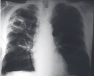
lobes of the right lung.(9) The blood workup revealed
the following: globular volume, 36.8%; hemo-globin, 12.4 g/dL; leukocytes, 9,130 × 103/µL, with
5% rods; platelet count, 294,000 × 103/µL; serum
creatinine, 0.9 mg/dL; aspartate aminotransferase, 18 mg/dL; alanine aminotransferase, 16 mg/dL; adenosine triphosphate (international normalized ratio), 1.09; anti-HBc, negative; anti-HCV, nega-tive; serological test for syphilis (Venereal Disease Research Laboratory test), negative; and anti-HIV, negative.
After the initial evaluation in the emergency room, the patient was admitted for investigation. The additional laboratory tests performed presented the following results: antineutrophil cytoplasmic antibody, negative; cryoglobulin, negative; antinu-clear factor, nonreactive; erythrocyte sedimentation rate, 86 mm in the first hour, and lactate dehydro-genase, 165 U/L. A computed tomography scan of the chest revealed cavitated and centrilobular opacities—presenting a “tree-in-bud” pattern— predominantly in the upper portions of the right lung,(10) suggesting a chronic granulomatous process
(Figure 3). Sputum smear microscopy (Ziehl-Neelsen staining) was positive in three consecutive samples, and the patient was classified as a weak reactor on the purified protein derivative test.
The patient was diagnosed with active pulmonary tuberculosis on post-admission day 4. Respiratory isolation was maintained, treatment with regimen I—rifampicin (600 mg/day), isoniazid (400 mg/day), and pyrazinamide (2,000 mg/day)—was instituted, and the patient remained hospitalized. A few days after the initiation of treatment, the patient devel-oped persistent arthralgia in the upper and lower limbs. In addition, the rash was still present, with periods of improvement and periods of exacerba-tion, sometimes accompanied by pruritus.
Consequently, the hypothesis of hypersensitivity vasculitis secondary to tuberculosis persisted. A skin lesion biopsy revealed cutaneous leukocytoclastic vasculitis. Therefore, the diagnosis of active pulmo-nary tuberculosis accompanied by hypersensitivity vasculitis secondary to tuberculosis was confirmed.
The treatment with regimen I was maintained. Analgesia with tramadol (50 mg/day) and dipyrone (1 g/day) was introduced, as was dexchlorphe-niramine (2 mg/day) for relief of the pruritus. The patient showed improvement of the arthralgia and a decrease in the number of skin lesions. We opted limbs (Figure 1). The patient also reported
throb-bing frontal headache, present 5 days before the onset of arthralgia, which improved spontaneously in a short period. He had lost 4 kg since the onset of these symptoms. The patient had been a smoker (one pack of cigarettes per day) for 35 years and a former drinker (three liters of hard liquor per day for 33 years). He had given up both habits 8 years prior. Physical examination revealed the patient to be in good general health. In addition, it revealed a maculopapular rash on the abdomen, the upper limbs, and, principally, the lower limbs. A chest X-ray (Figure 2), performed in a previous outpatient visit, revealed cavitation in the middle and upper
Figure 1 - Maculopapular rash on the lower leg.
Cutaneous leukocytoclastic vasculitis accompanied by pulmonary tuberculosis
J Bras Pneumol. 2008;34(9):745-748
747
sion of adhesion molecules. The involvement is usually cutaneous and limited. It can be idiopathic or secondary to infections, neoplasms, collagenosis, or the use of medications. However, in order to diagnose this type of vasculitis, it is necessary to rule out systemic manifestations and glomerulopa-thies.(1,2,11)
Since pulmonary tuberculosis is an infectious process, it can manifest as vasculitis, although this combination is rare.
In cases of cutaneous leukocytoclastic vasculitis, Mycobacterium tuberculosis is not found in the vessel wall, which differentiates it from cutaneous tuberculosis, in which microorganisms are seen in biopsy samples. Considering that the existence of circulating immune complexes in pulmonary tuber-culosis has previously been demonstrated, and that their levels are related to disease activity, the mech-anism of injury proposed for this type of vasculitis is deposition of immune complexes (formed by antibodies against antigens of the bacillus) in the vascular wall rather than direct aggression of the bacillus.(3,12,13)
Clinically, this type of vasculitis can appear as the first symptom of the disease or it can be part of the overall clinical profile. In our patient, the most pronounced profile was that of vasculitis, accompanied by systemic symptoms (fever, head-ache, arthralgia, and weight loss) that are common to all types of tuberculosis and that also occur in vasculitis. It is more common in young individuals whose immune system is unimpaired, and here we emphasize that our patient had no history of recur-rent infections suggestive of immunodeficiency, and that he tested negative for HIV. In addition, no gender-related differences in incidence have been reported.(4)
A confounding factor is the appearance of vascu-litis following tuberculosis treatment with rifampicin, since there are reports of cases demonstrating the relationship between the use of rifampicin and the onset of cutaneous leukocytoclastic vascu-litis. In cases of tuberculosis-related vasculitis, the skin lesions improve with the introduction of the treatment regimen, without the need for specific anti-inflammatory treatment, only medication for symptoms, as demonstrated in the case reported here, in which the skin lesions persisted for some days, showing gradual remission until complete resolution. There are host-related factors that facil-for outpatient follow-up treatment, and the patient
was discharged from the hospital after 20 days.
Discussion
Vasculitis can affect blood vessels of all sizes in any organ, and this results in a wide variety of signs and symptoms in the clinical presentation.(1,2)
There are different ways of classifying vasculitis. It can be classified as localized, such as isolated cutaneous vasculitis, or systemic, affecting various organs and systems. These can be designated as primary or idiopathic. They can also be designated as secondary when within the context of another pathological process, such as infections, including tuberculosis (as is the case of our patient), collagen-osis, neoplasms, and drug hypersensitivity. Currently, the most widely used classification system for vasculitis is the Chapel Hill Consensus Conference system.(1,2) According to this classification, cutaneous
leukocytoclastic vasculitis is classified as small vessel vasculitis. Histologically, the characteristic lesion is an angiocentric inflammatory process associated with leukocytoclasia (neutrophil fragmentation) and fibrinoid necrosis. It occurs more frequently in small blood vessels, and the pathophysiological mechanism involved is the deposition of immune complexes, usually immunoglobulin M and G, that activate the complement pathways, leading to the production of chemotactic factors and the
748 Carvalho M, Dominoni RL, Senchechen D, Fernandes AF, Burigo IP, Doubrawa E
J Bras Pneumol. 2008;34(9):745-748
References
1. Sneller MC, Langford CA, Fauci AS. The Vasculitis Syndromes. In: Kasper DL, Braunwald E, Fauci AS, Hauser SL, Longo DL, Jameson JL, editors. Harrison’s Principles of Internal Medicine. 16th ed. New York: McGraw-Hill; 2005. p.2002-14.
2. Jennette JC, Falk RJ. Small-vessel vasculitis. N Engl J Med. 1997;337(21):1512-23.
3. Parish WE, Rhodes EL. Bacterial antigens and aggregated gamma globulin in the lesions of nodular vasculitis. Br J Dermatol. 1967;79(3):131-47.
4. Mínguez P, Pintor E, Burón R, Díaz-Pollán B, Puche JJ, Pontes JC. Pulmonary tuberculosis presenting with cutaneous leukocytoclastic vasculitis. Infection. 2000;28(1):55-7. 5. Chan CH, Chong YW, Sun AJ, Hoheisel GB. Cutaneous
vasculitis associated with tuberculosis and its treatment. Tubercle. 1990;71(4):297-300.
6. Martinez V, Zeller V, Caumes E, Katlama C, Bricaire F. [Cutaneous vasculitis disclosing pulmonary tuberculosis][Article in French]. Ann Med Interne (Paris). 2000;151(8):664-6.
7. Iredale JP, Sankaran R, Wathen CG. Cutaneous vasculitis associated with rifampin therapy. Chest. 1989;96(1):215-6. 8. Han BG, Choi SO, Shin SJ, Kim HY, Jung SH, Lee KH. A case
of Henoch-Schönlein purpura in disseminated tuberculosis. Korean J Intern Med. 1995;10(1):54-9.
9. Bombarda S, Figueiredo CM, Funari MBG, Soares Jr J, Seiscento M, Terra-Filho M. Imagem em tuberculose pulmonar. J Pneumol. 2001;27(6):329-40.
10. Campos CA, Marchiori E, Rodrigues R. Tuberculose pulmonar: achados na tomografia computadorizada de alta resolução do tórax em pacientes com doença em atividade comprovada bacteriologicamente. J Pneumol. 2002;28(1):23-9. 11. Carlson JA, Ng BT, Chen KR. Cutaneous vasculitis update:
diagnostic criteria, classification, epidemiology, etiology, pathogenesis, evaluation and prognosis. Am J Dermatopathol. 2005;27(6):504-28.
12. Johnson NM, McNicol MW, Burton-Kee EJ, Mowbray JF. Circulating immune complexes in tuberculosis. Thorax. 1981;36(8):610-7.
13. Brostoff J, Lenzini L, Rottoli P, Rottoli L. Immune complexes in the spectrum of tuberculosis. Tubercle. 1981;62(3):169-73. 14. Nogueira AP. Motivos e tempo de internação e o tipo de
saída em hospitais de tuberculose do Estado de São Paulo, Brasil - 1981 a 1995. J Pneumol. 2001;27(3):123-9. 15. Ribeiro SA, Matsui TN. Hospitalização por tuberculose em
hospital universitário. J Pneumol. 2003;29(1):9-14. itate the onset of vasculitis, such as blood stasis,
hypercoagulability, and, in cases of tuberculosis, the degree of chronic inflammation, the rapid resolu-tion of which leads to remission of vasculitis.(5,7)
Another possible manifestation, though even rarer, is the combination of pulmonary tuberculosis and Henoch-Schönlein purpura: a type of renal involvement in the form of glomerulonephritis, as part of a bout of systemic vasculitis triggered by tuberculosis.(8)
Cutaneous leukocytoclastic vasculitis is treated by treating the underlying disease; in this case, tuberculosis, as previously mentioned. In vasculitis caused by medications (for example, rifampicin), it is recommended that the medication be discontinued and replaced by an alternative regimen, which results in clear lesion improvement. Antihistamines and nonsteroidal anti-inflammatory drugs can be used for symptomatic treatment. Severe skin mani-festations can be treated with corticosteroids, the use of which was not necessary in our patient.(2)
This case illustrates one of the multiple clinical presentations of tuberculosis, all of which make it difficult to rapidly identify and control the disease in order to break the transmission cycle.(14,15) It is of