Surgical treatment of 60 patients with pulmonary
malformations: What have we learned?*
Tratamento operatório de 60 pacientes com malformações pulmonares: O que aprendemos?
Altair da Silva Costa Júnior1, João Aléssio Juliano Perfeito2, Vicente Forte3
Abstract
Objective: To retrospectively analyze the medical charts of patients with pulmonary malformations submitted to surgical treatment and to investigate the clinical evolution prior to the definitive diagnosis. Methods: We analyzed the medical charts of patients with pulmonary malformations operated on at the São Paulo Hospital—Federal University of São Paulo/Paulista School of Medicine—from 1969 to 2004. Each medical chart was analyzed as to the following aspects: clinical profile; diagnosis; previous treatment; surgical treatment; and nosocomial complications. The inclusion criteria were having received a diagnosis of pulmonary malformation, having undergone pulmonary resection, and chart data being complete. Results: The analysis of the medical charts revealed that 60 patients diagnosed with pulmonary malformations—27 cases of bronchogenic cyst, 14 cases of congenital lobar emphysema, 10 cases of pulmonary sequestration, and 9 cases of cystic adenomatoid malformation—underwent surgery. Ages ranged from 4 days to 62 years (mean, 17.9 years). There was a predominance of males (55%). Ninety-two percent of the patients presented symptoms (mean duration, 15.37 months). Of the 60 patients undergoing surgery, 27 (45%) received preoperative home or hospital treatment with antibiotics. Regarding complications, we observed that morbidity was 23%, and mortality was 3.3%. Surgical times ranged from 1 to 8 h (mean, 3.2 h). Conclusions: Misdiagnosis or delayed diagnosis of pulmonary malformations resulted in unnecessary treatments and hospitalizations, as well as in frequent, recurrent infectious complications. We believe that the definitive treatment is surgery, which is curative and has low morbidity and mortality rates.
Keywords: Bronchogenic cyst; Bronchopulmonary sequestration; Cystic adenomatoid malformation of lung, congenital; Respiratory tract infections; Thoracic surgery.
Resumo
Objetivo: Analisar retrospectivamente os prontuários de pacientes com malformações pulmonares submetidos a tratamento operatório e verificar a evolução clínica até o diagnóstico definitivo. Métodos: Analisamos os prontuários dos pacientes com malformações pulmonares operados no Hospital São Paulo—Universidade Federal de São Paulo/Escola Paulista de Medicina—de 1969 a 2004. Cada prontuário foi anal-isado quanto aos seguintes aspectos: quadro clínico, diagnóstico, tratamento prévio, tratamento operatório e complicações hospitalares. Os critérios de inclusão foram os seguintes: ter diagnóstico de malformação pulmonar, ter sido submetido à ressecção pulmonar e ter prontuário com dados completos. Resultados: A análise dos prontuários revelou que 60 pacientes com diagnóstico de malformações pulmonares foram operados—27 casos de cisto broncogênico, 14 de ensifema lobar congênito, 10 de seqüestro pulmonar e 9 de malformação adenomatóide cística. A idade variou de 4 dias a 62 anos (média de 17,9 anos). Houve predominância do sexo masculino (55%). Noventa e dois por cento dos pacientes apresentavam sintomas (média de duração, 15,37 meses). Dos 60 pacientes operados, 27 (45%) receberam tratamento domi-ciliar ou hospitalar com antibiótico antes da operação. Quanto às complicações, observamos morbidade de 23% e mortalidade de 3,3%. A duração dos procedimentos operatórios realizados em nossos pacientes variou de 1 a 8 h (média, 3,2 h). Conclusões: A falha ou atraso no diagnóstico das malformações pulmonares resultou em tratamentos e hospitalizações desnecessárias e em complicações infecciosas recor-rentes e freqüentes. Acreditamos que o tratamento definitivo é a operação, a qual é curativa e tem baixa morbidade e mortalidade.
Descritores: Cisto broncogênico; Seqüestro broncopulmonar; Malformação adenomatóide cística congênita do pulmão; Infecções respiratórias; Cirurgia torácica.
* Study carried out in the Thoracic Surgery Section of the Department of Surgery of the Universidade Federal de São Paulo/Escola Paulista de Medicina – UNIFESP/ EPM, Federal University of São Paulo/Paulista School of Medicine – São Paulo, Brazil.
1. Attending Physician in the Thoracic Surgery Section of the Department of Surgery. Universidade Federal de São Paulo/Escola Paulista de Medicina – UNIFESP/ EPM, Federal University of São Paulo/Paulista School of Medicine – São Paulo, Brazil.
2. Adjunct Professor in the Thoracic Surgery Section of the Department of Surgery. Universidade Federal de São Paulo/Escola Paulista de Medicina – UNIFESP/ EPM, Federal University of São Paulo/Paulista School of Medicine – São Paulo, Brazil.
3. Tenured Adjunct Professor in the Thoracic Surgery Section of the Department of Surgery. Universidade Federal de São Paulo/Escola Paulista de Medicina – UNIFESP/EPM, Federal University of São Paulo/Paulista School of Medicine – São Paulo, Brazil.
Correspondence to: Altair da Silva Costa Júnior. Disciplina de Cirurgia Torácica, Rua Napoleão de Barros, 715, 4º andar, Vila Clementino, CEP 04024-002, São Paulo, SP, Brasil.
Tel 11 5576-4295. Fax 11 5572-0448. E-mail: [email protected] Financial support: None.
divided into two categories: preoperative clinical data (age at surgery, gender, and infection prior to the definitive treatment, as well as presence and duration of symptoms); and postoperative data (extubation in the operating room, need for inten-sive care unit—ICU—care, surgical times, type of procedure performed, length of hospital stay, and complications).
This study was approved by the Ethics in Research Committee of the institution (CEP 0496/04).
Results
The analysis of the medical charts revealed that 60 patients diagnosed with pulmonary malforma-tions—27 cases of bronchogenic cyst, 14 cases of congenital lobar emphysema, 10 cases of pulmonary sequestration, and 9 cases of cystic adenomatoid malformation—underwent surgery.
Preoperative clinical data
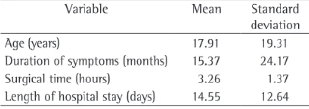
Ages ranged from 4 days to 62 years, with a mean of 17.9 years (Table 1). The incidence was highest in the first year of life (28.3% of the patients). Of the 60 patients, 32 (53.3%) underwent surgery at age 13 or younger (Figure 1).
There was a predominance of males: 33 (55%) males 27 (45%) females.
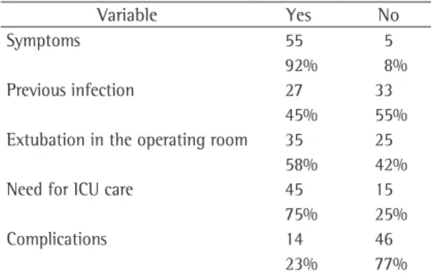
We observed that most (92%) of the patients presented symptoms that have little specificity, such as cough, dyspnea, or thoracic discomfort, but that were reason enough for these patients to seek medical attention (Table 2). The duration of symptoms was also evaluated. Despite the lack of specificity of the complaints, the duration reached up to 36 months in 3 patients, with a mean of 15.37 months (Table 1).
Introduction
Air-filled spaces in the lung were first described by Fontanus in 1638, whereas congenital cystic lung diseases were described in 1687 by Bartholinus and Marcellus Malpighius in the book “Opera Omnia”. Congenital chest lesions are rare, and their clinical
expression remains uncertain.(1)
It is clear that the majority of alveolar morpho-genesis occurs in the postnatal period. Morphometric studies estimate that only 8% (between 20 and 50 million) of the alveoli we possess in adulthood are present at birth. After the age of 8 years, the alveolar units continue to grow and develop, although more slowly. This growth and development is character-ized by the increase in alveolar size (hypertrophy), known as compensatory lung growth. At the age of 9, there are approximately 280 million alveoli, a
number close to that found in adulthood.(2,3)
The incidence of lung malformations range form 30 to 42 cases for every 100,000 inhabitants per year—from 0.06 to 2.2% of the patients admitted to
general hospitals.(4,5) The forms of presentation of
lung malformations vary. They can be very severe, including perinatal death, or remain totally asymp-tomatic until adulthood. It is important to recognize these conditions so that appropriate treatment (usually surgical) can be instituted, thereby avoiding unnecessary treatments.
The objective of the present study was to retro-spectively analyze the medical charts of patients with pulmonary malformations submitted to surgical treatment and to investigate the clinical evolution prior to the definitive therapeutic option.
Methods
We retrospectively analyzed the medical charts of patients undergoing surgery for the correction of pulmonary malformations between 1969 and 2004 at the São Paulo Hospital—Federal University of São Paulo/Paulista School of Medicine—located in São Paulo, Brazil. The inclusion criteria were having received a diagnosis of pulmonary malformation (confirmed by anatomopathological examination), having undergone pulmonary resection, and having a chart in which the data were complete.
Each medical chart was analyzed as to the following aspects: clinical profile; diagnosis; previous treatment; surgical treatment; and noso-comial complications. The data evaluated were
Table 1 - Age, duration of symptoms, surgical times, and length of hospital stay of the patients studied (n = 60).
Variable Mean Standard
deviation
Age (years) 17.91 19.31
Duration of symptoms (months) 15.37 24.17
Surgical time (hours) 3.26 1.37
period in the ICU (Table 2). Surgical times ranged from 1 to 8 h, with a mean of 3.2 h (Table 1).
The procedures performed were as follows: pulmonary lobectomy (in 58%); cystectomy (in 13%); pulmonary segmentectomy (in 13%); sequestrectomy (in 10%); bilobectomy (in 3%); and pneumonectomy (in 3%). Length of hospital stay ranged from 3 to 70 days, with a mean of 14.55 days (Table 1).
Regarding complications, we observed that morbidity was 23% (14 patients, 4 of whom presented more than one complication). The types of complication were pneumonia (6 cases), atel-ectasis (6 cases), pleural empyema (3 cases), and sepsis (2 cases). Death occurred in 2 cases. One patient presented a retained clot and needed to undergo a second operation. The mortality rate was 3.3%—2 patients with malformations who under-went surgery before the age of 1 year, at different time points, and who developed atelectasis, pneu-monia, and sepsis (one of them with hospital stay of 70 days).
Discussion
Pulmonary malformations have been known to physicians for more than three centuries. However, no consensus has yet been reached regarding how and when to treat these patients. Even if we consider that the incidence of pulmonary malfor-mations is low, it is up to twice as high as the incidence of lung neoplasms—17.6 patients for every
100,000 inhabitants in the state of São Paulo.(6)
The alterations leading to malformations remain unknown. They are thought to occur between the second and sixteenth weeks of gestation, although
there is still controversy in the literature.(1,5,7)
When we think of congenital lung diseases, we relate them to asymptomatic patients. However, a review of the literature revealed that this does not reflect reality. In fact, malformations present
symp-toms in most patients.(1,8-12) In a small percentage of
patients, the disease will be identified in imaging studies performed routinely or based on another non-respiratory indication. Patients with pulmo-nary malformations will present symptoms at some point, typically before the age of 13. In the present study, 92% of the patients were symptomatic at the time of diagnosis. Our results are similar to those reported in other studies in the literature, in which A total of 27 (45%) of the patients received
preoperative home or in-hospital treatment with antibiotics (Table 2). Recurrent pulmonary infection (pneumonia) was found in 18 (30%) of the patients. In 7 (11.7%) of the patients, more than five treat-ments were reported, and, in 11 (18.3%), two to four treatments were reported. One (1.7%) of the patients was treated with antibiotics on ten separate occasions, nine of which involved hospital admis-sion, without ever being diagnosed with pulmonary malformation.
Postoperative data
Extubation at the end of the procedure, while the patient was still in the operating room, was performed in 58% of the cases. A total of 75% of the patients spent the immediate postoperative recovery
Table 2 - Incidence of symptoms, previous infection, extubation in the operating room, need for intensive care unit care, and complications (n = 60).
Variable Yes No
Symptoms 55 5
92% 8%
Previous infection 27 33
45% 55%
Extubation in the operating room 35 25
58% 42%
Need for ICU care 45 15
75% 25%
Complications 14 46
23% 77%
ICU: intensive care unit.
100.0 80.0 60.0 40.0 20.0 0.0
(%)
0 to 1 2 to 8
9 to 12 13 to 19 20 to 29 30 to 39 40 to 49 50 to 59
>60
28.3 16.7
8.3 13.3 5.0 10.0 10.0
3.3 5.0
correct preoperative diagnosis is made in only 30%
of the patients.(8) Failure to diagnose malformations
results in unnecessary treatments, recurrent infec-tious complications, and frequent hospitalizations. Even with a delayed diagnosis, all patients can be cured with surgical treatment. We believe that surgical treatment should not be performed in the acute phase of the respiratory infection, except in
specific situations, with clinical treatment failure.(1)
Surgical treatment is indicated due to risk of infection, interference with lung growth (ipsilateral or contralateral), heart failure, and progression of the pulmonary lesions to malignancy. During preg-nancy, we can opt for clinical observation or prenatal interventions in fetuses with pleural effusion or hyperinflated cysts, which are usually treated with thoracocentesis. If necessary, intrauterine lobec-tomy can be used. In the postnatal period, the best treatment is malformation resection, which typically
has an excellent prognosis.(1,11,20,21)
In patients having experienced a great number of pulmonary infections prior to surgical treat-ment, the surgery itself is more difficult due to the inflammation around the malformation. There is a higher probability of finding pleural adherences and fibrotic tissue around the lesion, and, there-fore, surgical times can be longer. This probability is lower in asymptomatic patients. The reason for this difference in surgical times would be the longer duration of the disease, with more infections and inflammations in the lesion itself or in the adja-cent parenchyma. With more inflammations and adherences, resection tends to be slower and more cautious, and, therefore, surgical times are longer. In the literature, there have been reports of difficulties during surgery in patients who were symptomatic
in the preoperative period.(10,16,22) The degree of
difficulty during surgery is higher in symptomatic patients than in asymptomatic ones, and surgical treatment should therefore be performed while
patients are still asymptomatic.(1,23)
Surgical treatment is recommended, and some authors recommend that lobectomy become
the standard surgery.(23) Currently, lobectomy is
preferred only in cases of malformations affecting the entire lobe. Whenever possible and safe, only the malformed area of the lung is resected, by anatom-ical or nonanatomanatom-ical segmentectomy, as has been
reported in the literature.(24,25) However, lobectomy,
as was used in the present study, is still the type most such patients (65-100%) became symptomatic
by the age of 15.(8-12) Although the approach in
asymptomatic patients remains a point of contro-versy, these lesions have been increasingly resected
early due to the potential risk of complications.(13,14)
Even in asymptomatic patients, there is a risk of complications, and respiratory infection is the most common.
The prognosis of this disorder is unpredictable, and 45% of the patients with pulmonary malfor-mations have been reported to have some type of complications prior to the definitive treatment. These complications would include hemoptysis, pneumothorax, esophageal compression, pleuritis,
cyst infection, and pneumonia.(15,16) Other
compli-cations that can occur are the development of malignant lesions (lung carcinoma or sarcoma), pneumothorax, hemoptysis, and hemothorax. Since pulmonary resection will eventually be necessary, it is best to perform the resection before such
compli-cations occur.(13,17) One female patient in our sample
underwent resection of an adenocarcinoma in the wall of the congenital cyst.
In our series, 27 (45%) of the patients needed home or hospital treatment due to previous pulmo-nary infections or to misdiagnosis. Those infections were characterized as pneumonia. One of those patients was hospitalized nine times prior to receiving a diagnosis of malformation. One group of authors showed that diagnosis was delayed for up to 7 years, with a mean of 1.6 years; the patients remained asymptomatic in this period, and all
chest X-rays were abnormal.(18) A review involving
320 children diagnosed with pneumonia empha-sized the clinical-radiographic correlation with the evaluation of three professionals: a pediatric radiol-ogist, a pediatric pulmonolradiol-ogist, and an emergency room pediatrician. The concordance among the
observers was only 41%.(19) In Brazil, the diagnosis
of pneumonia, especially that known as “onset of pneumonia”, has evidently become commonplace. Many of the patients who present cough, rhinor-rhea, and fever, as well as an abnormal X-ray, are (or have been) treated as having pneumonia. However, the X-ray finding could correspond to a malforma-tion, such as bronchogenic cyst, and the clinical profile could correspond to that of an upper airway infection.
up to 45% of the patients,(16) an incidence that is
higher than the postoperative morbidity.
There is no consensus in the world literature regarding the ideal age at which surgical treatment should be performed after birth. There are many reasons why such surgery should be performed in adults rather than in children, and vice versa. However, it is known that lung growth through hyperplasia (actual increase in the number of alveoli) occurs up to the age of 8 years. Up to the age of 9 years, there is lung growth in volume and size,
but not in the absolute number of alveoli.(1) In a
study published in 1987, 230 patients submitted to pneumonectomy were monitored for up to 30 years. At the time of surgery, the patients were divided by age bracket. In the group undergoing surgery between birth and 5 years of age, 96% maintained
total lung capacity even after pneumonectomy.(30)
Other authors recommend that surgical treatment of patients diagnosed with pulmonary malforma-tions be performed at between 3 and 6 months of
age so that lung growth can occur.(13,29,30)
We conclude that pulmonary malformations were underdiagnosed at our facility. Sometimes, several conditions were confused with pulmonary infections, and, sometimes, they were treated as pneumonia. The diagnostic hypothesis of congen-ital pulmonary abnormality should be, at least, considered by the physician, especially in patients with recurrent pneumonia. We reiterate that misdiagnosis or delayed diagnosis of pulmonary malformations resulted in unnecessary treatments and hospitalizations, as well as in frequent, recur-rent infectious complications. We believe that the definitive treatment is surgery. There is a tendency toward resection being as conservative and as safe as possible. Currently, the indication for lobectomy is limited to patients in whom the entire lobe is affected. In patients who are still asymptomatic but present imaging evidence of pulmonary malfor-mation, surgery should be indicated even without confirmation of the diagnosis. If possible, we prefer to perform surgical treatment when the patient is ≥ 1 year of age. We emphasize that the surgical treatment is curative, with low rates of morbidity and mortality.
Further studies are necessary in order to eluci-date controversies and principally to enlighten the general medical population about the importance of the correct diagnosis of these diseases.
of resection most widely used for the treatment of
pulmonary malformations.(9,26-28)
In our sample, 58% of the patients were extubated at the end of the procedure in the oper-ating room. A total of 75% required ICU care in the immediate postoperative period. The children required greater care in the postoperative period, which might have been a precaution rather than a real need. For patients with this type of congenital pulmonary lesion, usually young and without other comorbidities, if there are no complications during the procedure, postanesthesia recovery in the imme-diate postoperative period is sufficient, and there is
no need for ICU admission.(13,14,28)
Length of hospital stay has been a constant cause for concern, and the mean hospital stay in the present study was 14.55 days. It is important to emphasize that this retrospective study was long and subject to flaws. During the three decades of this study, medicine has been through many changes. Currently, diagnostic investigation is pref-erably performed in outpatient clinics, reducing the length of hospital stays.
A total of 23% of the patients presented complications, and mortality was 3.3%. Among the complications, atelectasis prevailed (in 5 chil-dren and 1 adult), especially in the patients younger than 2 years, which is explained by the fact that the airway diameter is too small for the secretions, which are sometimes as viscous as those in adults. When the patients were divided by age bracket, morbidity and mortality were found to be higher in the chil-dren younger than 1 year (58% of such patients had atelectasis and pneumonia; 11.8% died). It is worthy of note that, in patients in this age bracket, indications for surgery should be pondered when-ever permitted by the clinical profile. If surgical treatment can be postponed until the child is 1 year of age or older, morbidity and mortality rates will be lower. We have found similar incidences in the literature, with morbidity being 40% and mortality being around 10%. Among the most common complications reported are prolonged drainage, empyema, pneumonia, and infection at the incision
site.(11,16,29) In the present study, the morbidity and
16. Sarper A, Ayten A, Golbasi I, Demircan A, Isin E. Bronchogenic cyst. Tex Heart Inst J. 2003;30(2):105-8.
17. Endo C, Imai T, Nakagawa H, Ebina A, Kaimori M. Bronchioloalveolar carcinoma arising in a bronchogenic cyst. Ann Thorac Surg. 2000;69(3):933-5.
18. Gustafson RA, Murray GF, Warden HE, Hill RC, Rozar GE. Intralobar sequestration. A missed diagnosis. Ann Thorac Surg. 1989;47(6):841-7.
19. Sarria E, Lima JB, Fischer GB, Barreto SS, Flôres JA, Sukiennik R. Concordância no diagnóstico radiológico das infecções respiratórias agudas baixas em crianças. J Pediatr (Rio J). 2003;79(6):497-503.
20. Canals-Riazuelo J, Boix Ochoa J, Peiro JL, Ezzedine M, Cobos Barroso N, Liñan Cortés S, et al. [Congenital lobar emphysema: report of 38 cases][Article in Spanish]. Cir Pediatr. 1994;7(2):97-101.
21. Conran RM, Stocker JT. Extralobar sequestration with frequently associated congenital cystic adenomatoid malformation, type 2: report of 50 cases. Pediatr Dev Pathol. 1999;2(5):454-63.
22. Aktoğu S, Yuncu G, Halilçolar H, Ermete S, Buduneli T. Bronchogenic cysts: clinicopathological presentation and treatment. Eur Respir J. 1996;9(10):2017-21.
23. Ribet ME, Copin MC, Gosselin BH. Bronchogenic cysts of the lung. Ann Thorac Surg. 1996;61(6):1636-40.
24. Browdie D, Todd D, Agnew R, Rosen W, Beardmore H. The use of “nonanatomic” pulmonary resection in infants with extensive congenital adenomatoid malformation of the lung. J Thorac Cardiovasc Surg. 1993;105(4):732-6.
25. van Koningsbruggen S, Ahrens F, Brockmann M, Michalk D, Rietschel E. Congenital cystic adenomatoid malformation type 4. Pediatr Pulmonol. 2001;32(6):471-5.
26. Leape LL, Longino LA. Infantile lobar emphysema. Pediatrics. 1964;34:246-55.
27. Murray GF. Congenital lobar emphysema. Surg Gynecol Obstet. 1967;124(3):611-25.
28. Ozçelik U, Göçmen A, Kiper N, Doğru D, Dilber E, Yalçin EG. Congenital lobar emphysema: evaluation and long-term follow-up of thirty cases at a single center. Pediatr Pulmonol. 2003;35(5):384-91.
29. Kim YT, Kim JS, Park JD, Kang CH, Sung SW, Kim JH. Treatment of congenital cystic adenomatoid malformation-does resection in the early postnatal period increase surgical risk? Eur J Cardiothorac Surg. 2005;27(4):658-61.
30. Laros CD, Westermann CJ. Dilatation, compensatory growth, or both after pneumonectomy during childhood and adolescence. A thirty-year follow-up study. J Thorac Cardiovasc Surg. 1987;93(4):570-6.
References
1. Costa Jr AS. Análise retrospectiva do tratamento operatório das malformações torácicas (pulmonares e linfáticas). [dissertation]. São Paulo: Universidade Federal de São Paulo; 2006.
2. Ruiz Jr RL, Carvalho LR, Catâneo AJ. Crescimento pulmonar compensatório (CPC): massa corpórea, conteúdo protéico e massa pulmonar em ratos subnutridos trilobectomizados. Acta Cir Bras. 2004;19(2):146-52.
3. Catâneo AJM, Catâneo DC. Crescimento pulmonar compensatório em implante lobar autólogo pós-pneumonectomia em cães. Acta Cir Bras. 2005;20(5):368-74.
4. Juan ES. Tratamento dos cistos congênitos do pulmão. [thesis for tenure]. São Paulo: Universidade de São Paulo; 1954.
5. Skandalakis JE, Gray SW, Symbas P. The trachea and the lungs. In: J.E. Skandalakis JE, Gray SW, editors. Embryology for surgeons: the embryological basis for the treatment of congenital anomalies. 2nd ed. Baltimore: Williams & Wilkins; 1994. p. 414-450.
6. Zamboni M. Epidemiologia do cancêr do pulmão. J Pneumol. 2002;28(1):41-7.
7. Landing BH. Five syndromes (malformation complexes) of pulmonary symmetry, congenital heart disease, and multiple spleens. Pediatr Pathol. 1984;2(2):148-51.
8. Ramenofsky ML, Leape LL, McCauley RG. Bronchogenic cyst. J Pediatr Surg. 1979;14(3):219-24.
9. Snyder ME, Luck SR, Hernandez R, Sherman JO, Raffensperger JG. Diagnostic dilemmas of mediastinal cysts. J Pediatr Surg. 1985;20(6):810-5.
10. St-Georges R, Deslauriers J, Duranceau A, Vaillancourt R, Deschamps C, Beauchamp G, et al. Clinical spectrum of bronchogenic cysts of the mediastinum and lung in the adult. Ann Thorac Surg. 1991;52(1):6-13.
11. Nobuhara KK, Gorski YC, La Quaglia MP, Shamberger RC. Bronchogenic cysts and esophageal duplications: common origins and treatment. J Pediatr Surg. 1997;32(10):1408-13.
12. Louie HW, Martin SM, Mulder DG. Pulmonary sequestration: 17-year experience at UCLA. Am Surg. 1993;59(12):801-5. 13. Laberge JM, Puligandla P, Flageole H. Asymptomatic
congenital lung malformations. Semin Pediatr Surg. 2005;14(1):16-33.
14. Shanmugam G. Adult congenital lung disease. Eur J Cardiothorac Surg. 2005;28(3):483-9.