One - staged reconstruction of bladder exstrophy in male
patients: long - term follow-up outcomes
_______________________________________________
Amilcar Martins Giron
1, Marcos Figueiredo Mello
1, Paulo Afonso Carvalho
1, Paulo Renato Marcelo
Moscardi
1, Roberto Iglesias Lopes
1, Miguel Srougi
11 Divisão de Urologia do Departamento de Cirurgia, Universidade de São Paulo, SP, Brasil
ABSTRACT
ARTICLE
INFO
______________________________________________________________ ______________________
Keywords:
Bladder Exstrophy; Reconstructive Surgical Procedures; Male; Patients
Int Braz J Urol. 2017; 43: 155-62
_____________________
Submitted for publication: December 01, 2015
_____________________
Accepted after revision: March 13, 2016
___________________
Published as Ahead of Print: October 18, 2016
INTRODUCTION
Classic bladder exstrophy is a rare malfor-mation of the genitourinary tract and its incidence is around 1 case to 30.000 to 40.000 live births (1).
The surgical management of bladder exstrophy has evolved during the last years, with the stan-dard treatment until the late 1950s being urinary diversion with ureterosigmoidostomy. Afterwards, in 1970s it evolved to a staged repair, with early
Introduction: The surgical correction of bladder exstrophy remains challenging. In our institution, the repair has evolved from a staged repair to one-stage reconstruction. The one-stage reconstruction includes; bladder closure, Cantwell-Ransley neourethroplasty and abdominoplasty using groin flaps, without the need of pelvic ostheotomies. Repair of urinary continence (UC) and vesicoureteral reflux (VUR) is done after development of the infant.
Objective: To present our experience of our modified one-stage reconstruction of bla-dder exstrophy in male patients.
Materials and Methods: Medical records of male patients submitted to one-stage re-construction of bladder exstrophy were analyzed retrospectively. Fifteen exstrophy bladder patients with mean age 4.2±7 years were treated at our institution between 1999-2013.
Results: Eleven patients were referred to us after previous surgery. Sixteen procedu-res were performed; one patient had complete wound dehiscence and needed another reconstruction (6.7%). Mean follow up was 10.3±4.5 years. No patient has had a loss of renal function. Postoperative complications: four patients (26.6%) presented small fistulas, one presented penile rotation. Eleven patients (73.3%) patients underwent bladder-neck surgery. Five (33.3%) required bladder augmentation. Three cases (20%) needed subsequent treatment of VUR.
At the time of our review nine (60%) patients achieved UC, two (13.3 %) patient without additional procedure. A mean of 3±1.1 procedures (2-5) was accomplished per children.
pelvic ring approximation and abdominal wall, bladder and posterior urethral closure performed as a first stage, followed by second stage neoure-throplasty (modified Cantwell-Ransley technique) and finally a bladder neck surgical reinforcement such as Yong-Dees-Leadbetter procedure (2). The modern staged repair (MSRE) technique involves bladder closure shortly after birth, followed by epispadias repair at age 6-12 months and bladder neck reconstruction at age 4-5 years when it is thought that the child can cooperate with attemp-ting continence.
In 1990s, Mitchell introduced the concept of one-stage reconstruction of extrophy, where all the aforementioned procedures were performed as a single surgery comprehensive approach (3). The concept of this approach was to decrease the number of surgical procedures required to achieve continence as well as achieve early bladder neck resistance and bladder cycling (4). This technique presented good results, although it has some dra-wbacks such as risk of penile tissue loss and ne-cessity of ostheotomies in older children or after failed repair.
Placing the posterior urethra and bla-dder deep into the pelvis in combination with a tension-free closure and adequate postoperative management prevent complications and are now consensus among pediatric urologists (5). The two well-described techniques: MSRE (6) and one-sta-ge reconstruction of bladder exstrophy advocates
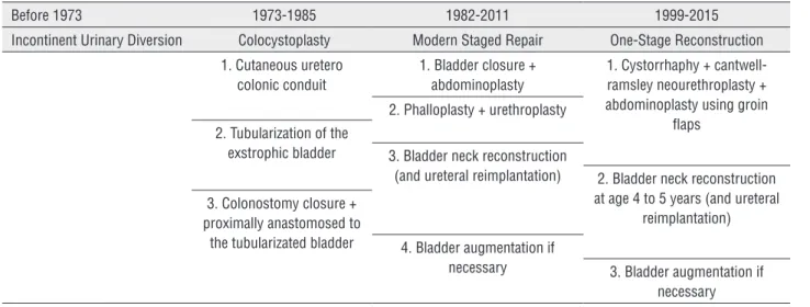
the observance of these fundamental principles (4). In our institution, the repair of bladder exstrophy has evolved from a staged repair to one-stage reconstruction (Table-1). However, we describe our one-stage reconstruction of bladder exstrophy, performed in University of São Paulo, since late 1990s as a single comprehensive sur-gery that was adapted to our environment since it was common to receive older children with pre-vious failed repairs. In this procedure, we perform bladder closure and positioned it deep in the pel-vis, Cantwell-Ransley neourethroplasty and abdo-minoplasty using groin flaps, without the need of pelvic ostheotomies. Urinary continence (UC) and vesicoureteral reflux (VUR) are addressed later, at toilet training age.
OBJECTIVE
To present our experience of our modified one-stage reconstruction of bladder exstrophy in male patients.
MATERIALS AND METHODS
Medical records of male patients submitted to one-stage reconstruction of bladder exstrophy were analyzed retrospectively. Fifteen exstrophy bladder patients (16 procedures) with mean age 4.2±7 years (45 days to 22 years) were treated at our institution between September 1999 and
Table 1 - Treatment of bladder exstrophy in male patients (time table).
Before 1973 1973-1985 1982-2011 1999-2015
Incontinent Urinary Diversion Colocystoplasty Modern Staged Repair One-Stage Reconstruction 1. Cutaneous uretero
colonic conduit
1. Bladder closure + abdominoplasty
1. Cystorrhaphy + cantwell-ramsley neourethroplasty + abdominoplasty using groin
flaps 2. Phalloplasty + urethroplasty
2. Tubularization of the
exstrophic bladder 3. Bladder neck reconstruction
(and ureteral reimplantation) 2. Bladder neck reconstruction at age 4 to 5 years (and ureteral
reimplantation) 3. Colonostomy closure +
proximally anastomosed to
the tubularizated bladder 4. Bladder augmentation if
October 2013. Nine patients were referred to us after previous failed bladder closure elsewhere. Additionally, five patients had undergone other surgical procedures: inguinal herniorrhaphy in two and urinary diversion in three cases (two colonic conduits, one bilateral cutaneous ureterostomy).
At time of bladder exstrophy repair, pa-tients that were referred to us after previous failed bladder closure elsewhere had a mean age of 6.5 years (2 months to 22 years) and children without previous attempts of repair had a mean age of 9 months (6 to 18 months). At time of data analysis, the group with previous surgery had mean age of 17.7 years (6 to 34 yeas) and the naïve surgical group had a mean age of 9.5 years (3 to 18 years).
All patients were treated with cystorrhaphy, Cantwell-Ransley neourethroplasty, and abdomino-plasty using groin flaps to close the abdominal wall defect, without osteotomies. Cystorrhaphy consists in bladder closure in two planes and placing the posterior urethra and bladder deep into the pelvis performing a tension-free closure of the bladder; bladder neck surgery was only performed at time of toilet training. Cantwell-Ransley neourethroplasty begins with extensive dissection of the epispadias but without complete penile disassembly providing easy access to the intersymphyseal ligament, which is deeply incised, dissection of each neurovascular bundle and the urethral plate with its spongiosal tissue, preserving the glans; then the urethral plate is tubularized as shown in Figure-1A. The corpo-ra are rotated medially by approximately 90º and maintained in this new configuration by a proxi-mal caverno-cavernostomy; this new anastomosis between the corpora keeps the urethra in its ventral position and gives the penis a dangling position when flaccid (Figure-1B).
Abdominal wall repair consists in using hy-pogastric skin and rectus abdominis and obliquus externus abdominis muscle aponeurosis flaps (the-se groin flaps are rotated to the midline resulting in a very strong abdominal wall). Groin flaps are made of the rectus anterior aponeuroses rotated medially, flipped over, and sutured with prolene su-tures to close the defect (Figure-1C) (7). By rotating the facial flaps medially, complete reinforcement of the abdominal wall to the level of the pubic bone
is achieved.
All received broad-spectrum intravenous antibiotics (3rd generation cephalosporins)
intra--operatively and continued postoperatively (1st
generation cephalosporin) and analgesics and anti--inflammatory drugs as needed for 1-2 weeks. A urethral catheter was left for 7-10 days and two plastic catheters were used for drainage of subcuta-neous tissue (Figure-1D).
Successful primary closure was defined as an acceptable functional outcome with no wound dehiscence or fistula; failed closure was defined as wound dehiscence.
Considered variables were length of surgery, length of hospital stay, complications related to one-stage reconstruction and urinary continence.
Bladder neck reconstruction was performed at age 4 to 5 years when it is thought that the child can cooperate with attempting continence. Addi-tional procedures were needed in many cases to achieve the treatment goals (urinary continence, normal genital cosmetic and preservation of upper urinary tract). Patients were considered continent if dry after the age of toilet training (3 to 5 ye-ars), and for those under this age, if dry for inter-vals between 2 to 3 hours. They were considered incontinent if any urinary leakage was observed between voiding or catheterizations from either the catheterizable channel or the urethra (for toi-let trained children) or if they could not achieve continence intervals of ≥2 hours (for toddlers and infants). Patients who were using diapers were in-cluded in the incontinent group.
RESULTS
Mean operative time was 325±61.3 minu-tes (240 to 420 min) and mean hospital stay was 13.2±5.8 days (6 to 23 days). Successful closure was achieved in 14 patients (93.3%) performing a single procedure; one patient had complete wound dehis-cence and needed another reconstruction (6.7%) - this patient had previous bladder closure elsewhe-re. None had ischemic loss of the glans or corporal bodies. Other four patients (26.6%) presented small fistulas and one (6.7%) presented penile rotation as a complication related to one-stage reconstruction. Mean follow-up was 10.3±4.5 years (2y8mos to 16y).
Nine patients (60%) are continent at pre-sent: seven voids spontaneously and two are on clean intermittent catheterization. From the group with previous bladder closure elsewhere, five (55%) achieved continence and from the group of naive patients, four (66.7%) are continent. All patients have normal kidneys on US and normal serum creatinine. Seven patients (46.6%) showed vesicoureteral reflux on voiding cystourethrogra-phy performed after urinary tract infections.
Additional procedures were needed to achieve the treatment goals, including upper urinary tract protection, urinary continence and satisfactory cosmetic results. Eleven patients (73.3%) patients underwent bladder neck surgery Figure 1 – Steps of one-stage reconstruction: A) Closure of the bladder and tubularized urethra; B) Penile closure;
C) Groin flaps; D) Final aspect
C D
(nine pts. underwent Young Dees Leadbetter and two was submitted to bladder neck injection of Durasphere), and seven out of eleven achieved continence. Five (33.3%) required augmentation ileocystoplasty and Mitrofanoff stoma to facili-tate CIC. There are two out of five patients with bladder augmentation that are still incontinent. VUR needed subsequent treatment in three ca-ses (20%) (ureteral reimplantation). Three boys (20%) required inguinal herniorrhaphy during follow-up (two unilateral and one bilateral). A mean of 3±1.1 procedures (2-5) was accompli-shed per children. Six patients have still pen-ding procedures (three awaits for augmentation cystoplasty associated with bladder neck
re-construction and three cases need urinary fistu-la repair) (Table-2).
Patients that were referred to us after previous failed bladder closure elsewhere nee-ded more procedures then children without pre-vious bladder closure, (mean of 3.3±1.1; mean of 2.5±0.8 procedures per children, respective-ly). The most common additional procedure was bladder neck surgery. Eight patients (88%) from the group with previous bladder closure perfor-med elsewhere needed the procedure, while three patients (50%) from the naive treatment group needed this procedure. Moreover, at present, five out of nine (55%) patients with previous failed bladder closure are continent and four out of six
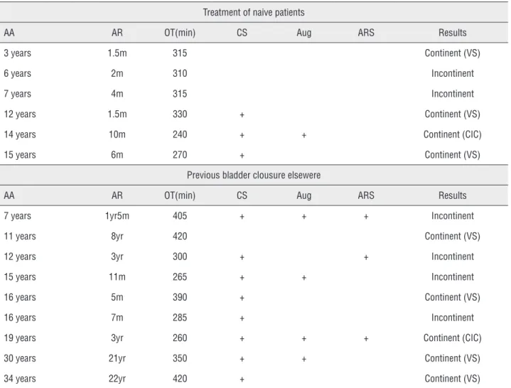
Table 2 - Patient characteristics and results of operation with one-stage reconstruction.
Treatment of naive patients
AA AR OT(min) CS Aug ARS Results
3 years 1.5m 315 Continent (VS)
6 years 2m 310 Incontinent
7 years 4m 315 Incontinent
12 years 1.5m 330 + Continent (VS)
14 years 10m 240 + + Continent (CIC)
15 years 6m 270 + Continent (VS)
Previous bladder clousure elsewere
AA AR OT(min) CS Aug ARS Results
7 years 1yr5m 405 + + + Incontinent
11 years 8yr 420 Continent (VS)
12 years 3yr 300 + + Incontinent
15 years 11m 265 + + Incontinent
16 years 5m 390 + Continent (VS)
16 years 7m 285 + Incontinent
19 years 3yr 260 + + + Continent (CIC)
30 years 21yr 350 + + Continent (VS)
34 years 22yr 420 + Continent (VS)
(67%) of those without previous bladder closure are continent.
DISCUSSION
The management of children with blad-der exstrophy remains a challenge and despite the choice of approach (staged versus one-stage reconstruction), patients have to undergo several procedures in order to attain goals of surgical treatment such as: urinary continence, preserva-tion of upper urinary tract and genital funcpreserva-tion and cosmesis.
Stjernqvist et al. showed a median of 12 procedures to achieve good results in bladder exstrophy staged approach (MSRE) (8). There are few experiences with one-stage reconstruction: Gargollo et al. had a mean of 4 (range 1 to 31) procedures to achieve satisfactory results (9). Ebert et al. reported mean of 2.95 (range 1 to 8) surgeries in patients who underwent single stage repair, with only 13.6% requiring more than 4 surgeries, about half of these patients were re-ferred after failed reconstruction elsewhere (10). In our series, around 73.3% (11 out of 15) of pa-tients were referred after failed reconstruction, and a mean of 3±1.1 procedures (range 2-5) per children was observed, and we still have six pa-tients waiting for additional surgeries.
The incidence of urinary continence af-ter bladder exstrophy repair is variable (12% to 83%) (11-15). Various factors interfere with re-sults analysis. There is no standard definition for continence and as a result studies address continence in a non-uniform way. Patient age at bladder closure, the type of closure performed, the number and type of procedures required to establish continence, the need for concomitant bladder augmentation and the need for clean in-termittent catheterization is not reported in most papers. Again, there are few cases treated with one-stage reconstruction. Mitchell and collea-gues showed 74% (17 of 23 patients) of dayti-me continence. Overall, 2 of 10 boys (20%) with bladder exstrophy achieved primary daytime continence with one-stage reconstruction alone and without the need for further bladder neck re-construction (16). In parallel, we showed 60% of
our cohort (9 of 15 patients) continent, but only two (13.3%) achieved continence with one-stage reconstruction alone. This result indicates that one-stage reconstruction alone was mostly not able to give continence and bladder neck surgery is usually necessary.
The incidence of progressive or seve-re hydronephrosis and/or seve-renal scarring ranges from 0% to 30% after one-stage reconstruction. Later surgical repair of vesicoureteral reflux was necessary in 0-50% of patients (17). In our series, three (20%) patients developed vesicoureteral re-flux that required treatment.
The limitations of our study are that the experience with one-stage reconstruction was relatively small and not all patients underwent complete treatment to evaluate the efficacy of this procedure. However, we do believe it has advantages over the traditional approaches to bladder exstrophy. Based in our experience, one--staged reconstruction without osteotomy is fea-sible at any age, even after previous failed proce-dures, reducing the surgical steps and facilitating closure of the structures. It helps minimize the total number of surgeries. Improved urethral re-sistance may increase bladder capacity in young patients and restores bladder cycling, which re-sults in the expansion of even very small recons-tructed bladders with poor bladder plates (18, 19). While increased outlet resistance may allow for an increase in bladder capacity, a recognized consequence is elevated bladder pressure, which may lead to upper tract changes. It enables con-comitant abdominoplasty, with good cosmetic results. Primary or secondary bladder neck re-construction is required for optimal continence. In our pool of patients treated with primary re-pair, the need for bladder augmentation is still significant, but complications are less frequent than in the staged procedures.
exstrophy-epis-padias complex, as it decreases tension across the abdominal wall, reduces the pubic diastasis, and helps restore the pelvic ring and floor to the nor-mal anatomical configuration. However, in our series, it was not necessary to perform pelvic os-theotomies and bone mobilization, as we opted to use groin flaps for the abdominoplasty. It is necessary to wait for rectus anterior sheets con-sistency, which occurs usually after 45-60 days of life. The main reason for us to adopt this ap-proach was that most of our patients were refer-red after failed attempt and these older children are less amenable to collaborate with traction ne-eded after ostheotomies.
Another advantage of our technique in comparison to Mitchell’s one-stage reconstruc-tion is that we do not perform complete penile disassembly, reducing risks of penile ischemia and loss (Figure-1). In our series, ischemic loss of the glans or corporal bodies was not observed.
CONCLUSIONS
Most patients with bladder exstrophy will require multiple operations to achieve normal voiding and provide cosmetically acceptable and functional genitalia. One-stage reconstruction minimizes the number of surgical procedures required to achieve the treatment goals (urinary continence, normal genital cosmesis and preser-vation of upper urinary tract). The advantages of using groin flaps over current techniques for complete repair are the small risk for penile tis-sue loss and the avoidance of pelvic ostheoto-mies. The major drawback of this technique is the necessity to correct the bladder exstrophy defect after 45-60 days of life (wait to rectus anterior sheets consistency) and also a theoretical risk of malperfusion and loss of flaps, which was not observed in this study.
CONFLICT OF INTEREST
None declared.
REFERENCES
1. Wiesel A, Queisser-Luft A, Clementi M, Bianca S, Stoll C; EUROSCAN Study Group. Prenatal detection of congenital renal malformations by fetal ultrasonographic examination: an analysis of 709,030 births in 12 European countries. Eur J Med Genet. 2005;48:131-44.
2. Jeffs RD. Functional closure of bladder exstrophy. Birth Defects Orig Artic Ser. 1977;13:171-3.
3. Grady RW, Mitchell ME. Complete primary repair of exstrophy. Surgical technique. Urol Clin North Am. 2000;27:569-78, xi. 4. Grady RW, Mitchell ME. Complete primary repair of
exstrophy. J Urol. 1999;162:1415-20.
5. Grady RW, Mitchell ME. Surgical techniques for one-stage reconstruction of the exstrophyeepispadias complex. In: Wein AJ, Kavoussi LR, Novick AC, Partin AW, Peters CA, editors. Campbell-Walsh urology. Philadelphia: Saunders Elsevier Publishers; 2007; pp. 3553-72.
6. Gearhart JP, Mathews R. Exstrophyeepispadias complex. In: Wein AJ, Kavoussi LR, Novick AC, Partin AW, Peters CA, editors. Campbell-Walsh urology. Philadelphia: Saunders Elsevier Publishers; 2007; pp. 3497-553.
7. Giron AM, Lopes RI, Guarniero R, Passerotti C, Srougi M. One-stage external iliac fixation device and bilateral fascial and groin flaps facilitate abdominal wall closure after posterior sagittal iliac osteotomy in cloacal exstrophy. Eur J Pediatr Surg. 2011;21:377-80.
8. Stjernqvist K, Kockum CC. Bladder exstrophy: psychological impact during childhood. J Urol. 1999;162:2125-9.
9. Gargollo PC, Borer JG, Diamond DA, Hendren WH, Rosoklija I, Grant R, et al. Prospective followup in patients after complete primary repair of bladder exstrophy. J Urol. 2008;180:1665-70.
10. Ebert A, Scheuering S, Schott G, Roesch WH. Psychosocial and psychosexual development in childhood and adolescence within the exstrophy-epispadias complex. J Urol. 2005;174:1094-8.
11. Capolicchio G, McLorie GA, Farhat W, Merguerian PA, Bägli DJ, Khoury AE. A population based analysis of continence outcomes and bladder exstrophy. J Urol. 2001;165:2418-21. 12. Hollowell JG, Ransley PG. Surgical management of
incontinence in bladder exstrophy. Br J Urol. 1991;68:543-8. 13. Lottmann HB, Melin Y, Cendron M, Lombrail P, Beze-Beyrie
P, Cendron J. Bladder exstrophy: evaluation of factors leading to continence with spontaneous voiding after staged reconstruction. J Urol. 1997;158:1041-4.
15. Shaw MB, Rink RC, Kaefer M, Cain MP, Casale AJ. Continence and classic bladder exstrophy treated with staged repair. J Urol. 2004;172:1450-3.
16. Shnorhavorian M, Grady RW, Andersen A, Joyner BD, Mitchell ME. Long-term followup of complete primary repair of exstrophy: the Seattle experience. J Urol. 2008;180:1615-9.
17. Husmann DA. Surgery Insight: advantages and pitfalls of surgical techniques for the correction of bladder exstrophy. Nat Clin Pract Urol. 2006;3:95-100.
18. Mitchell ME. Bladder exstrophy repair: complete primary repair of exstrophy. Urology. 2005;65:5-8.
19. Gearhart JP, Jeffs RD. Bladder exstrophy: increase in capacity following epispadias repair. J Urol. 1989;142:525-6.
20. Inouye BM, Lue K, Abdelwahab M, Di Carlo HN, Young EE, Tourchi A, et al. Newborn exstrophy closure without osteotomy: Is there a role? J Pediatr Urol. 2016;12:51.e1-4.
_______________________ Correspondence address:
Marcos F. Mello, MD Divisão de Urologia do Departamento de Cirurgia,