SURGICAL MANAGEMENT OF THE NEUROGENIC
BLADDER AND BOWEL
GERALD C. MINGIN, LAURENCE S. BASKIN
Pediatric Urology, Children’s Hospital, University of California, San Francisco, California, USA
ABSTRACT
Spina bifida and myelodysplasia are associated with neurogenic abnormalities of the bladder and bowel function. All children with myelodysplasia require an evaluation of their urinary tract with ultrasound and urodynamics to confirm normal bladder and kidney function. Patients with anatomical and functional abnormalities require treatment, the mainstay being intermittent catheterization and anticholinergic medication. The treatment goals for patients with a neurogenic bladder are the preservation of the upper urinary tract, bladder and bowel continence, independence, autonomy, and facilitation of self-esteem. A minority of children will not respond to conservative therapy and will ultimately require surgical intervention. This review will discuss the surgical options for bladder augmentation, bladder neck reconstruction and closure, as well as the methods for the creation of continent catheterizable stomas. The timing, indications, and description for each procedure will be addressed. Finally, the antegrade continence enema procedure will be described for the management of refractory fecal incontinence.
Key words: bladder; bowel; neural tube defects; bladder, neurogenic; therapeutic; surgery
Int Braz J Urol. 2003; 29: 53-61
INTRODUCTION
The most common etiology of patients with a neurogenic bladder is myelodysplasia, which oc-curs in approximately 1 in 1,000 births in the United States (1). Myelodysplasia is a spectrum of diseases ranging from severe motor and neurogenic abnormali-ties affecting ambulation, bowel and bladder func-tion, to more minor defects such as an asymptomatic tethered chord.
All patients with myelomeningocele should have an evaluation of their urinary tract in order to confirm normal bladder and kidney function. This is especially true in ambulatory patients, where 95% will have evidence of neurogenic bladder (2). Urologic treatment of myelomeningocele includes defining the status of the bladder and upper tracts by renal blad-der ultrasound and urodynamics. In patients with a
aim of this paper is to review the surgical treatment of patients with neurogenic bladder and bowel.
TIMING OF SURGERY
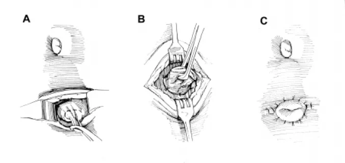
The most important aspect in the treatment of patients with myelodysplasia is preserving renal function. Urodynamic evaluation reveals that approxi-mately 27% of patients have poor outlet resistance alone, and can be managed with observation. Sixty-two percent have failure to coordinate their bladder and sphincter, with hypertonicity/spasticity and poor emptying. These children will require intermittent catheterization and anticholinergic medications. The remaining 10% have flaccid bladders and sphincter dysinergia, which may develop spasticity or hyperto-nicity and require intermittent catheterization and an-ticholinergic medications (5). Regardless of medical management, a few patients in the neonatal and early years may have recurrent infection or increasing hydronephrosis. In these cases, vesicostomy is a use-ful surgical adjunct. We advocate the classic Blocksom technique (Figure-1). Vesicostomy allows the neuro-genic bladder to cycle while keeping the pressures low. During a bladder contraction with inappropriate
sphinc-ter contraction (in the absence of a vesicostomy) the increased pressure will pop off into a refluxing kid-ney, or alternatively cause increasing hydronephro-sis. Reconstruction with closure of the vesicostomy and augmentation is typically performed when the child begins school, when the patient and the family are ready to accept the responsibilities of intermit-tent catheterization.
Patients that continue to have stable renal function without recurrent infection are monitored until they start school. At that time careful assess-ment of bowel and bladder continence is performed. There are some variations in the exact age when con-tinence is sought after depending on developmental issues, family support, and patient and family com-pliance. Usually we recommend attaining continence between 5 and 7 years of age.
PREOPERATIVE PLANNING
We have been strong advocates in the initia-tion of neonatal intermittent catheterizainitia-tion for pa-tients with myelomeningocele (6). We feel that the concept of intermittent catheterization is well
cepted by parents with a newborn baby presenting spina bifida. The psychological consequences of the early initiation of catheterization facilitates its latter acceptance. Since eventual continence is almost al-ways dependent on catheterization, we do not hesi-tate to introduce the concept at birth. Parents and chil-dren who are catheterizing early are better prepared to accept and handle reconstruction. Early institution of catheterization also improves bladder compliance and decreases the need for augmentation (3,6).
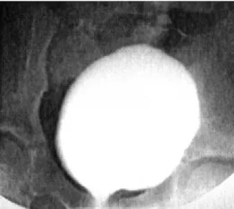
When the patient and his/her family are pre-pared to accept the requirements associated with con-tinence, an assessment of the bladder and urinary sphincter is required. Two key questions must be an-swered. First: is the bladder volume acceptable and the urinary sphincter competent, or does the child re-quire augmentation and/or an outlet resistance pro-cedure to achieve continence (7)? For example, a patient with marginal capacity may have an outlet that is insufficient to achieve continence. With augmen-tation, however, the compliance will improve and the outlet then becomes sufficient to keep the patient dry. The status of the patient’s ambulation is also impor-tant. Ambulatory patients with myelodysplasia have increased intra-abdominal pressures, and therefore require greater outlet resistance to achieve continence. Prior to surgical correction of incontinence, urodynamic evaluation with fluoroscopic imaging is recommended. Bladder capacity prior to leakage is compared to expected capacity (7). Sphincter func-tion is assessed by radiologic evaluafunc-tion of the blad-der neck. An open bladblad-der neck throughout filling is unlikely to provide continence, and will require sur-gical correction (Figure-2). Since emptying in patients with a neurogenic bladder will require intermittent catheterization, preoperative assessment determines whether catheterization will be performed through the urethra or an abdominal stoma will be necessary. It has been our bias that patients who require extensive bladder neck reconstruction be provided with an ab-dominal stoma to assure catheterization. In achiev-ing continence (defined as the ability to wear normal undergarments without protection), we have found that even properly performed bladder neck reconstruc-tion can make routine catheterizareconstruc-tion less than 100% reliable. Since catheterization is critical for the
patient’s safety, an abdominal stoma reduces the risk of the patient being unable to empty his/her reservoir. Preoperative assessment also includes docu-menting the presence of vesicoureteral reflux, which can be detected during urodynamics. Although aug-mentation may itself lead to reflux resolution, it has been our preference to reimplant refluxing ureters at the time of augmentation (8). This can be done safely with little increase in the operative time, leading to complete resolution of reflux (9).
BLADDER AUGMENTATION
Historically, many different segments of bowel have been used for bladder augmentation. The ideal segment should be lined with urothelium, mak-ing ureterocystoplasty an excellent choice. However, it is unusual for a patient to have a nonfunctional seg-ment amenable to this type of reconstruction. There have been multiple attempts to maintain urothelial lined structures, such as autoaugmentation or autoaugmentation with demucosalized sigmoid, ileal, or gastric patches (10-14). Evidence exists that would not support placing intestinal stroma in contact with urothelium (15). This may create abnormal cellular signaling between cell types that are normally not in contact with each other, leading to cellular
typic changes. For example, intestinal stromal influ-ences of the bowel are likely to change the urothelium to an intestinal phenotype overcoming the purpose of composite augmentations (15).
Intensive work is also currently in progress with tissue matrices, either homotypic acellular or het-erotypic intestinal scaffolds (16,17). Autologous cul-tured urothelium may improve the outcomes, but so far, tissue engineering has not proven to be clinically successful.
The most commonly used segment for aug-mentation is ileum. Classic clamshell augaug-mentation cystoplasty has been utilized successfully both in adults and in children. It is important to isolate the segment at least 10 cm from the ileocecal valve, so as not to interfere with bowel control, especially in patients with myelodysplasia who already have bowel continence issues. Detubularization on the anti-mesenteric border provides a low-pressure res-ervoir and increases capacity when fashioned into either a U or W- shape (18). The bladder plate needs to be widely opened to prevent an hourglass defor-mity when anastomosing the patch to the bladder (Fig-ure-3). Sigmoid augmentation has also been success-ful, although there are reports of increased rhythmic contractions when this segment is used (19). In se-lect cases, gastric augmentation has been quite suc-cessful. Proper patient selection, which includes neu-rogenic patients who are insensate, is critical to avoid the clinical syndrome of hematuria/dysuria. Gastric augmentation has the advantage of less mucous pro-duction and less changes in metabolic parameters (20). Patients with ileal augmentation are subject to metabolic acidosis, mucous production, and uroli-thiasis, as well as a risk of cancer at the anastomotic site (21,22). We have recently shown that metabolic changes do not have an adverse affect on linear growth or bone density in children undergoing augmentation (23). Finally, patients with any type of intestinal aug-mentation are at risk for perforation, especially if compliance with catheterization is sub-optimal (24,25). Any patient undergoing anaugmentation should wear a medical alert band as a reminder of the risk of bladder perforation. This will serveto warn medical personal that abdominal pain should initiate a work-up to ensure an intact augmentation cystoplasty.
BLADDER NECK CONTINENCE
RECONSTRUCTION
In children with poor urethral outlet resis-tance, bladder neck reconstruction may be necessary to obtain continence. The literature is filled with a myriad of papers describing different methods to in-crease urethral resistance, while at the same time al-lowing ease of catheterization. In our experience, none of these procedures is fully proven.
In females, our choice has been a fascial sling placed just distal to the bladder neck, with the con-comitant creation of an abdominal stoma (Figure-4). The sling can be placed abdominally, which is our choice, if a concomitant augmentation is performed. To safely identify the space between the urethra and the vagina (urethra and rectum in males), the poste-rior midline approach behind the bladder is used (26). In patients with adequate bladder compliance, the sling can be placed by a vaginal approach. Recently, human cadaveric fascia and xenotropic sling mate-rial has become available, avoiding the need for an abdominal incision and autologous sling harvest (27). We have been successful in catheterizing the urethral sling in the majority of our female patients. Occa-sionally, we have utilized the Mitrofanoff principle providing an abdominal stoma for catheterization.
In males, we have not found one operation that can be applied to every patient. Our choices in-clude either the Young-Dees Leadbetter (Figure-5), or a modified Kropp bladder neck tube (Figure-6) (28). The Young-Dees Leadbetter procedure is per-formed by reimplantation of the ureter to a higher position in the bladder, and tubularization of the mu-cosa over a 5F feeding tube. In both theYoung-Dees and Kropp procedures, augmentation cystoplasty is typically performed at the same setting with a catheterizable abdominal stoma thus avoiding any issues with catheterization.
ARTIFICIAL URINARY SPHINCTER
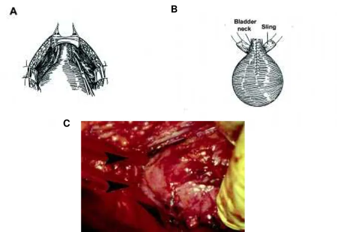
Figure 3 - Anastomosis of the bowel segment to an open bladder plate. A) Diagram of the open bladder. B) Intra-operative photo of the clamed shelled bladder, with arrows pointing to each bladder half. (From Hinman F Jr: Atlas of Pediatric Urology. Philadelphia, W.B. Saunders Co., 1994, with permission).
Figure 4 - Placement of the fascial sling around the bladder neck. A) Diagram showing incision of the endopelvic fascia. B) Diagram showing the fascial sling around the bladder neck. C) Intra-operative photo with arrows indicating the position of a vessel loop around the bladder neck. (From Elder JS: Sling Procedure. Atlas of the Urologic Clinics of North America, 2000, with permission).
B
B
sphincter include infection, which requires removal, as well as the need for replacing pump reservoir and tubing should malfunction occur. The average lifespan of the artificial sphincter is 8 years, all but guarantee-ing eventually replacement. In prepubertal children, the cuff must be placed at the bladder neck, while post-pubertally it can be placed around the bulbar urethra.
BLADDER NECK CLOSURE
Bladder neck closure is a last resort in those patients who have undergone alternative procedures
with refractory incontinence. The procedure can be performed retropubically, transvesically, or transvaginally in girls. This procedure is done in combination with the creation of an abdominal stoma, and can be difficult in patients with previous bladder neck surgery. The amount of scarring often requires opening the bladder to gain access to the bladder neck. The key to a successful operation is wide mobilization of the bladder neck, and interpo-sition of healthy tissue between the closed bladder neck and urethral stump. Omentum is the preferred tissue. Our long term results illustrate the difficulty of the procedure (29).
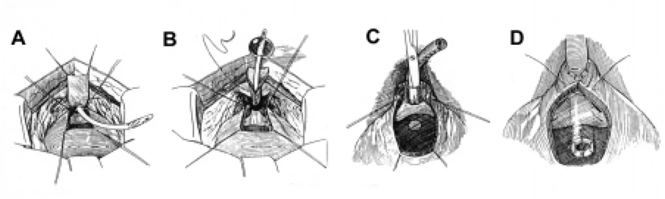
Figure 5 - Diagram of the Young-Dees Leadbetter bladder neck reconstruction. A) The bladder is opened and the bladder mucosa denuded, leaving a strip of mucosa. B)The mucosal strip is anastomosed over a catheter. C) and D) The bladder muscle is reaproximated in pants-over-vest fashion. (From Gearhart JP: Modified Young-Dees Leadbetter Procedure for Urinary Incontinence in the Non-Neurogenic Bladder. Atlas of the Urologic Clinics of North America, 2000, with permission).
CATHETERIZABLE STOMAS
All continent catheterizable stomas employ a flap valve (Mitrofanoff) principal to maintain conti-nence. Continence is achieved as the bladder fills and the intravesical pressure is transmitted to the conduit (30). We prefer the appendix. The proximal end is brought out to the abdominal wall, either through the lower quadrant or through the umbilicus, and is spatu-lated and anastamosed to a U-shaped skin flap created to prevent stenosis. If appendix is not available, ileum
Figure 7 - Diagram of the classic Monti procedure. A) and B) A 2 cm ileal segment is isolated and then opened on its anti-mesenteric border. B) Detubularization on the antimesenteric side of the segment. C) and D) The flap is then transversely tubularized over a catheter. (From Monti PR: New techniques for construction of efferent conduits based on the Mitrofanoff principle. Urology 1997, 49:112-5, with permission).
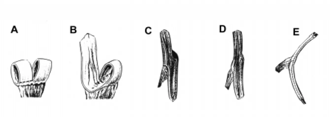
and colon can also be fashioned into a conduit utiliz-ing the Monti procedure (31). In the classic Monti pro-cedure (Figure-7), a 2 cm segment of ileum or colon is isolated and opened on the antimesenteric border and retubularized; if the length is inadequate, 2 segments can be fashioned and anastamosed to each other. Al-ternatively, a single segment can be split in the middle, opened from opposite ends, and rolled over a feeding tube to create a spiral Monti (Figure-8). This provides adequate length in most cases, avoiding the need to harvest 2 separate intestinal segments.
A newer technique, which is gaining wide-spread acceptance is augmentation with combined ab-dominal stoma that is devised from an eccentric U-shaped ileal patch (Macedo procedure, Figure-9) (32). The advantage of the Macedo procedure is that a sepa-rate segment of bowel is not required for the Monti procedure.
NEUROGENIC BOWEL – ACE MALONE
In children who have failed conservative man-agement of their fecal incontinence, the same Mitrofanoff and Monti principals can be applied to the creation of an antegrade continence enema (ACE) (33). The appendix can be brought out through the umbilicus or the lower quadrant; if unavailable, a 2 cm segment of ileum or colon can be substituted for the appendix. The appendix or tubularized bowel is imbricated or reimplanted into the cecum or large bowel in an antirefluxing fashion to prevent leakage of stool or flatus.
CONCLUSION
In patients with myelodysplasia, the primary goal is preserving upper tract function with concomi-tant attainment of urinary and fecal continence. In order to accomplish these goals, each child requires evaluation, with the institution of intermittent cath-eterization and/or medical therapy from early child-hood. If the child develops recurrent infection or in-creasing hydronephrosis early on, a vesicostomy is
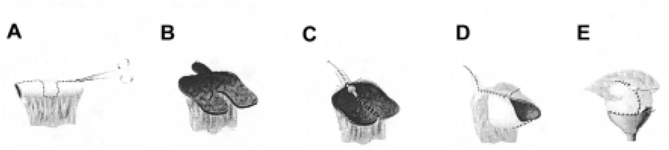
Figure 9 - Diagram of the Macedo procedure. A) A 3 cm segment of ileum is first isolated. It is then opened on the anti-mesenteric border, taking care to isolate a 3 cm segment from the anterior part of the ileum. B), C) and D) The 3 cm segment and ileal plate are then tubularized. E) The continence valve mechanism is created by embedding the tube over a serous-lined extramural tunnel. (From Macedo A, Srougi M: A continent ileum-based reservoir. BJU Int. 2000; 85:160-2).
needed until the child is old enough for reconstruc-tion. Otherwise, patients should be followed until school age when continence issues can be addressed. Only when the patient and his/her family are ready and understand the consequences of surgery, should augmentation, bladder neck reconstruction, or the creation of a continent catheterizable stoma be per-formed.
REFERENCES
1. Bauer SB: Neurogenic bladder dysfunction. Pediatric Clin North Am. 1987; 34:1121-32.
2. Mevorach RA, Bogaert GA, Baskin LS, Lazzaretti CC, Edwards MS, Kogan BA: Lower urinary tract func-tion in ambulatory children with spina bifida. Br J Urol. 1996; 77:593-6.
3. Sutherland RS, Mevorach RA, Baskin LS, Kogan BA: Spinal dysraphism in children: an overview and an ap-proach to prevent complications. Urology 1995; 46:294-304.
4. Teichman JM, Scherz HC, Kim KD, Cho DHP, MG, Kaplan GW: An alternative approach to myelodyspla-sia management: aggressive observation and prompt intervention. J Urol. 1994; 152:807-11.
5. Baskin LS, Kogan BA, Benard F: Treatment of infants with neurogenic bladder dysfunction using anticholin-ergic drugs and intermittent catheterization. Br J Urol. 1990; 66:532-4.
7. Houle AM, Gilmour RF, Churchill BM, Gaumond M, Bissonnette B: What volume can a child normally store in the bladder at a safe pressure? J Urol. 1993; 149:561-4. 8. Nasrallah PH, Alilabad HA: Bladder augmentation in
patients with neurogenic bladder and vesicoureteral reflux. J Urol. 1991; 146:563.
9. Nomura S, Ishido T, Tanaka K, Komiya A: Augmen-tation ileocystoplasty in patients with neurogenic blad-der due to spinal cord injury or spina bifida. Spinal Cord. 2002; 40:30-3.
10. Cartwright PC, Snow BW: Bladder autoaugmentation: early clinical experience. J Urol. 1989; 142:505-8. 11. Cartwright PC: Bladder autoaugmentation: partial
de-trusor excision to augment the bladder without use of bowel. J Urol. 142:1050-3.
12. Dewan PA: Autoaugmentation demucosalized enterocystoplasty. World J Urol. 1998. 16:255-61. 13. Duel BP, Gonzalez R, Barthold JS: Alternative
tech-nique for augmentation cystoplasty. J Urol. 1998; 159:998-1005.
14. Nguyen DH, Mitchell ME, Horowitz MB, DJ Carr MC: Demucosalized augmentation gastrocystoplasty with bladder autoaugmentation in pediatric patients. J Urol. 1996; 156:206-9.
15. Li YW, Liu WH, et al.: Plasticity of the urothelial phe-notype: effects of gastro- intestinal mesenchyme/stroma and implications for urinary tract reconstruction. Dif-ferentiation 2000; 66:126.
16. Sutherland RS, Baskin LS, Hayward SW: Regenera-tion of bladder urothelium, smooth muscle, blood ves-sels and nerves into an accellular tissue matrix. J Urol. 1996; 156:571-7.
17. Kropp BP, Rippy MK, Badylahak ST: Characteriza-tion of small intestine submucosa regenerated canine detrusor: an assessment of reinnervation, in vitro com-pliance and contractility. J Urol. 1996; 156:599-607. 18. Hinman F: Selection of intestinal segments for
blad-der substitution: physical and physiological character-istics. J Urol. 1988; 139:519-23.
19. Goldwasser B, Webster GD: Augmentation and sub-stitution enterocystoplasty. J Urol. 1986; 135:215-24. 20. Bogaert GA, Mevorach RA, Kim A, Kogan BA: The physiology of gastrocystoplasty: once a stomach, al-ways a stomach. J Urol. 1995; 153:1977-80.
21. Mitchell ME, Piser JA: Intestinocystoplasty and total bladder replacement in children and young adults: fol-low-up in 129 cases. J Urol. 1987; 138:579-84. 22. Eraklus AJ, Folkman MJ: Adenocarcinoma at the site
of ureterosigmoidostomies for exstrophy of the blad-der. J Pediatric Surg. 1978; 13:730-4.
23. Mingin GC, Nguyen HT, Mathias RS, Shephered JA: Growth and metabolic consequences of bladder aug-mentation in children with myelomeningocele and bladder exstrophy. Pediatrics 2002; 110:1193-8. 24. Sheiner JR, Kaplan GW: Spontaneous bladder rupture
following enterocystoplasty. J Urol. 1988; 140:1157-8. 25. Rink RC, Woodbury PW, Mitchell ME: Bladder per-foration following enterocystoplasty. J Urol. 1988; 139:234.
26. de Badiola F, Gosalbez R, Ruiz E, Sosa A, Labbie A: The posterior approach for bladder neck dissection. Br J Urol. 2000; 85:59.
27. Grossklaus DF, Shappell SB, Adams MC, Brock JW, Pope JC: Small intestinal submucosa as a urethral cov-erage layer. J Urol. 2001; 166:636-9.
28. Kropp KA: Kropp Urethral Lengthening. Atlas, Urol Clin North Am. 2001; 9:81.
29. Nguyen HT, Baskin LS: Is bladder neck closure the final surgery in children with severe urinary inconti-nence. Presented at the Society for Pediatric Urology, Orlando, Florida, 2002.
30. Mitrofanoff P: Trans-appendicular continent cys-tostomy in the management of the neurogenic bladder [in French]. Chir Pediatr. 1980; 21:297.
31. Monti PR, Lara RE, Dutra MA, Carvalho JR: New tech-niques for construction of efferent conduits based on the Mitrofanoff principle. Urology 1997; 49:112-5. 32. Macedo A Jr, Srougi M: A continent catheterizable
ileal based reservoir. BJU Int. 2000; 85:160-2. 33. Malone PS, Ransely PG, Kiely EM: Preliminary
re-port: the antegrade continence enema. Lancet 1990; 336:1217-8.
Received: August 7, 2002 Accepted: August 30, 2002
Correspondence address:
Dr. Laurence S. Baskin Section of Pediatric Urology
University of California, San Francisco 533 Parnassus Avenue, U575
San Francisco, California, 94143-0738, USA Fax: + 1 415 476-8849