Prostate cancer detection using multiparametric 3 - tesla
MRI and fusion biopsy: preliminary results
_______________________________________________
Thais Caldara Mussi
1, Rodrigo Gobbo Garcia
2, Marcos Roberto Gomes de Queiroz
2, Gustavo Caserta
Lemos
3, Ronaldo Hueb Baroni
11 Departamento de Radiologia e Diagnóstico por Imagem do Hospital Israelita Albert Einstein, São Paulo,
SP, Brasil; 2 Departamento de Intervenção Guiada por Imagem do Hospital Israelita Albert Einstein, São Paulo, SP, Brasil; 3 Departamento de Urologia do Hospital Israelita Albert Einstein, São Paulo, SP, Brasil
ABSTRACT
ARTICLE
INFO
______________________________________________________________ ______________________
Objective: To evaluate the diagnostic efficacy of transrectal ultrasonography (US) bi-opsy with imaging fusion using multiparametric (mp) magnetic resonance imaging (MRI) in patients with suspicion of prostate cancer (PCa), with an emphasis on clini-cally significant tumors according to histological criteria.
Materials and Methods: A total of 189 consecutive US/MRI fusion biopsies were per-formed obtaining systematic and guided samples of suspicious areas on mpMRI using a 3 Tesla magnet without endorectal coil. Clinical significance for prostate cancer was established based on Epstein criteria.
Results: In our casuistic, the average Gleason score was 7 and the average PSA was 5.0ng/mL. Of the 189 patients that received US/MRI biopsies, 110 (58.2%) were positive for PCa. Of those cases, 88 (80%) were clinically significant, accounting for 46.6% of all patients. We divided the MRI findings into 5 Likert scales of probability of having clinically significant PCa. The positivity of US/MRI biopsy for clinically significant PCa was 0%, 17.6% 23.5%, 53.4% and 84.4% for Likert scores 1, 2, 3, 4 and 5, respectively. There was a statistically significant difference in terms of biopsy results between dif-ferent levels of suspicion on mpMRI and also when biopsy results were divided into groups of clinically non-significant versus clinically significant between different lev-els of suspicion on mpMRI (p-value <0.05 in both analyzes).
Conclusion: We found that there is a significant difference in cancer detection using US/MRI fusion biopsy between low-probability and intermediate/high probability Lik-ert scores using mpMRI.
Keywords:
Prostatic Neoplasms; Magnetic Resonance Imaging; Biopsy; Prostate
Int Braz J Urol. 2016; 42: 897-905
_____________________
Submitted for publication: April 09, 2015
_____________________
Accepted after revision: April 18, 2016
INTRODUCTION
Prostate cancer (PCa) is the most common male malignancy in United States, excluding skin cancers, and the second most common cause of male cancer-related death (1). Diagnosis of PCa increased with the use of prostate-specific agent (PSA) as a blood test for screening. The diagno-sis of PCa is made with a histological sample of
systematic transrectal ultrasound-guided biopsy (TRUSGB) of the prostate, which is indicated with increased values of PSA blood-test and/or altered digital rectal examination (DRE) (2-4).
2.6ng/mL,) (5, 6), DRE also has low sensitivity (37%) (7), and, when positive, prostate biopsy has up to a 54% underestimation of the Gleason score when compared with prostate specimen (2, 4, 7-12).
PCa screening with a PSA blood test still raises intense debate. The European Randomized Study of Screening for Prostate Cancer (ERSPC) showed a 20% decrease in mortality related to PCa in patients screened with PSA but with consi-derable rates of overdiagnosis and overtreatment (4.5% of the cases) (3). Others studies have shown rates ranging from 22% to 56% of overdiagnosed PCa (9, 13). Due to this, many cases of indolent and non-aggressive cancers have been discovered and treated, increasing morbidity with impacts on quality of life without changing mortality (2, 9) (14). But overdiagnosis does not have to lead to overtreatment, and active surveillance (AS), in pa-tients with low-risk tumors, can be the modality of choice for patients until early signs of disease progression (1, 3, 4, 13).
Because of the diagnostic limitations of PCa mentioned above, other tools are needed to improve detection, localization and sampling of PCa (7). Advances in 3-Tesla multiparametric (mp) magnetic resonance imaging (MRI) have improved the detection of PCa prior to biopsy (15-18).
The use of mpMRI to guide biopsy has sho-wn to increase the diagnosis of intermediate/high risk PCa and decrease the diagnosis of low-risk tumors (2).
The objective of this study was to evaluate the diagnostic efficacy of mpMRI with different levels of suspicion in detecting PCa, using TRUS-GB with US/MRI real-time imaging fusion, with an emphasis on the detection of clinically signifi-cant tumors according to histological criteria.
MATERIALS AND METHODS
This retrospective study was approved by the ethical committee of our institution and a wai-ver of informed consent was obtained. We perfor-med a database search for patients who received prostate mpMRI for the detection of clinically sig-nificant PCa, followed by TRUSG with real-time imaging fusion of US and MRI images, between
August 2013 and September 2014. Inclusion crite-ria were patients who underwent prostate mpMRI and prostate biopsy with US/MRI imaging fusion, both in our institution. Exclusion criteria were: incomplete or poor quality MRI, interval grea-ter than 6 months between MRI and biopsy, and unavailability of histopathological report. Since the aim of our study was to compare the overall mpMRI results with histological analysis based on clinically significant disease, which is classified using all samples together, we also included pa-tients whose additional samples were not identi-fied separately.
All patients underwent mpMRI on a 3 Tesla scanner (Magnetom Trio, Siemens Healthcare, Er-langen, Germany) using a phased-array coil. The mpMRI protocol is described in Table-1.
MpMRI images were read independently by two radiologists (in cases of discrepancies a consensus agreement was achieved), and scored using a 5-point Likert scale of probability of ha-ving clinically significant PCa, based on the PI--RADS version 1 classification proposed by the European Society of Urogenital Radiology (1-cli-nically significant disease is very unlikely to be present, 2-clinically significant disease is unlike-ly to be present, 3-clinicalunlike-ly significant disease is equivocal, 4-clinically significant disease is likely to be present and 5-clinically significant disease is highly likely to be present) (16, 17).
A certified pathologist evaluated the biopsy specimens. Clinical significance for PCa was established based on Epstein criteria and included any Gleason pattern 4 or higher, or Gleason 3+3 disease with more than 50% of cancer in any core and/or more than 3 positives cores (19).
Statistical analysis was made using the Shapiro-Wilk test to decide between mean (and standard deviation-SD) or median (and first and third interquartile intervals-IQs) for age, PSA levels and prostate weight. To study the associa-tion between suspicion on mpMRI and Gleason score we used the Spearman correlation coeffi-cient, and because the variables had more than two categories, we performed subanalysis with Chi-square partitions. Finally, to calculate the association between suspicion level on mpMRI with PSA level and with Gleason score we used the Kruskal-Wallis test.
RESULTS
A total of 195 consecutive patients were included in the study; six patients were excluded due to an interval greater than 6 months between MRI and biopsy leaving a final casuistic of 189 patients for analysis. The mean age was 58.12 ye-ars old (SD±9.16), median serum PSA was 5.0ng/ mL (IQs 3.6-7.1) and median prostate volume was 45cc (IQs 34-62). The mean of additional frag-ments on suspicious areas was three (range: two to four). Of 189 patients, 153 had never received
a prostate biopsy and 36 had received at least one with negative results. MpMRI was considered suspicious for PCa in 103 patients (Likert 4 or 5), equivocal in 68 (Likert 3) and low level of suspi-cion in 18 (Likert 1 or 2) (Figure-1).
Of the 189 patients who performed US/MRI biopsies, 110 (58.2%) had positive biopsy for PCa. Of those cases, 88 (80%) were clinically significant, accounting for 46.6% of all patients (Figure-2). The overall distribution of US/MRI biopsy for negative biopsy, positive biopsy with clinically non-significant cancer, and positive biopsy with clinically significant cancer was, respectively, 1/0/0 in Likert 1, 10/4/3 in Likert 2, 42/10/16 in Likert 3, 21/6/31 in Likert 4, and 5/2/38 in Likert 5, resulting in positive indications for clinically significant prostate cancer of 0%, 17.6%, 23.5%, 53.4% and 84.4% in Likert scores 1, 2, 3, 4 and 5, respectively (Table-2).
There was a statistically significant diffe-rence in the level of suspicion on mpMRI (very low, low and equivocal probability versus inter-mediate and high probability, or Likert 1, 2 and 3 versus Likert 4 and 5) compared with biopsy re-sults in terms of clinically significant disease (ne-gative biopsy and non-significant positive biopsy versus significant positive biopsy). This was also true when we included the “equivocal” category on mpMRI as positive (Likert 1 and 2 versus Likert 3, 4 and 5) (p<0.001 in both analyses) (Table-3).
We observed that most patients with a Gle-ason score of 6 were considered to have an equi-vocal level of suspicion for PCa on mpMRI, while
Table 1 - mpMRI prostate protocol.
Sequence Thickness (mm) Spacing (mm)
T2 FSE axial with fat sat 6 1
T1 GRE axial “in-phase” and “opposed-phase” 6 1
T2 FSE sagittal 3 0.3
T2 FSE axial high resolution 3 0.3
T2 FSE 3D coronal volumetric isotropic 1
-Diffusion (b50, 400 and 800) 3 0.3
T1 GRE VIBE pre-contrast 3
-T1 GRE VIBE dynamic post-contrast 3
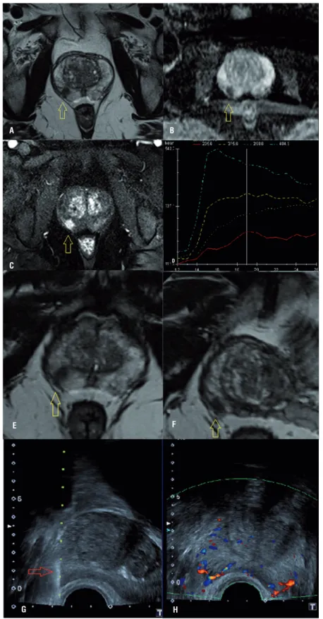
-Figure 1 - MpMRI example of a suspicion lesion on the right lobe of peripheral zone of the prostate: A) a round lesion on T2 weighted-image; B) ADC map shows diffusion restriction; C) dynamic-contrast-imaging with hypervascularization and D) washout (blue line). Patient received US/MRI-guided biopsy where we can see the lesion on MRI (E and F) and in real time on US (G), helping to target the lesion (H).
A
C
E F
B
Table 2 - Suspicion level on mpMRI and biopsy results.
Biopsy
Negative Positive
non-significant
Positive significant
Suspicion on mpMRI
Very low 1 0 0
Low 10 4 3
Equivocal 42 10 16
Moderate 21 6 31
High 5 2 38
p-value <0.001
Figure 2 - A suspicious lesion on mpMRI (images B and D) submitted to a US-MRI fusion biopsy. The lesion was not seen on US (arrow in A) and the biopsy was performed based on mpMRI (arrow in C). The biopsy result was Gleason 3+4 in all tree fragments of this area.
A
C
B
most patients with Gleason scores of 7, 8 and 9 were considered as moderate or high suspicion for PCa, as shown in Figure-2. The association betwe-en suspicion on mpMRI and Gleason score was moderately positive, with a coeffi cient of 0.435 (p<.001) (Figure-3).
In the three positive cases of clinically sig-nifi cant cancer that we classifi ed as Likert 2 (fal-se negative), two had only one positive fragment on biopsy (5% and 10% of Gleason 3+4 and 4+3, respectively), and the third case had 5% of the fragments committed with Gleason 3+4. On the other hand, in the fi ve negative cases of clinically signifi cant cancer that we had classifi ed as
Li-kert 5 (false positive), one lesion seen on MRI was diagnosed as a leiomyoma, two lesions were acute prostatitis, one lesion was chronic prostatitis and one was a small lesion (5mm) in a large prostate (125cc), which, we believe, might have resulted in undersampling during the biopsy.
DISCUSSION
Our results show that mpMRI, performed on a 3 Tesla scanner without endorectal coil, has the ability to stratify the risk of detection of clinically signifi cant prostate cancer on US/MR fusion biopsy, therefore increasing the likelihood
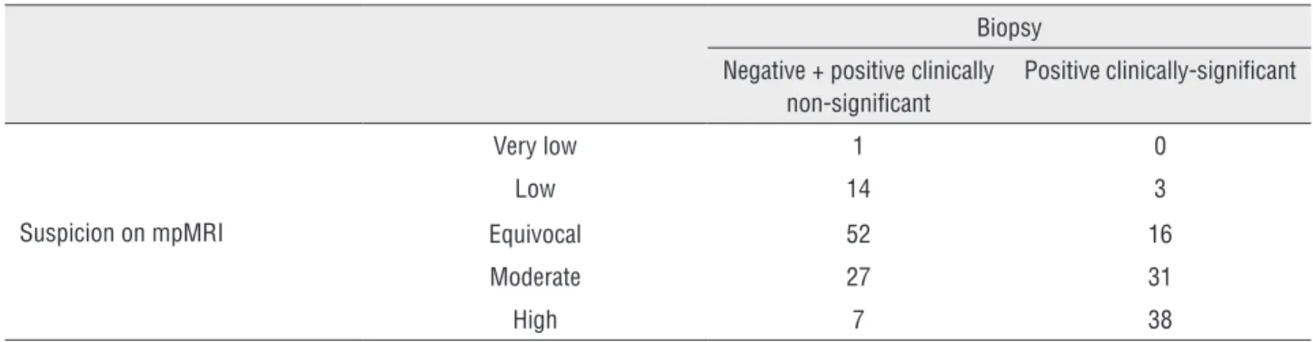
Table 3 - Suspicion level on mpMRI and biopsy results according clinical relevance.
Biopsy
Negative + positive clinically non-signifi cant
Positive clinically-signifi cant
Suspicion on mpMRI
Very low 1 0
Low 14 3
Equivocal 52 16
Moderate 27 31
High 7 38
p-value <0.001
of positivity of the procedure and decreasing unnecessary biopsies.
The prostate is the only solid organ that has the diagnosis of tumor made by non-target sampling biopsy (20). Other studies have shown that mpMRI has the capability to detect suspicious areas for PCa and to target the US-guided biopsy of lesions seen on mpMRI (8, 15-17, 21, 22), and have already suggested higher identification ra-tes than random biopsy (23). It has been shown that mpMRI increases the detection of clinically significant PCa (including those located in the an-terior region of the prostate, usually blinded on systematic biopsy), without increasing diagnosis of clinically insignificant disease (8, 18, 24).
Performing mpMRI in patients with clini-cal-laboratorial profiles suspicious for PCa could prevent unnecessary systematic biopsies and a delay in diagnosis and treatment (7).
Currently, transrectal systematic US-gui-ded biopsy is the modality of choice for pros-tate biopsy, however it is limited in that it can miss and undersample existing tumor (7, 15, 25). The use of mpMRI prior to biopsy has resulted in the development of new methods to increase the detection of clinically significant PCa: 1) in--bore MRI-guided biopsy, which is expensive and time-consuming; 2) cognitive fusion, where the lesion location is estimated by the operator; 3) US/MRI-fusion-guided biopsy, in which the pre--biopsy mpMRI is fused in real-time by a navi-gation system, allowing additional sampling of suspicious lesions through direct visualization during the procedure (8, 15, 25-27).
With the aim to make recommendations for conduct, interpretation, and reporting of pros-tate mpMRI for PCa detection and localization, a European Consensus Meeting was performed and a 5-point scale was suggested to indicate the pro-bability of malignancy (PI-RADS system) (16). We used a 5-point subjective scale of probability (Likert scale) based on the PI-RADS classifica-tion, but in which the overall impression of the imaging findings is the most important aspect of grading. There are many mpMRI prostate stu-dies in the literature using Likert, PI-RADS and also comparing both classification methods (28, 29). Rosenkrantz et al. compared the systematic
model proposed by the European Consensus (PI--RADS) with the probability score (Likert) and showed that radiologists performed well locali-zing PCa with both methods, however tumors in the central gland had better correlation with the specimen using the Likert scale (30). PI-RADS is a promising method and used by many radiolo-gists, but its implementation is still a cause for debate. Because of that we used a scoring sys-tem that we believe adds value of a standardized method (such as PI-RADS) but also relies on the radiologist’s experience and learning curve.
US/MRI-fusion-guided biopsy increases the detection of clinically significant PCa (es-pecially with higher suspicion level on mpMRI) when compared with systematic biopsy, positive-ly impacting treatment decisions and outcomes (8, 20). Thompson et al. correlated the biopsy fin-dings with PI-RADS scores on mpMRI, and found high negative predictive value and moderate po-sitive predictive value for the detection of PCa, demonstrating a potential screening test to guide biopsy decisions (12). Porpiglia et al. showed that mpMRI had better accuracy to diagnose PCa in patients with a negative biopsy than promising biomarkers (PSA3 and p2PSA) (11).
Our results show that 3 Tesla mpMRI wi-thout an endorectal coil is a non-invasive techni-que that helps to detect clinically significant PCa, with high concordance between the probability of clinically significant disease on mpMRI and biopsy results.
It is known that mpMRI has limited sen-sitivity for the detection of lesions smaller than 5mm (20, 27), possibly explaining the two cases of clinically significant tumors (Gleason score 7) that were classified as low suspicion on MRI and came up with one positive fragment on the biopsy.
result of the samples. MpMRI cases were read independently and a final report was reached by consensus agreement, but interobserver variability was not evaluated. Also, we used the biopsy as reference test, and could miss or misclassify some tumors as compared to the prostatectomy specimen; however, we believe that using a prostatectomy specimen would substantially limit the casuistic, and we would have to consider the effect of a reference standard on our population (biopsy for negative, prostatectomy for positive cases). Finally, because it was a retrospective project with the aim of studying mpMRI performance, we did not perform a follow-up on patients and record the number of complications related to biopsy.
CONCLUSIONS
In conclusion, we found that mpMRI per-formed on a 3-Tesla scanner without an endorec-tal coil and using a Likert scale has significant correlation with biopsy results in terms of can-cer detection and clinical significance. This stu-dy highlights the potential use of this method in clinical practice to manage patients with clinical suspicion of PCa, decreasing unnecessary biopsies and overdetection of clinically non-significant tumors, and increasing the diagnosis of clinically significant cancers.
CONFLICT OF INTEREST
None declared.
REFERENCES
1. Society AC. American Cancer Society. Cancer Facts & Figures 2014. Atlanta: American Cancer Society; 2014.
2. Pokorny MR, de Rooij M, Duncan E, Schröder FH, Parkinson R, Barentsz JO, et al. Prospective study of diagnostic accuracy comparing prostate câncer detection by transrectal ultrasound-guided biopsy versus magnetic resonance (MR) imaging with subsequent MR-guided biopsy in men without previous prostate biopsies. Eur Urol. 2014;66:22-9.
3. Heijnsdijk EA, Wever EM, Auvinen A, Hugosson J, Ciatto S, Nelen V, et al. Quality-of-life effects of prostate-specific antigen screening. N Engl J Med. 2012;367:595-605.
4. Bains LJ, Studer UE, Froehlich JM, Giannarini G, Triantafyllou M, Fleischmann A, et al. Diffusion-weighted magnetic resonance imaging detects significant prostate cancer with high probability. J Urol. 2014;192:737-42.
5. Holmström B, Johansson M, Bergh A, Stenman UH, Hallmans G, Stattin P. Prostate specific antigen for early detection of prostate cancer: longitudinal study. BMJ. 2009;339:b3537.
6. Punglia RS, D’Amico AV, Catalona WJ, Roehl KA, Kuntz KM. Effect of verification bias on screening for prostate cancer by measurement of prostate-specific antigen. N Engl J Med. 2003;349:335-42.
7. Hoeks CM, Barentsz JO, Hambrock T, Yakar D, Somford DM, Heijmink SW, et al. Prostate cancer: multiparametric MR imaging for detection, localization, and staging. Radiology. 2011;261:46-66.
8. Siddiqui MM, Rais-Bahrami S, Truong H, Stamatakis L, Vourganti S, Nix J, et al. Magnetic resonance imaging/ ultrasound-fusion biopsy significantly upgrades prostate cancer versus systematic 12-core transrectal ultrasound biopsy. Eur Urol. 2013;64:713-9.
9. Dall’Era MA, Cooperberg MR, Chan JM, Davies BJ, Albertsen PC, Klotz LH, et al. Active surveillance for early-stage prostate cancer: review of the current literature. Cancer. 2008;112:1650-9.
10. Kobus T, Vos PC, Hambrock T, De Rooij M, Hulsbergen-Van de Kaa CA, Barentsz JO, et al. Prostate cancer aggressiveness: in vivo assessment of MR spectroscopy and diffusion-weighted imaging at 3 T. Radiology. 2012;265:457-67. 11. Porpiglia F, Russo F, Manfredi M, Mele F, Fiori C, Bollito
E, et al. The roles of multiparametric magnetic resonance imaging, PCA3 and prostate health index-which is the best predictor of prostate cancer after a negative biopsy? J Urol. 2014;192:60-6.
12. Thompson JE, Moses D, Shnier R, Brenner P, Delprado W, Ponsky L, et al. Multiparametric magnetic resonance imaging guided diagnostic biopsy detects significant prostate cancer and could reduce unnecessary biopsies and over detection: a prospective study. J Urol. 2014;192:67-74.
13. Loeb S, Bjurlin MA, Nicholson J, Tammela TL, Penson DF, Carter HB, et al. Overdiagnosis and overtreatment of prostate cancer. Eur Urol. 2014;65:1046-55.
14. Turkbey B, Mani H, Shah V, Rastinehad AR, Bernardo M, Pohida T, et al. Multiparametric 3T prostate magnetic resonance imaging to detect cancer: histopathological correlation using prostatectomy specimens processed in customized magnetic resonance imaging based molds. J Urol. 2011;186:1818-24.
16. Dickinson L, Ahmed HU, Allen C, Barentsz JO, Carey B, Futterer JJ, et al. Magnetic resonance imaging for the detection, localisation, and characterisation of prostate cancer: recommendations from a European consensus meeting. Eur Urol. 2011;59:477-94.
17. Dickinson L, Ahmed HU, Allen C, Barentsz JO, Carey B, Futterer JJ, et al. Scoring systems used for the interpretation and reporting of multiparametric MRI for prostate cancer detection, localization, and characterization: could standardization lead to improved utilization of imaging within the diagnostic pathway? J Magn Reson Imaging. 2013;37:48-58.
18. Johnson K. First Trial of Targeted MRI vs Standard Prostate Biopsy. 2014. Available at www.medscape.com/ viewarticle/823847 Accessed on july, 07 2014.
19. Epstein JI, Walsh PC, Carmichael M, Brendler CB. Pathologic and clinical findings to predict tumor extent of nonpalpable (stage T1c) prostate cancer. JAMA. 1994;271:368-74. 20. Rastinehad AR, Turkbey B, Salami SS, Yaskiv O, George
AK, Fakhoury M, et al. Improving detection of clinically significant prostate cancer: magnetic resonance imaging/ transrectal ultrasound fusion guided prostate biopsy. J Urol. 2014;191:1749-54.
21. Kirkham AP, Emberton M, Allen C. How good is MRI at detecting and characterising cancer within the prostate? Eur Urol. 2006;50:1163-74.
22. Salami SS, Vira MA, Turkbey B, Fakhoury M, Yaskiv O, Villani R, et al. Multiparametric magnetic resonance imaging outperforms the Prostate Cancer Prevention Trial risk calculator in predicting clinically significant prostate cancer. Cancer. 2014;120:2876-82.
23. Porpiglia F, Russo F, Manfredi M, Mele F, Fiori C, Regge D. Preoperative prostate biopsy and multiparametric magnetic resonance imaging: reliability in detecting prostate cancer. Int Braz J Urol. 2015;41:124-33.
24. Volkin D, Turkbey B, Hoang AN, Rais-Bahrami S, Yerram N, Walton-Diaz A, et al. Multiparametric magnetic resonance imaging (MRI) and subsequent MRI/ultrasonography fusion-guided biopsy increase the detection of anteriorly located prostate cancers. BJU Int. 2014;114:E43-9.
25. van de Ven WJ, Barentsz JO. Prostate cancer: MRI/US-guided biopsy--a viable alternative to TRUS-guidance. Nat Rev Urol. 2013;10:559-60.
26. Singh AK, Kruecker J, Xu S, Glossop N, Guion P, Ullman K, et al. Initial clinical experience with real-time transrectal ultrasonography-magnetic resonance imaging fusion-guided prostate biopsy. BJU Int. 2008;101:841-5.
27. Rastinehad AR, Baccala AA Jr, Chung PH, Proano JM, Kruecker J, Xu S, et al. D’Amico risk stratification correlates with degree of suspicion of prostate cancer on multiparametric magnetic resonance imaging. J Urol. 2011;185:815-20.
28. Renard-Penna R, Mozer P, Cornud F, Barry-Delongchamps N, Bruguière E, Portalez D, et al. Prostate Imaging Reporting and Data System and Likert Scoring System: Multiparametric MR Imaging Validation Study to Screen Patients for Initial Biopsy. Radiology. 2015;275:458-68.
29. Thompson JE, Moses D, Shnier R, Brenner P, Delprado W, Ponsky L, et al. Multiparametric magnetic resonance imaging guided diagnostic biopsy detects significant prostate cancer and could reduce unnecessary biopsies and over detection: a prospective study. J Urol. 2014;192:67-74.
30. Rosenkrantz AB, Kim S, Lim RP, Hindman N, Deng FM, Babb JS, et al. Prostate cancer localization using multiparametric MR imaging: comparison of Prostate Imaging Reporting and Data System (PI-RADS) and Likert scales. Radiology. 2013;269:482-92.