ABSTRACT
Objective: To evaluate the effects of exposure to emissions from sugar cane burning on inlammatory mechanisms in tissues of the trachea and lung parenchyma in Wistar rats after different periods of exposure. Methods: This was an experimental open randomized study. The animals were divided into four groups: a control group (CG) underwent standard laboratory conditions, and three experimental groups were exposed to emissions from sugar cane burning over different periods of time, in days—1 (EG1), 7 (EG7), and 21 (EG21). After euthanasia with 200 mg/kg of ketamine/xylazine, fragments of trachea and lung were collected and ixed in 10% formalin. Histological analyses were performed with H&E and picrosirius red staining. Results: No inlammatory iniltrates were found in the tissues of CG rats. The histological examination of tissues of the trachea and lung parenchyma revealed that the inlammatory process was signiicantly more intense in EG7 than in the CG (p < 0.05 and p < 0.01, respectively). In comparison with the CG and EG1, angiogenesis in the lung parenchyma and collagen deposition in tracheal tissues were signiicantly greater only in EG21 (p < 0.001 and p < 0.01, respectively). Conclusions: In this sample, emissions from sugar cane burning induced acute focal and diffuse inlammation in the lamina propria of tracheal tissues, with no loss of ciliated epithelial tissue. In the lung parenchyma of the animals in the experimental groups, there was interstitial and alveolar edema, together with polymorphonuclear cell iniltrates.
Keywords: Saccharum; Smoke; Inlammation; Respiratory system.
Effects of emissions from sugar cane burning
on the trachea and lungs of Wistar rats
Verena Sampaio Barbosa Matos1, Felipe da Silva Gomes2,
Tarcio Macena Oliveira2, Renata da Silva Schulz1, Lídia Cristina Villela Ribeiro3, Astria Dias Ferrão Gonzales3,4, Januário Mourão Lima4,
Marcos Lázaro da Silva Guerreiro3,4
Correspondence to:
Verena Sampaio Barbosa Matos. Avenida Luis Viana, 6775, Paralela Salvador, CEP 41745-130, Salvador, BA, Brasil. Tel.: 55 71 3206-8015. E-mail: [email protected]
Financial support: None.
INTRODUCTION
Sugar cane, Saccharum oficinarum, is widely cultivated in Brazil for the production of ethanol and sugar, being essential to the country’s economy.(1,2) Harvesting of sugar cane, when performed manually, is preceded by burning of sugar cane ields to remove dry leaves, facilitate cutting, and reduce the risk of bites and stings by venomous animals.(3-7)
Sugar cane combustion releases a large quantity of particulate matter (PM), in addition to gases, such as ozone, carbon monoxide, nitric oxide, sulfur oxide, formaldehyde, benzopyrene, and polycyclic aromatic hydrocarbons,(6-8) all of which contribute to air pollution and adversely affect human health.(2,9,10)
The PM and toxic compounds generated by sugar cane burning are harmful to the respiratory tract, because as they are inhaled and deposited in the lower airways, they are phagocytosed by alveolar macrophages, which release cytotoxic cytokines, thus inducing inlammation. (4,8) The stress caused by smoke can trigger a series of cellular reactions that aim to restore stability; however, when this stress is chronic, it causes irreversible cellular damage. (11) Coarse PM from smoke damages the upper airways, which are directly exposed to the external environment,
whereas ine PM from smoke accumulates in the bronchi and bronchioles, leading to permanent damage and ibrosis.(12,13) Alveolar structures are susceptible to responses of an inlammatory nature and can cause pathological reactions with obstructive and restrictive consequences,(11,12,14-16) generally associated with the process of tissue remodeling.(14)
Although the risks from exposure to smoke, such as tobacco smoke or emissions from fossil fuel burning, are known, there have been few studies on this topic. Therefore, the objective of the present investigation was to use an experimental model to evaluate the effects of exposure to emissions from sugar cane burning on inlammatory mechanisms in tissues of the trachea and lung parenchyma after different periods of exposure.
METHODS
This was an experimental open randomized study. In the study, 28 male Wistar rats weighing 250-300 g were housed in cages with sawdust bedding, maintained on a 12/12-h light/dark cycle at 25-28°C, and provided free access to standard rodent chow and iltered water. The animals were divided into four groups: a control group (CG) of 4 animals underwent standard laboratory
1. Centro Universitário Jorge Amado – Unijorge – Salvador (BA) Brasil.
2. Faculdade Regional da Bahia, Salvador
(BA) Brasil.
3. Universidade do Estado da Bahia – UNEB – Salvador (BA) Brasil.
4. Programa de Mestrado Proissional em
Bioenergia, Faculdade de Tecnologia e Ciências da Bahia, Salvador (BA) Brasil.
Submitted: 12 September 2016.
Accepted: 5 January 2017.
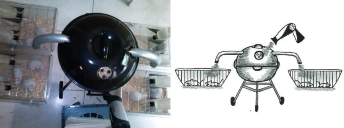
conditions for 24 h; and three experimental groups of 8 animals each were exposed to emissions from sugar cane straw burning for consecutive periods of 1, 7, and 21 days (EG1, EG7, and EG21, respectively). For the purposes of the study, a combustion chamber with a portable air extraction device was built where 200 g of sugar cane straw were burnt, generating a continuous stream of smoke that was piped into the cages of the experimental animals for 2 h daily at the same time of day (Figure 1).
The animals were euthanized by i.p. administration of 5% ketamine/xylazine diluted with 2 mL of physiological saline. After this procedure, the trachea was exposed and cannulated, a laparotomy was performed to separate the organs, and the abdominal aorta and inferior vena cava were sectioned. The trachea was then occluded by suture to maintain the lungs at functional residual capacity. A trans-sternal thoracotomy was performed through the diaphragm to remove the trachea and lungs. The collected organs were washed with physiological saline for macroscopic examination and were ixed in 10% buffered formalin.
Tissue fragments obtained from the material were processed conventionally and embedded in parafin to prepare slides containing 5-μm sections. The sections were stained with H&E and picrosirius red for analysis under light microscopy (BX51; Olympus Optical, Tokyo, Japan) at a magniication of ×100. A digital camera (C-7070; Olympus) was used to obtain photomicrographs. To perform a semi-quantitative microscopic analysis, the histological changes found were classiied as mild, moderate, or marked.
For a morphometric analysis, three photomicrographs were obtained of non-overlapping ields of the tissues studied. In the tracheal sections, areas containing hyaline cartilage, lamina propria, and ciliated epithelium were selected. In the lung parenchyma, bronchioles, alveoli, and blood vessels were evaluated. The nuclear area was quantiied (in pixels) to evaluate the inlammatory process, using Adobe Photoshop CS5 software (Adobe Systems Inc.; San Jose, CA, USA). Collagen deposition was quantiied by picrosirius red staining, positive staining corresponding to the presence
of type I collagen. Angiogenesis was assessed by counting the number of blood vessels per quadrant of each panoramic photomicrograph, using BioEstat 5.3 software.
A statistical analysis was carried out using the Shapiro-Wilk normality test. After conirmation of normality, ANOVA with Tukey’s post hoc test was used. In cases of non-normality, the Kruskal-Wallis test and Dunn’s post hoc test were used. Results were expressed as group means and standard deviations and as box plots. The level of signiicance was set at 5%.
The study was approved by the Animal Research Ethics Committee of the Faculdade Adventista da Bahia (Protocol no. 013/2014).
RESULTS
Macroscopic examination of the trachea revealed no changes in tissue color or integrity in any of the groups. The lungs of EG7 and EG21 rats showed focal and diffuse macroscopic changes in different lobes, these changes being related to tissue color and texture.
The histological indings for each group are described below.
In the CG, there were no inlammatory iniltrates or structural tissue changes in the trachea (Figure 2). The lung parenchyma showed no alveolar, septal, or bronchiolar changes and was of normal appearance (Figure 3).
In EG1, a mild inlammatory iniltrate was present in the trachea in 87.5% of the cases. However, in 12.5% of the samples analyzed, the inlammatory response was intense and predominantly focal (Figure 2). In the lung parenchyma, an inlammatory process, consisting of 50% of polymorphonuclear cells, was present in 75% of the cases, and the diffuse form predominated over the focal one. Iniltrates were found in perivascular areas (ranging from mild to moderate) and in peribronchiolar areas. In addition, mild interstitial edema was observed, and there was no collagen deposition (Figure 3).
In EG7, inlammatory iniltrates of varying intensity— mild (in 62.5% of the cases), moderate (in 50.0%),
and intense (in 25.0%)—were present in the tracheal tissue (Figure 2). The onset of collagen deposition was demonstrated by the increased acidophilic reaction in the trachea after picrosirius red staining (Figure 2). Microscopic examination of the lung parenchyma revealed an inlammatory process in 87.5% of the cases and a polymorphonuclear cell iniltrate in 62.5%. A mild to intense diffuse inlammatory process was present in 75% of the cases. A perivascular iniltrate pattern was present in 62.5% of the specimens. In addition, perivascular and peribronchiolar collagen deposition was found (Figure 3).
In EG21, a diffuse inlammatory iniltrate was observed in 75% of the cases, being of mild (in 25%) to moderate
(in 50%) intensity. No intense inlammatory iniltrates were found (Figure 2). After picrosirius red staining, there was increased acidophilia, indicating the presence of collagen (Figure 2). Histological examination of the lung parenchyma revealed mild to intense diffuse inlammatory iniltrates, with a predominance of mononuclear cells, in 100% of the cases. There was mild perivascular and peribronchiolar inlammation in 50% and 25% of the cases, respectively. Necrosis was observed in 37.5% of the cases and angiogenesis was observed in 100% (p < 0.001) when EG21 was compared with the CG and EG1; in addition, perivascular and peribronchiolar collagen deposition was seen, as was alveolar collagen deposition (Figure 3).
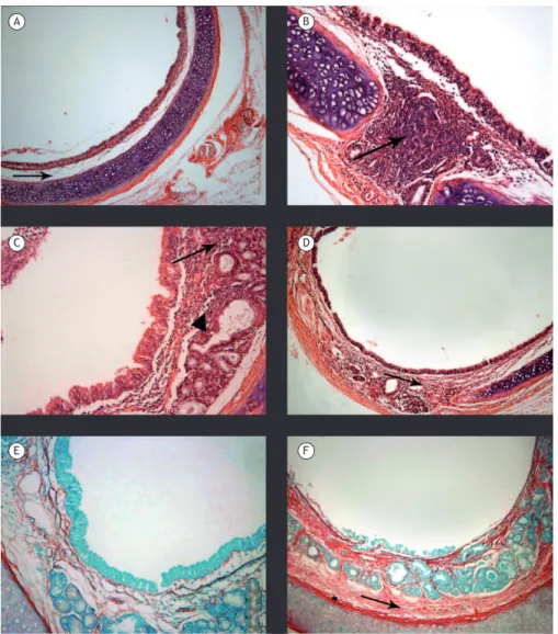
Figure 2. Photomicrographs of tracheal sections from animals in the different groups studied (H&E and picrosirius red; magniication, ×100). A (control group), Photomicrograph showing hyaline cartilage of normal appearance, with preservation of ciliated epithelium and no inlammatory iniltrates in the lamina propria (arrow). B (group exposed to emissions from sugar cane straw burning for 1 day), Photomicrograph showing intense focal inlammatory iniltrate in the lamina propria (arrow). C (7-day exposure group), Photomicrograph showing moderate diffuse acute inlammatory iniltrate in the lamina propria of the mucosa (arrow) and in the submucosal region adjacent to the seromucous glands (triangle), with ciliary preservation. D (21 day-exposure group), Photomicrograph showing mild to moderate diffuse inlammatory iniltrate in the lamina propria of the mucosa (arrow). E (7-day exposure group), Photomicrograph showing mild staining for collagen. F (21-day exposure group), Photomicrograph showing increased acidophilia of the connective tissue, indicating collagen deposition (arrow).
A
C
E
B
D
Morphometric and statistical analysis (Figure 4) demonstrated the presence of an inlammatory process in the tracheal tissue of experimental group rats. The mean nuclear area (in pixels) was 379.78 ± 105.65 in the CG, 650.36 ± 147.74 in EG1, 899.18 ± 183.65 in EG7, and 751.96 ± 143.64 in EG21. In comparison with the CG, EG7 showed a more signiicant inlammatory response (p < 0.05). The data obtained from the morphometric analysis are complementary to the indings of the semi-quantitative analysis.
Morphometry after picrosirius red staining revealed no collagen deposition in the CG and showed a slight progressive, but not statistically signiicant,
increase in acidophilia in EG1 and EG7. However, tracheal tissue collagen deposition was found to be greater in EG21 than in the CG and EG1 (p < 0.01 for both; Figure 4).
Morphometric analysis of the lung parenchyma was performed by determining the mean nuclear area (in pixels). In the CG, the mean nuclear area was 893.13 ± 51.89, which is within normal values. In EG1, polymorphonuclear and mononuclear cellularity started to increase, and the mean nuclear area was 1,373.66 ± 155.43; in EG7, the mean nuclear area reached 2,280.98 ± 744.80 (p < 0.01 in the intergroup comparison). However, the mean nuclear area decreased Figure 3. Photomicrographs of tissue sections from animals in the different groups studied (H&E and picrosirius red; magniication, ×100). A (control group), Photomicrograph showing terminal bronchioles (triangle), respiratory bronchioles (arrow), alveolar ducts, and bronchus-associated lymphoid tissue (star) without tissue changes. B (group exposed to emissions from sugar cane straw burning for 1 day), Photomicrograph showing focal perivascular (arrows) and peribronchiolar (triangle) inlammatory iniltrates and diffuse iniltrate in the lung parenchyma (circle). C (7-day exposure group), Photomicrograph showing changes in alveolar architecture (circle) and intense, diffuse peribronchiolar and perivascular inlammatory iniltrates (arrows). D (21-day exposure group), Photomicrograph showing moderate diffuse alveolar inlammatory iniltrate, together with alveolar edema and septal losses. E (7-day exposure group), Photomicrograph showing perivascular (arrows) and peribronchiolar (triangle) collagen deposition. F (21-day exposure group), Photomicrograph showing perivascular and peribronchiolar collagen deposition and alveolar collagen deposition (circle).
A
C
E
B
D
to 1,251.31 ± 231.75 in EG21, which is suggestive of tissue repair (Figure 5).
Morphometric analysis showed no angiogenesis in the CG, EG1, or EG7. In contrast, in EG21, angiogenesis was detected in 100% of the cases (Figure 5).
DISCUSSION
Our results show that exposure of Wistar rats to emissions from sugar cane burning over different periods of exposure was associated with increased inlammation in tracheal and lung tissues. Focal and diffuse inlammatory polymorphonuclear cell iniltrates were found in the acute phase in the trachea of experimental group rats, especially of EG7 rats. No loss of ciliated epithelium was observed in any of the
experimental groups relative to the CG. Tissue ibrosis in the trachea, corresponding to the early stages of the chronic phase, was conirmed in EG21. In the lung parenchyma, alveolar, vascular, and bronchiolar changes were observed in the experimental groups relative to the CG. The time criteria adopted for designating the inlammatory response phases were based on a study of Wistar rats that were administered bleomycin sulfate, in which the inlammatory response was characterized as acute (from day 1 to day 7 after the insult); subacute (from day 7 to day 14 after the insult); and resolving (from day 15 to day 30 after the insult).(17)
An experimental study, in which rats received intratracheally-instilled fine PM, observed lung inflammation characterized by macrophage and
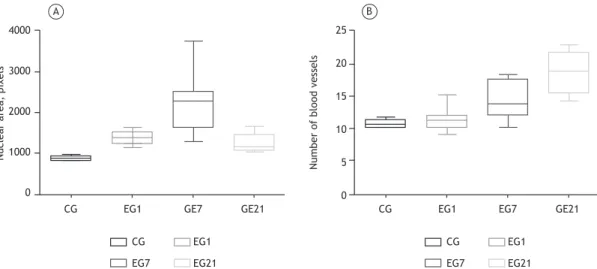
Figure 5. Morphometric analysis of the lung parenchyma in the control group (CG) and in the groups exposed to emissions from sugar cane straw burning for 1 day (EG1), for 7 days (EG7), and for 21 days (EG21). In A, nuclear area analysis showing the presence of an inlammatory iniltrate in all groups. EG7 showed statistically signiicant differences relative to the CG, EG1, and EG21 (p < 0.01 for all). There was a reduction in inlammation in EG21, suggestive of initiation of tissue repair. In B, quantiication of the number of blood vessels. There was an increase in EG7, but angiogenesis was conirmed only in EG21 with signiicant differences relative to the CG and EG1 (p < 0.001 for both).
1500
1000
500
0
1500
1000
500
0
A B
CG
CG EG1
EG1
GE7 GE21
EG7 EG21
CG
CG EG1
EG1
EG7 GE21
EG7 EG21
Nuclear area, pixels
Collagen deposition, pixels
4000
3000
2000
1000
0
CG
CG EG1
EG1
GE7 GE21
EG7 EG21
25
20
15
10
5
0 CG
CG EG1
EG1
EG7 GE21
EG7 EG21
A B
Number of blood vessels
Nuclear area, pixels
neutrophil iniltrates, demonstrating that cytokines (IL-12 and IFN-γ) play a key role in injury severity(18); similarly, an experimental rabbit study found an increased recruitment of macrophages and polymorphonuclear cells in the lung parenchyma.(19) In the present study, we found inlammatory changes consisting of polymorphonuclear cells and alveolar macrophages in the lung tissue of rats as early as in EG1.
Tracheal instillation of low doses of PM from sugar cane burning produced changes in the respiratory tract by reducing the thickening of the connective tissue and increasing the production of proinlammatory cytokines and chemokines.(8) In the present study, morphometry conirmed that emissions from sugar cane burning are able to induce signiicant and progressive necrotic inlammatory processes, even after short periods of exposure. Alveolar macrophages (after phagocytosis), as well as lung epithelial cells, respond to exposure to PM by increasing inlammatory mediator production, which can lead to certain mechanisms, such as leukocyte proliferation and activation, apoptosis, and endothelial repair.(20) These changes are explained by the elements present in smoke; these data conirm our indings, especially in EG7.
It is known that the size of particles emitted from biomass burning has a negative impact on the airways.(21,22) The size of inhaled PM, in terms of varying particle granularity, determines the clinical manifestations in the body. Coarse PM (< 10 µm) is retained in the upper airways and can be removed by ciliary activity; thin PM (< 2.5 µm) and ultraine particles/nanoparticles (< 0.1 µm) are usually the result of incomplete oxidation of carbon.(22-24) Fine and ultraine PM have the ability to reach the alveoli and be phagocytosed by alveolar macrophages, having greater deleterious effects, such as changes in lung mechanics, alveolar collapse, and oxidative stress, in rats.(23-26) Chronic exposure to ine particles is strongly associated with higher rates of chronic respiratory disease.(3)
After direct exposure of mice to dung biomass smoke for 7 consecutive days, a signiicant increase in cellularity, especially in macrophage cellularity, was observed in the lungs.(27) Exposure to emissions from sugar cane burning produced inlammatory iniltrates of varying intensity in the animals of the present study, and, in EG7, there was a more signiicant inlammatory iniltrate in both tissues of the trachea and lung parenchyma, given that there were areas of more severe inlammation.
Damage to the lung parenchyma after chronic exposure to dung biomass smoke and cigarette smoke includes moderate to marked interstitial inlammation, bronchial and perivascular inlammation, vascular congestion, alveolar destruction, an increase in the number of macrophages, and an increase in vascular wall thickness.(3) In the present study, although the exposure was acute only, it was possible to observe that the experimental groups showed a growing change in alveolar structure (septal loss) relative to the CG, with PM being phagocytosed especially in EG1. Areas suggestive of necrosis were identiied especially in EG7 and EG21, and angiogenesis was conirmed in EG21.
Rabbits chronically exposed to emissions from biomass and cigarette burning show perivascular inlammation, peribronchiolar inlammation, parenchymal iniltrate, and ibrosis, with respiratory epithelial proliferation and emphysematous changes.(28) Using picrosirius red staining, we demonstrated that, in EG7 and EG21, there were areas of tracheal tissue collagen deposition, areas of peribronchiolar and vascular collagen deposition, and diffuse areas of lung parenchymal collagen deposition, which suggest that continued exposure can lead to a tissue ibrotic process.
On the basis of morphological criteria, we found that the changes in alveolar structure in EG7 and the rupture of interalveolar septa in EG21 were consistent with the indings of a rat study that showed that 24.6% of the cigarette smoke-exposed animals had increased alveolar spaces relative to controls, which were not exposed.(29)
Therefore, the above data allow us to state that acute exposure to emissions from sugar cane burning is able to induce severe damage to the respiratory system. The components of emissions from sugar cane burning trigger polymorphonuclear cell inlammatory processes in the trachea and also induce inlammatory iniltrates and interstitial and alveolar edema in the lung parenchyma of Wistar rats. Changes in alveolar architecture and angiogenesis are also found.
Further research targeting prolonged exposure and determination of proinlammatory marker levels is needed to demonstrate the potential damage caused by chronic exposure to the components of emissions from sugar cane burning, since workers in sugar cane ields and the neighboring population are exposed to these emissions for long periods of their life.
REFERENCES
1. Brasil. Ministério da Agricultura, Pecuária e Abastecimento. Anuário
Estatístico da Agroenergia 2014. Brasília: o Ministério; 2015.
2. Lelieveld J. Evans JS, Fnais M, Giannadaki D, Pozzer A. The contribution of outdoor air pollution sources to premature mortality on a global scale. Nature. 2015;525(7569):367-71. https://doi. org/10.1038/nature15371
3. Dogan OT, Elagoz S, Ozsahin SL, Epozturk K, Tuncer E, Akkurt I. Pulmonary toxicity of chronic exposure to tobacco and biomass smoke in rats. Clinics (Sao Paulo). 2011;66(6):1081-7. https://doi. org/10.1590/S1807-59322011000600027
4. Ribeiro H. Sugar cane burning in Brazil: respiratory health effects [Article in Portuguese]. Rev Saude Publica. 2008;42(2):370-6. https://
doi.org/10.1590/S0034-89102008005000009
5. Brasil. Ministério da Agricultura, Pecuária e Abastecimento [homepage on the Internet]. Brasília: o Ministério [cited 2016 Feb 10]. Sapcana - Sistema de Acompanhamento de Produção Canavieira. [Adobe Acrobat document, 3p.]. Available from: http://www.agricultura. gov.br/assuntos/sustentabilidade/agroenergia/acompanhamento-da-producao-sucroalcooleira/arquivos-2014-2015/16-04-2014.pdf/view
6. The WHO Pages. Wildland/Vegetation Fires. Ann Burns Fire Disasters [serial on the Internet]. 2000 [cited 2016 Feb 10] 13(3):178 [about 6 p.]. Available from: www.medbc.com/annals/review/vol_13/ num_3/text/vol13n3p178.htm
dos cortadores de cana-de-açúcar em Macatuba, São Paulo. Saude Soc. 2010;9(1)48-63. http://dx.doi.org/10.1590/ S0104-12902010000100005 https://doi.org/10.1590/S0104-12902010000100005
8. Bittar OT. Estudo dos efeitos do material particulado proveniente
da biomassa de cana-de-açúcar sobre o aparelho respiratório e circulatório de ratos Wistar combinados ou não com a infecção estailocócica [thesis]. Campinas: Universidade Estadual de Campinas; 2013.
9. Hutton G. The economics of health and climate change: key evidence for decision making. Global Health. 2011;7:18. https://doi. org/10.1186/1744-8603-7-18
10. National Aeronautics and Space Administration [homepage on the
Internet]. Washington, DC: the Administration; c2015 [updated 2015 Jul 30; cited 2016 Feb 10]. A Story of Ozone: The Earth’s natural sunscreen. Available from: https://www.nasa.gov/content/goddard/ a-story-of-ozone-earths-natural-sunscreen
11. Prado GF, Zanetta DM, Arbex MA, Braga AL, Pereira LA, de Marchi MR, et al. Burnt sugarcane harvesting: Particulate matter exposure and the effects on lung function, oxidative stress, and urinary 1-hydroxypyrene. Sci Total Environ. 2012;437:200-8. https://doi. org/10.1016/j.scitotenv.2012.07.069
12. Arbex MA, Santos Ude P, Martins LC, Saldiva PH, Pereira LA, Braga AL. Air pollution and the respiratory system. J Bras Pneumol. 2012;38(5):643-55. https://doi.org/10.1590/S1806-37132012000500015
13. Mazzole-Rocha F, Carvalho GM, Lanzetti M, Valença SS, Silva LF,
Saldiva PH et al. Respiratory toxicity of repeated exposure to particles produced by trafic and sugar cane burning. Respir Physiol Neurobiol. 2014;191:106-13. https://doi.org/10.1016/j.resp.2013.11.004 14. William AH, Wallace A. Simpson J, Hirani N. Acute lung injury. In:
Hasleton P, Flieder DB, editors. Spencer’s Pathology of the Lung. 6th ed. Cambridge: Cambridge University Press; 2013.p. 342-60. 15. Paraiso ML, Gouveia N. Health risks due to pre-harvesting sugarcane
burning in São Paulo State, Brazil. Rev Bras Epidemiol. 2015;18(3):691-701. https://doi.org/10.1590/1980-5497201500030014
16. Silva R, Oyarzún M, Olloquequi J. Pathogenic mechanisms in chronic obstructive pulmonary disease due to biomass smoke exposure. Arch Bronconeumol. 2015;51(6)285-92. https://doi.org/10.1016/j. arbr.2015.04.013
17. Rossari JR. Fibrose pulmonar induzida por bleomicina intratraqueal em ratos Wistar: uso do interferon--2b em um modelo experimental de síndrome da distração respiratória aguda [dissertation]. Porto Alegre: Universidade Federal do Rio Grande do Sul; 2004.
18. Ding LR, Wang K, Fahmy B, Shen HH, Cormier S. Airborne ine
particle matter induced pulmonary inlammation as well as oxidative stress in neonate rats. Chin Med J (Engl). 2010;123(20):2895-900.
19. Miyata R, Bai N, Vincent R, Sin DD, Van Eeden SF. Statins reduce ambient particulate matter-induced lung inlammation by promoting the clearance of particulate matter, < 10 μm from lung tissues. Chest. 2013;143(2):452-60. https://doi.org/10.1378/chest.12-1237
20. Poma A, Limongi T, Pisan C, Granato V, Picozzi P. Genotoxicity
induced by ine urban air particulate matter in the macrophages cell line RAW 264.7. Toxicol In Vitro. 2006;20(6):1023-9. https://doi. org/10.1016/j.tiv.2006.01.014
21. Bovo F, Wisniewski P. Efeitos de material particulado sobre mecanismos imunológicos. Biosaude. 2009;11(2):93-100.
22. Donaldson K, Stone V. Current hypotheses on the mechanisms of
toxicity of ultraine particles. Ann Ist Super Sanita. 2003;39(3):405-10.
23. Pope CA, Muhlestein JB, Anderson JL, Cannon JB, Hales NM,
Meredith KG, et al Short-Term Exposure to Fine Particulate Matter Air Pollution Is Preferentially Associated With the Risk of ST-Segment Elevation Acute Coronary Events. J Am Heart Assoc. 2015;4(12). pii: e002506. https://doi.org/10.1161/JAHA.115.002506
24. Colombini MP. Exposição aguda ao material particulado total em suspensão proveniente de diferentes fontes e suas repercussões nas respostas inlamatórias, sistêmica e local, em ratos [thesis]. São Paulo. Faculdade de Medicina, Universidade de São Paulo; 2007. 25. Mazzoli-Rocha F, Magalhães CB, Malm O, Saldiva PH, Zin WA, Faffe
DS. Comparative respiratory toxicity of particles produced by trafic and sugar cane burning. Environ Res. 2008;108(1):35-41. https://doi. org/10.1016/j.envres.2008.05.004
26. Riva DR, Magalhães CB, Lopes AA, Lanças T, Mauad T, Malm O, et al. Low dose of ine particulate matter (PM2.5) can induce acute oxidative stress, inlammation and pulmonary impairment in healthy mice. Inhal Toxicol. 2011;23(5):257-67. https://doi.org/10.3109/0895 8378.2011.566290
27. Mehra D, Geraghty PM, Hardigan AA, Foronjy R. A comparison of the inlammatory and proteolytic effects of dung biomass and cigarette smoke exposure in the lung. PLoS One. 2012;7(12):e52889. https:// doi.org/10.1371/journal.pone.0052889
28. Fidan F, Unlu M, Sezer M, Sahin O, Tokyol C, Esme H. Acute effects of environmental tobacco smoke and dried dung smoke on lung histopathology in rabbits. Pathology. 2006;38(1):53-7. https://doi. org/10.1080/00313020500459615
29. Zheng H, Liu Y, Huang T, Fang Z, Li G, He S. Development and
characterization of a rat model of chronic obstructive pulmonary disease (COPD) induced by sidestream cigarette smoke. Toxicol Lett. 2009;189(3):225-34. https://doi.org/10.1016/j.toxlet.2009.06.850