ISSN 1806-3713 © 2017 Sociedade Brasileira de Pneumologia e Tisiologia
http://dx.doi.org/10.1590/S1806-37562017000000024
Opaque hemithorax
Edson Marchiori1, Bruno Hochhegger2, Gláucia Zanetti1
1. Universidade Federal do Rio de Janeiro, Rio de Janeiro (RJ) Brasil.
2. Universidade Federal de Ciências da Saúde de Porto Alegre, Porto Alegre (RS) Brasil. CLINICAL HISTORY
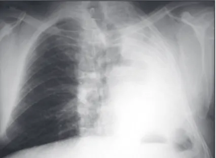
A 69-year-old male patient presented with complaints of cough and progressive dyspnea. A chest X-ray showed
complete opaciication of the left hemithorax (Figure 1).
DISCUSSION
Complete opaciication of a hemithorax on chest X-ray is termed opaque hemithorax (OH) and is a common inding in emergency room patients. Attending physicians encountering an OH should be able to make an immediate
decision regarding the most appropriate approach.
The differential diagnosis of an OH is primarily based on the volume of the affected hemithorax, as determined by the position of the mediastinum (the position of the trachea providing the best reference for that):
• increased hemithoracic volume—mediastinal shift to the unaffected side
• reduced hemithoracic volume—mediastinal shift to the affected side
• normal hemithoracic volume—no mediastinal shift
The differential diagnosis of an OH with increased volume includes large pleural effusions (which constitute the
most frequent cause of OH) and large thoracic masses, especially in children. In most cases, these conditions can be easily differentiated by ultrasound or CT.
The differential diagnosis of an OH with reduced volume includes pulmonary agenesis, pneumonectomy, and atelectasis. Bronchial obstruction by a foreign body (in children) or an endobronchial tumor (in adults) is the
most common cause of atelectasis.
There are cases in which an OH presents with normal volume. In children, such cases are primarily due to extensive pneumonia affecting the entire lung parenchyma. In adults, however, such cases are primarily due to bronchial carcinoma, accompanied by pleural effusion
and atelectasis.
Our patient presented with opaciication of the left hemithorax with a marked mediastinal shift to the left. The absence of history of surgery or surgical scar on the chest wall, ruled out a previous pneumonectomy. A
previous normal chest X-ray ruled out pulmonary agenesis.
Therefore, a diagnosis of atelectasis was made, and a bronchoscopy revealed a tumor resulting in complete left main bronchial obstruction.
Figure 1. Anteroposterior chest X-ray showing diffuse opaciication of the left hemithorax (opaque hemithorax). Note mediastinal structures, particularly the trachea, shifted to the left. Note also that the heart cannot be seen overlapping the vertebral bodies. These indings characterize an opaque hemithorax whose volume is reduced.
RECOMMENDED READING
1. Fraser RS, Müller NL, Colman NC, Pare PD, editors. Diagnosis of Diseases of the Chest. 4th ed. Philadelphia: WB Saunders Company; 1999. J Bras Pneumol. 2017;43(3):161-161