Basilic vein cystic adventitial disease: case report
Doença cística da adventícia na veia basílica: relato de caso
Rafael Sampaio Vasconcelos1
*
, Cesar Augusto Cherubim Filho1, Felipe Mavignier Pereira França1, Eduardo de Lucca D’allacqua1, Marcelo Bellini Dalio1, Edwaldo Edner Joviliano1
Abstract
Cystic adventitial disease is a rare entity that most often involves the popliteal artery. It rarely occurs in veins. Its etiology is unknown. Clinically, it presents with ischemia, thrombosis or pain, depending on the vessel afected. Here we present the case of a young male with a nodule in the left arm. Magnetic resonance angiography showed a cystic lesion in contact with the basilic vein, with homogenous content without post-contrast enhancement. he lesion was resected en bloc together with the venous segment involved. he results of microscopic analysis were suggestive of basilic vein cystic adventitial disease.
Keywords: cystic adventitial disease; basilic vein; magnetic resonance angiography.
Resumo
A doença cística da adventícia é uma entidade rara que acomete principalmente a artéria poplítea. A ocorrência em veias é muito rara, e sua etiologia é desconhecida. Clinicamente, apresenta-se como isquemia, trombose ou dor a depender do território acometido. Apresentamos o caso de um paciente masculino jovem referindo nódulo no braço esquerdo. A angiorressonância magnética do membro mostrou lesão cística em contato com a veia basílica, com conteúdo homogêneo e sem realce pós-contraste. Foi realizada ressecção da lesão em bloco com o segmento venoso envolvido. O estudo anatomopatológico foi sugestivo de cisto de adventícia de veia basílica.
Palavras-chave: doença cística da adventícia; veia basílica; angiorressonância magnética.
1 Universidade de São Paulo – USP, Faculdade de Medicina de Ribeirão Preto, Hospital das Clínicas, Departamento de Cirurgia e Anatomia, Divisão de Cirurgia Vascular e Endovascular, Ribeirão Preto, SP, Brazil.
Financial support: his study was partially funded by Fundação FAEPA.
Conlicts of interest: No conlicts of interest declared concerning the publication of this article. Submitted: May 06, 2016. Accepted: June 25, 2016.
INTRODUCTION
Cystic adventitial disease is a rare entity1 that is
characterized by cystic degeneration of the adventitial layer, with mucoid cystic contents.2,3 The majority of
reports in the literature describe cases in arteries.4,5
Cystic adventitial disease of veins is extremely rare.1 We were unable to locate any reports of cases
of cystic adventitial disease involving veins of the upper limbs. The objective of this article is to report on a rare case of cystic adventitial disease of the basilic vein that was successfully treated by resection en bloc together with the segment of vein involved. The patient authorized publication of the case via signature of an Informed Consent Form.
CASE DESCRIPTION
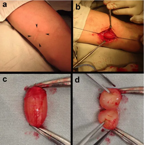
A 34-year-old, white, male patient presented complaining of swelling on the medial surface of his left arm with onset 2 years previously. He sought
care at a primary care service, which raised a hypothesis of subcutaneous tissue lipoma and made an attempt at resection under local anesthesia. During the procedure, it was observed that the lesion was intimately related to the basilic vein and the decision was taken to abort the attempt and refer the patient to our vascular service. At the initial consultation the patient complained of localized pain. A physical
examination detected a well-deined nodule with ibroeleastic consistency on the medial surface of
the left arm, measuring approximately 2.5 x 3.0 cm, provoking mild pain on palpation and with no signs
of inlammation (Figure 1a). The wound left by the
prior incision was visible and had a satisfactory appearance. We conducted an investigation using magnetic resonance angiography of the upper limb, which showed a cystic lesion in contact with the wall of the basilic vein, causing compression of the vessel. The lesion was smooth-walled, with homogenous content and was not highlighted after administration
of contrast (Figure 2). There was no thrombosis of
Figure 1. Images showing the preoperative appearance (a); the cystic lesion dissected and irmly adhering to the basilic vein, which
the basilic vein and the other veins in the limb were clearly visible with contrast.
The patient was treated surgically, with dissection of the lesion and control of the basilic vein. The cyst
did not exhibit signiicant adherence to the adjacent
tissue layers, but was intimately related to the basilic vein, with no plane of cleavage between the lesion
and the vein (Figure 1b). It was decided to resect
the lesion together with the segment of the basilic vein that was involved. The stumps of the basilic vein were treated with simple ligature. Macroscopic anatomopathological examination revealed a cyst with thin walls formed by dense connective tissue
and illed with colloid material (Figures 2c and 2d).
A microscopic analysis revealed contents with sparse
distribution of cells, consisting of mature ibroblasts free from atypia, loose connective tissue ibers and
sparse capillaries. These findings confirmed the hypothesis of a cyst of the adventitial layer of the basilic vein. The patient recovered with no complaints or complications.
DISCUSSION
Cystic adventitial disease is a rare entity of unknown etiology.1 Four theories have been proposed
in attempts to explain its origins: the ganglion theory
(synovial cells implanted in the adventitial layer), the traumatic theory (degeneration caused by local traumas), the developmental theory (implants occur
during embryogenesis), and the systemic disease theory
(secondary to systemic connective tissue disease).1,6
The majority of the reports in the literature describe cases in arteries.4 Cystic adventitial disease involving
veins is extremely rare.6,7 Francis et al. described three
cases involving veins in the iliofemoral region, where the condition is most commonly found.8 A recent
review by Desy & Spinner did not include any case reports in which this condition was described in veins of the upper limbs.1 This degree of rarity means that
clinical suspicion is generally late.
The condition’s clinical presentation varies depending on the territory involved.9-12 In arteries, it may manifest
clinically with ischemia of a limb or pain caused by localized compression.13 In veins, it manifests as
local pain or venous thrombosis.7 As happened in the
case described here, it may be confused with more common causes of nodules in subcutaneous tissues, such as lipomas, adenomegaly, sebaceous cysts, and
ibromas.1 Diagnostic suspicion of cystic adventitial
disease in veins should by aroused when nodular lesions are found along the paths of veins. Diagnostic
conirmation will generally require an imaging exam.
In view of the low cost, wide availability and no need for injected contrast, ultrasonography is generally
the irst examination used. The lesion will appear as a well-deined nodule, with anechoic content. Computed tomography angiography offers adequate visualization of the lesion, but is expensive, requires
injection of contrast and emits ionizing radiation. Magnetic resonance angiography is an examination that offers better definition of anatomic planes, facilitates surgical planning and enables differential diagnosis to rule out articular cysts.1 However, it is
also expensive and requires infusion of gadolinium
as a contrast medium. In the case described here, we used magnetic resonance angiography because of the advantages mentioned above.
Treatment options include clinical follow-up, percutaneous aspiration guided by imaging, angioplasty, with and without stents, simple resection of the cyst and resection of the cyst with vascular reconstruction.1,14
In their recent review, Desy & Spinner stated that the most common treatment approach is resection of the lesion, with or without resection en bloc with the vessel involved.1 They also stated that after resection,
the need for vascular reconstruction with autologous vein or synthetic material should be evaluated. Since the patient in the present case exhibited symptoms of localized compression, resection of the lesion was proposed. During the procedure it was found that the
anatomic planes were well-deined, making dissection possible without dificulty. However, because of the
cyst’s intimate relation with the basilic vein, it was not possible to resect it without affecting the vein.
Since the patient’s supericial and deep vein systems
were patent, we opted for resection en bloc with the basilic vein. There is generally a considerable functional reserve of venous drainage in the upper limb and so the basilic vein can be resected without
causing sequelae. Another treatment method that
has been described is aspiration of the content of the cyst under ultrasonographic guidance.15 This method
offers the advantage of being less invasive, but is not applicable to all cases. The content of the cyst is generally viscous and cannot always be aspirated with a needle.16 Endovascular treatment has not
proven effective with this condition,17 and we did not
ind reports of this treatment method for adventitial
cysts of veins.
CONCLUSIONS
differential diagnosis of nodular lesions in this region. Resection of the lesion en bloc with the basilic vein segment involved produced good results.
REFERENCES
1. Desy NM, Spinner RJ. The etiology and management of cystic adventitial disease. J Vasc Surg. 2014;60(1):235-45. http://dx.doi. org/10.1016/j.jvs.2014.04.014.
2. Wu X, Jiang B, Lun Y, et al. Venous occlusion due to cystic adventitial degeneration of the common femoral vein. Vasa. 2013;42(6):461-4. http://dx.doi.org/10.1024/0301-1526/a000318. PMid:24220125.
3. Chen Y, Sun R, Shao J, Li Y, Liu C. A contemporary review of venous adventitial cystic disease and three case reports. Phlebology. 2015;30(1):11-6. http://dx.doi.org/10.1177/0268355513516948. PMid:24357449.
4. Lejay A, Ohana M, Delay C, et al. Cystic adventitial pathology as an entity in peripheral arterial disease. J Cardiovasc Surg. 2016;57(2):282-91. PMid:26471959.
5. Nasser M, Pivetta LGA, Teixeira JL Fo, Rocha ES, Botta AE. Critical limb ischemia in a young patient with cystic disease of the popliteal artery. J Vasc Bras. 2012;11(2):144-9. http://dx.doi.org/10.1590/ S1677-54492012000200012.
6. Jones DW, Rezayat C, Winchester P, Karwowski JK. Adventitial cystic disease of the femoral vein in a 5-year-old boy mimicking deep venous thrombosis. J Vasc Surg. 2012;55(2):522-4. http:// dx.doi.org/10.1016/j.jvs.2011.06.117. PMid:21917399.
7. Kim YK, Chun HJ, Hwang JK, et al. Adventitial cystic disease of the common femoral vein presenting as deep vein thrombosis. Asian J Surg. 2013;23:1-4. PMid:23978427.
8. Dix FP, McDonald M, Obomighie J, et al. Cystic adventitial disease of the femoral vein presenting as deep vein thrombosis: A case report and review of the literature. J Vasc Surg. 2006;44(4):871-4. http://dx.doi.org/10.1016/j.jvs.2006.05.034. PMid:17012010.
9. Morizumi S, Suematsu Y, Gon S, Shimizu T, Iwai T. Adventitial cystic disease of the femoral vein. Ann Vasc Surg. 2010;24(8):1135.e5-7. http://dx.doi.org/10.1016/j.avsg.2010.03.007. PMid:20599347.
10. Albernaz DTS, Albernaz LFL, Eggers EE. Doença cística da artéria poplítea: relato de caso. J Vasc Bras. 2010;9(3):168-72. http:// dx.doi.org/10.1590/S1677-54492010000300013.
11. Carrilho C, Mesquita A. Doença cístíca da adventícia da artéria poplítea: diagnóstico e tratamento – a propósito de um caso clínico. Angiol e Cir Vasc. 2013;9(1):1-4.
12. Romiti M, Silvano D. Microembolia por degeneração cística da adventícia da artéria poplítea. Cir Vasc e Angiol. 1991;7(1):14-6.
13. Scott MF, Gavin T, Levin S. Venous cystic adventitial disease presenting as an enlarging groin mass. Ann Vasc Surg. 2014;28(2):489.e15-8. http://dx.doi.org/10.1016/j.avsg.2013.04.019. PMid:24075153.
14. Johnson JM, Kiankhooy A, Bertges DJ, Morris CS. Percutaneous image-guided aspiration and sclerosis of adventitial cystic disease of the femoral vein. Cardiovasc Intervent Radiol. 2009;32(4):812-6. http://dx.doi.org/10.1007/s00270-009-9581-z. PMid:19449068.
15. Kauffman P, Kuzniec S, Sacilotto R, Teivelis MP, Wolosker N, Tachibana A. Doença cística adventicial da artéria poplítea: causa infrequente de claudicação intermitente. Einstein. 2014;12(3):358-60. http:// dx.doi.org/10.1590/S1679-45082014RC2818. PMid:25167336.
16. Keo HH, Baumgartner I, Schmidli J, Do D-D. Sustained remission 11 years after percutaneous ultrasound-guided aspiration for cystic adventitial degeneration in the popliteal artery. J Endovasc Ther. 2007;14(2):264-5. http://dx.doi.org/10.1583/1545-1550(2007)14[264:SR YAPU]2.0.CO;2. PMid:17484540.
17. Rai S, Davies RSM, Vohra RK. Failure of endovascular stenting for popliteal cystic disease. Ann Vasc Surg. 2009;23(3):410.e1-5. http://dx.doi.org/10.1016/j.avsg.2008.01.014. PMid:18513486.
*
Correspondence
Rafael Sampaio Vasconcelos Hospital das Clínicas de Ribeirão Preto Departamento de Cirurgia e Anatomia Av. Bandeirantes, 3900 CEP 14040-900 - Ribeirão Preto (SP), Brazil Tel.: +55 (16) 3602-2593 / +55 (85) 99925-2120 E-mail: [email protected]
Author information
RSV, CACF, FMPF and EDLD - Resident physicians (Surgery), Divisão de Cirurgia Vascular e Endovascular, Departamento de Cirurgia e Anatomia, Hospital das Clínicas, Faculdade de Medicina de Ribeirão Preto, Universidade de São Paulo (USP). MBD - Primary physician, Divisão de Cirurgia Vascular e Endovascular, Departamento de Cirurgia e Anatomia, Hospital das Clínicas, Faculdade de Medicina de Ribeirão Preto, Universidade de São Paulo (USP). EEJ - Associate professor and chief, Divisão de Cirurgia Vascular e Endovascular, Departamento de Cirurgia e Anatomia, Hospital das Clínicas, Faculdade de Medicina de Ribeirão Preto, Universidade de São Paulo (USP).
Author contributions
Conception and design: RSV, MBD Analysis and interpretation: RSV Data collection: CACF, FMPF, EDLD Writing the article: RSV, MBD Critical revision of the article: EEJ Final approval of the article*: RSV, CACF, FMPF, EDLD, MBD, EEJ Statistical analysis: N/A. Overall responsibility: RVS