J Bras Pneumol. 2008;34(3):185-188
185
Case Report
* Study carried out in the Department of Thoracic Surgery of the Hospital do Câncer/Instituto Nacional de Câncer/Ministério da Saúde – HC/INCA/MS, Cancer Hospital/National Cancer Institute/Ministry of Health – Rio de Janeiro, Brazil.
1. Thoracic Surgeon. Hospital do Câncer/Instituto Nacional de Câncer – HC/INCA, Cancer Hospital/National Cancer Institute – Rio de Janeiro, Brazil.
2. Head of the Department of Thoracic Surgery. Hospital do Câncer/Instituto Nacional de Câncer – HC/INCA, Cancer Hospital/National Cancer Institute – Rio de Janeiro, Brazil.
3. Pulmonologist in the Department of Thoracic Surgery. Hospital do Câncer/Instituto Nacional de Câncer – HC/INCA, Cancer Hospital/National Cancer Institute – Rio de Janeiro, Brazil.
Correspondence to: Samuel Zuínglio de Biasi Cordeiro. Estrada Francisco da Cruz Nunes, 11.784, Bloco 3, apto 401, Itaipú, CEP 24340-000, Niterói, RJ, Brasil. Tel 55 21 2506-6185. E-mail: [email protected]
Submitted: 28 November 2006. Accepted, after review: 8 May 2007.
Giant cell tumor of the rib occupying the entire hemithorax*
Tumor de células gigantes costal ocupando todo o hemitórax
Samuel Zuínglio de Biasi Cordeiro1, Paulo de Biasi Cordeiro2,
Aureliano Mota Cavalcanti Sousa1, Deborah Cordeiro Lannes3,
Gustavo Soares de Moura Pierro1
Abstract
The authors report the case of a 28-year-old female patient with a giant cell tumor originating from the rib. The tumor, measuring 25 × 17 cm, occupied the entire hemithorax and caused atelectasis of the left lung. This tumor was a benign mesenchymal neoplasm, which rarely affects the ribs. A thoracotomy involving en bloc resection of the chest wall and tumor was performed. Despite the large dimensions of the tumor, complete resection was possible, and lung function was restored.
Keywords: Neoplasms; Giant cells; Mesoderm; Thoracotomy; Medical records.
Resumo
Os autores relatam o caso de uma paciente de 28 anos de idade portadora de tumor de células gigantes originário da costela. O tumor de grandes dimensões (25 × 17 cm) ocupava todo o hemitórax e causava atelectasia do pulmão esquerdo. Tratava-se de uma neoplasia mesenquimal benigna, a qual raramente acomete as costelas. Foi realizada toracotomia com ressecção em bloco da parede torácica e do tumor. O objetivo deste artigo é enfatizar que, apesar da grande dimensão do tumor, ele pôde ser completamente ressecado, e o pulmão foi reabilitado.
Descritores: Neoplasias; Células gigantes; Mesoderma; Toracotomia; Registros médicos.
Introduction
Giant cell tumor (GCT) is a neoplasm that is mesen-chymal in nature and is characterized by the proliferation of multinucleated giant cells, which are similar to osteoclasts, in the stroma of spindle-shaped mononuclear cells.(1)
This type of tumor was first described in 1818 by Sir Astley Cooper. In 1860, Nelaton described the clinical and histological characteristics of GCTs, highlighting the fact that they are locally aggressive.(2)
Accounting for 8.6% of all bone tumors, GCTs typically affect a single bone. There are few reports of multicentric tumors in the literature. The sites most commonly affected are the distal femoral epiphysis (36%) and proximal tibial epiphysis (17%), followed by the distal radius (15%) and proximal humerus (2%). Although rarely occurring in the axial skeleton, among the 2% of GCTs that affect that
region, most are sacral. It is even rarer for a GCT to appear in the ribs. In the literature, there are only reports of isolated cases (no case studies). Most often occurring in the third and fourth decades of life, GCTs are also slightly more common in females.(3)
Pain and an increase in local volume are the principal forms of presentation. Some patients present pathological fracture resulting from weakening of the cortical bone.
On a routine X-ray, GCT presents an initially eccentric lytic lesion, without a surrounding sclerotic halo, respecting the cortical borders. As the lesion grows, it can encompass the entire circumference of the bone, causing rupture of the cortical bone.(4)
186 Cordeiro SZB, Cordeiro PB, Sousa AMC, Lannes DC, Pierro GSM
J Bras Pneumol. 2008;34(3):185-188
the left hemithorax, and the Karnofsky perform-ance status was 90. The sequence of preoperative tests performed in order to elucidate the diagnosis is presented here.
A computed tomography scan of the chest revealed a heterogeneous mass accompanied by left pleural effusion.
Fine-needle aspiration and a cutting needle biopsy were performed. The results showed only a few atypical cells intermingled with macrophages and some inflammatory mediators. The histopatho-logical diagnosis was low-grade fibrohistiocytic lesion.
A bone scintigraphy study revealed areas of extreme fixation unrelated to secondary implants.
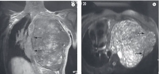
An echocardiogram showed a mass displacing the heart to the right, and a Doppler echocardio-graphic study revealed minimal mitral insufficiency. Magnetic resonance imaging of the chest revealed an extensive, mixed, and heterogeneous formation with well-defined borders. On T1-weighted images, a multiloculated mass appeared as a hyperintense signal corresponding to hemorrhagic foci and occu-pying the entire left hemithorax. This mass was compressing and displacing the mediastinal struc-tures and the diaphragm. There was a small area of extrathoracic involvement at the level of the axillary line (Figure 1).
performed before the largest diameter of the tumor exceeds 4 cm. In the case of tumors whose diameter is larger, a previous biopsy allows more appropriate surgical timing.(5)
Despite being benign, GCTs can present local recurrence in 20 to 40% of the cases. In 2% of cases, pulmonary metastasis appears even though the original tumor has not developed the histolog-ical characteristics of a malignancy. The pulmonary metastases present histological characteristics similar to those of the primary tumor and are benign in nature. Malignant GCTs account for less than 5% of the cases. The diagnosis is made based on the presence of areas within the malignant neoplasm that are typical of GCTs.
The treatment is segmental resection of the lesion with oncological margins of safety in the bone and soft tissues.(6)
Case report
A 28-year-old female patient presented with a 5-month history of chest pain and cough, initially dry and subsequently with mucoid expectoration. A chest X-ray showed opacification of the entire hemithorax. The patient had lost 2 kg in 5 months and had received a blood transfusion 6 months prior. The patient had a maternal and paternal history of cancer. In addition, breath sounds were absent in
a
Figure 1 - a) Magnetic resonance image showing a mass occupying the entire hemithorax and displacing the
mediastinum to the right, with a cleavage plane next to the mediastinum (arrows); and b) Cross-sectional magnetic resonance image showing the edge of the mass abutting the ribs (arrows).
Giant cell tumor of the rib occupying the entire hemithorax
J Bras Pneumol. 2008;34(3):185-188
187
tissues. The tumor was accompanied by extensive necrotic and hemorrhagic material. There was no angiolymphatic invasion. The lesion was completely excised, with tumor-free surgical margins.
The postoperative evolution was satisfactory, with mild re-expansion pulmonary edema, which regressed spontaneously. The patient was discharged from the hospital on postoperative day 8.
Outpatient follow-up evaluation showed that the patient was in very good general health and had gained weight. Left lung expansion was restored through respiratory therapy.
The patient remained asymptomatic 24 months after the surgical procedure.
Discussion
The occurrence of a GCT in the axial skeleton is considered rare, and few cases of GCT of the rib have been reported. In Japan, only 13 cases had been reported as of 2003.(7) The case described here
is the only one in 20 years of monitoring by the National Cancer Institute in Rio de Janeiro, which is a referral institution for tumors in the state of Rio de Janeiro.
The clinical, radiological, and histopatho-logical characteristics of GCTs are similar to those of various tumors and pseudotumors, such as the brown tumors seen in cases of hyperparathy-roidism.(8) Other lesions, such as aneurysmal bone
cyst, telangiectatic osteosarcoma, and chondroblas-toma, are considered in the differential diagnosis.(9)
The GCT cells are similar to osteoclasts (giant and The results of the blood tests, including the
deter-mination of ionized calcium levels, were normal, as were the arterial blood gas analysis findings.
An open lung biopsy, which was performed using a lateral minithoracotomy, revealed a histopatho-logical pattern consistent with GCT.
The patient was initially treated with radio-therapy for 5 weeks (total dose of 4600 cGy in 23 fractions). The response to the treatment was considered insufficient.
On the third week after radiotherapy was discon-tinued, it was decided that surgical resection should be performed.
Pulmonary function tests were conducted, and, in terms of pulmonary function, there was no impediment to performing the procedure. The procedure was performed under general inhalation anesthesia, and a peridural catheter was used.
Access was obtained through thoracotomy. En bloc resection of the intrathoracic tumor, together with the anterior portions of the fourth and fifth ribs, was performed, as were extrapleural dissection (releasing the tumor from the lung, mediastinum, and chest wall) and parietal reconstruction (using polypropylene mesh).
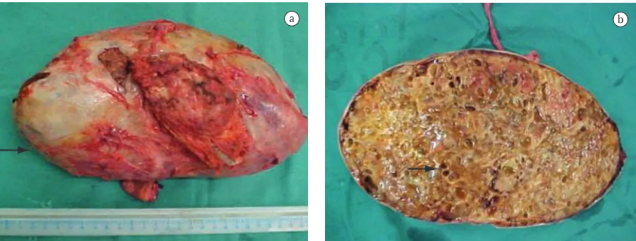
The surgical sample was a tumor mass weighing 2180 g. The tumor was surrounded by a firm, elastic, whitish capsule. The surface of the tumor was hard, multicystic, and yellowish. The material within the tumor was brownish and viscous, with areas of bone tissue (Figure 2). The histological find-ings confirmed the diagnosis of a GCT, measuring 25 × 17 cm, infiltrating the rib and adjacent soft
a b
Figure 2 - a) Surgical sample: solid, encapsulated tumor with a well-defined capsule (arrow); and b) Tumor section
188 Cordeiro SZB, Cordeiro PB, Sousa AMC, Lannes DC, Pierro GSM
J Bras Pneumol. 2008;34(3):185-188
following prolonged pneumothorax, contributed to the rapid regression.
Since metastases can be present, the evaluation of patients submitted to surgery should include diagnostic imaging of the surgical site and of the lungs, as was done in the case presented here, despite the fact that it was a benign disease.
References
1. Locher GW, Kaiser G. Giant-cell tumors and aneurysmal bone cysts of ribs in childhood. J Pediatr Surg. 1975;10(1):103-8. 2. Gholve PA, Hosalkar HS, Kreiger PA, Dormans JP. Giant cell
tumor of tendon sheath: largest single series in children. J Pediatr Orthop. 2007;27(1):67-74.
3. Cohen MC, Drut R, Garcia C, Kaschula RO. Mesenchymal hamartoma of the chest wall: a cooperative study with review of the literature. Pediatr Pathol. 1992;12(4):525-34. 4. Hanna RM, Kyriakos M, Quinn SF. Case report 757: Giant
cell tumor of rib. Skeletal Radiol. 1992;21(7):482-8. 5. Ascoli V, Facciolo F, Muda AO, Martelli M, Nardi F.
Chondroblastoma of the rib presenting as an intrathoracic mass. Report of a case with fine needle aspiration biopsy, immunocytochemistry and electron microscopy. Acta Cytol. 1992;36(3):423-9.
6. Nakayama K, Gu K, Yamauchi M, Okada K, Sasaki T, Yamada K. [Successful operation for two cases of an aneurysmal bone cyst of the rib] [Article in Japanese]. Nippon Geka Gakkai Zasshi. 1993;94(6):648-51.
7. Ogose A, Motoyama T, Hotta T, Emura I, Inoue Y, Morita T, et al. Clear cell chondrosarcomas arising from rare sites. Pathol Int. 1995;45(9):684-90.
8. Triantafillidou K, Zouloumis L, Karakinaris G, Kalimeras E, Iordanidis F. Brown tumors of the jaws associated with primary or secondary hyperparathyroidism. A clinical study and review of the literature. Am J Otolaryngol. 2006;27(4):281-6.
9. Tokitsu K, Tachibana S, Kawakami M, Orino T, Nakao K, Morita T, et al. [A case of aneurysmal bone cyst arising from the left 4th rib] [Article in Japanese]. Kyobu Geka. 1998;51(2):158-60.
10. Maitra A, Timmons CF, Siddiqui MT, Saboorian MH. Fine-needle aspiration biopsy features in a case of giant cell fibroblastoma of the chest wall. Arch Pathol Lab Med. 2001;125(8):1091-4.
11. Volmar KE, Sporn TA, Toloza EM, Martinez S, Dodd LG, Xie HB. Giant cell tumor of rib masquerading as thymoma: a diagnostic pitfall in needle core biopsy of the mediastinum. Arch Pathol Lab Med. 2004;128(4):452-5.
12. Kim L, Park IS, Han JY, Kim JM, Chu YC. Aspiration cytology of fibrosarcomatous variant of dermatofibrosarcoma protuberans with osteoclastlike giant cells in the chest wall: a case report. Acta Cytol. 2005;49(6):644-9.
13. Anazawa U, Hanaoka H, Shiraishi T, Morioka H, Morii T, Toyama Y. Similarities between giant cell tumor of bone, giant cell tumor of tendon sheath, and pigmented villonodular synovitis concerning ultrastructural cytochemical features of multinucleated giant cells and mononuclear stromal cells. Ultrastruct Pathol. 2006;30(3):151-8.
14. Briccoli A, Malaguti C, Iannetti C, Rocca M, Bertoni F. Giant cell tumor of the rib. Skeletal Radiol. 2003;32(2):107-10.
multinucleated), and the stroma consists of spindle-shaped mononuclear cells.
As for skeletal screening by bone scintigraphy, GCT presents intense local detection due to an increase in reactive osteoblastic activity, and the test should be performed in search of other bone lesions. In the case reported here, the foci of intense detection outside the thoracic area were explored and monitored by regular X-rays and physical exam-ination. During 24 months of follow-up evaluation, there were no changes in terms of clinical charac-teristics or tumor size. Locoregional tumor disease is best assessed using computed tomography and magnetic resonance imaging, which can facilitate the diagnosis and the determination of the extent of the lesion, as well as allowing the detection of local recurrence.
The histopathological diagnosis of chest wall tumors cannot be made based on the evaluation of samples collected through fine-needle aspira-tion.(10) As previously mentioned, a tissue biopsy
was performed using a cutting needle.(11) Open lung
biopsy is considered the most appropriate method for making a definitive diagnosis of GCT.(12,13) The
initial diagnosis was low-grade fibrohistiocytic tumor. However, the uncertainty of the histological findings led to the decision to perform an open lung biopsy.
Radiotherapy was recommended, since the tumor was initially considered nonresectable. The deci-sion to perform surgery was made after a detailed evaluation of the magnetic resonance imaging and was also based on the anatomopathological find-ings, which indicated a benign GCT. The open lung biopsy allowed the confirmation of the diagnosis of neoplasia, the elective treatment being radical tumor resection with free margins.(14)
During surgery, it is essential that oncologic margins of distance from the tumor be maintained and that en bloc resection of the tumor and chest wall, including the ribs involved, be performed. The anatomopathological examination of the sample confirmed the free margins.