Abstract
Submitted: May 02, 2016 0RGL¿FDWLRQ1RYHPEHU Accepted: November 27, 2016
Clinical trial for evaluation of
Ricinus
com m unis
and sodium hypochlorite as
denture cleanser
The development of opportunistic infections due to poor denture hygiene
Ricinus com m unis and
of candidiasis, antimicrobial activity, and participant satisfaction. Material and Methods: It was conducted a controlled clinical trial, randomized, double-blind, and crossover. Sixty-four denture wearers with (n=24) and without candidiasis (n=40) were instructed to brush (3 times/day) and immerse their dentures (20 min/day) in different storage solutions (S1 / S2: 0.25%
/ 0.5% sodium hypochlorite; S3: 10% R. com m unis; S4: Saline).The trial
period for each solution was seven days and a washout period of seven days was used before starting the use of another solution. The variables
surfaces of maxillary dentures was disclosed, photographed, and total and
was calculated. Remission of candidiasis was assessed by visual scale and score were attributed. Antimicrobial activity was assessed by the DNA-Checkerboard hybridization method. Patient satisfaction was measured using
effective solution in remission of candidiasis (50%), followed by S1 (46%). Concerning antimicrobial action, S1/S2 were similar and resulted in the
differences were found with patient’s satisfaction. Conclusions: 10% R.
com m unis
causing remission of candidiasis and reducing the formation of microbial colonies in denture surfaces. All solutions were approved by patients.
Ke yw or ds: Maurício Malheiros BADARÓ1
Marcela Moreira SALLES1 Vanessa Maria Fagundes LEITE1 Carolina Noronha Ferraz de ARRUDA1 Viviane de Cássia OLIVEIRA1
Cássio do NASCIMENTO1
Raphael Freitas de SOUZA1 Helena de Freitas de Oliveira PARANHOS1 Cláudia Helena SILVA-LOVATO1
http://dx.doi.org/10.1590/1678-7757-2016-0222
1Universidade de São Paulo, Faculdade de Odontologia de Ribeirão Preto, Departamento de Materiais Dentários e Prótese, Ribeirão Preto, SP, Brazil
Introduction
Complete denture is a potential microbial reservoir,
Several studies have examined the development of
microbial colonies in dentures and in the supporting
soft and hard oral tissues22,24,25,28. Species commonly
found in the oral microbiota of healthy individuals can cause chronic atrophic candidiasis and systemic
diseases such as bacterial endocarditis, intestinal
infection, chronic obstructive pulmonary disease and
aspiration pneumonia2,21.
Correct denture hygiene is essential to reduce
or eliminate pathogens15,22,23 and establish an
microbiota. Studies have shown that a combination of
maintenance of denture hygiene7,22.
Among chemical solutions, sodium hypochlorite (1% and 0.5% NaClO) is the most commonly used and
shows good bactericidal and fungicidal properties15,22,23.
However, these solutions may adversely affect
physical and mechanical properties of the denture4,17.
In addition, the unpleasant taste and odor of NaClO
may cause some discomfort for patients, although
there aren’t studies that have evaluated the extent
acceptance of antiseptic solutions by denture wearers
and, therefore, their usage on a regular basis could
be lower than shown in short-term trials29. Therefore,
studies using lower concentrations are needed. The method chosen for home prosthetic care
should be effective in removing organic and inorganic
debris, exhibit fungicidal and bactericidal properties,
be compatible with the structural material of the prosthesis, be non-toxic to users, have low cost,
and be easy to handle. Since most of the current
methods used for denture hygiene do not present
all these characteristics, numerous studies have
protocol2,4,7,10,16,19,20,22-24,26,27,29.
The R. com m unis solution has been studied as a potential denture cleaner, since it acts as a detergent
and has antimicrobial properties. Moreover, it does not
have toxic effects on oral tissues2,9,18-20. R. com m unis
derives from the castor plant (Ricinus com m unis; division Magn olioph y t a, class Magn oliopside, sub-class Rosidae, order Euforbiales, family Euforbiaceae), which is a vegetable native to the Middle East and the
northeastern Africa, but is commonly found in tropical
climate areas such as Brazil11,20. The presence of a
hydroxyl group, a single point of unsaturation and a carboxyl group – three highly reactive functional
groups in the ricinoleic acid present in the castor
oil composition – give R. com m unis important oil-chemical potential. It may be subjected to various chemical processes to obtain by-products used in
the pharmaceutical and cosmetic industry, in the
production of lubricants, polymers, biodiesel11,12, and
Although a few studies have focused on the use of R. com m unis incomplete dentures, the available results are promising2,4,19,20,22,23, particularly at a concentration
of 10%10,22,23. However, although controlled clinical
antimicrobial properties and patient acceptability are
inconclusive, and call for further investigation. Thus, the aim of this clinical study was to evaluate
the effectiveness of 10% Ricin u s com m u n is and 0.25% NaClO solution as denture cleaning agents.
The properties assessed include the ability to remove
properties and patient satisfaction. Results were
compared with 0.5% sodium hypochlorite and
R. com m unis, 0.25% sodium hypochlorite and 0.5% sodium hypochlorite denture cleansers would have the
candidiasis, as well as the same antimicrobial action. The second null hypothesis was that immersion in
10% R. com m unis would have the same acceptance as saline by the patients.
Methodology
This protocol was approved by the institutional
Ethics Committee (CAAE-0013.0.138.000-07) and
registered at ClinicalTrials.gov (NC T02407834;
U.S. National Institutes of Health). Regular patients from Ribeirão Preto Dental School were invited to
participate. Inclusion criteria were: having good
general health and motor coordination; wearing
conventional maxillary dentures fabricated with heat-activated acrylic resin and in use for 5 to 10 years;
(Additive index1). Exclusion criteria were: systemic
(e.g., uncontrolled diabetes; immunosuppressive
disorders; anemia; xerostomia); use of antibiotics,
antifungal agents or corticosteroids; having received chemotherapy or radiotherapy in the last four weeks
prior to enrollment in the study. Evidences for denture
adaptation problems, the need for reline, repair, or
a fractured denture also led to the exclusion of the participant.
Variables of quantitative response were effectiveness
of biofilm removal, remission of candidiasis and antimicrobial action. As a qualitative variable, the
acceptance of the solutions by the participants was
analyzed. Participants were instructed to brush their
dentures three times a day (after breakfast, lunch,
® , Itupeva,
SP, Brazil) and neutral liquid soap (Pleasant, Perol
Commercial and Industrial Ltda., Ribeirão Preto, SP,
Brazil), and to soak the dentures for 20 min, once a day, in 200 mL of the following solutions: S1: 0.25%
sodium hypochlorite (Inject Center, Ribeirão Preto,
SP, Brazil); S2: 0.5% sodium hypochlorite (Inject
Center); S3: 10% R. com m unis oil solution (Institute of Chemistry, University of São Paulo, São Carlos,
SP, Brazil); and S4: 0.85% saline solution (control;
sodium chloride P.A.; Labsynth Laboratory Products
Ltda., Diadema, SP, Brazil). All participants used each solution for seven days in a random sequence
(cross-over). Following each period of use, there was a
1-week washout period during which the patients used
dentures, in order to eliminate the residual effect of
previous treatment (carry over effect)22. Participants
were instructed to rinse dentures before insertion into
the oral cavity and keeping the dentures immersed in water overnight.
For the blinding of involved parts, the products
follows: Researcher P1 obtained a list of random
numbers (Excel 2013, Microsoft Brazil, Sao Paulo,
SP, Brazil), corresponding to the possible sequences of treatments. All possible sequences had the same
probability of being assigned. Researcher P2 received
the random numbers and distributed the products to
the participants according to the codes. Researcher P3 provided the hygiene instructions and applied the
questionnaire. Researchers P4 and P5 were responsible
and P7 obtained the photographs of the dentures,
collected the biofilm, and processed it by
DNA-Checkerboard method. Researcher P8 conducted
forwarded the data to researcher P9, who performed
the statistical analysis.
Baseline conditions were recorded for all
participants. The intaglio surfaces of the upper dentures
were dyed (1% neutral red) and photographed (Canon
a stand (CS-4 Copy Stand, Testrite Inst. Co., Inc.,
distance and controlling exposure time. Images were transferred to a computer, and total surface and
stained areas were measured (Software ImageTool
of the denture multiplied by 10016,26. Thereafter, the
researcher (P4 and P5) using a brush with neutral
liquid soap. All participants received cleaned dentures at the start of the experimental period. After each
experimental period, the intaglio surfaces of the
photographed and analyzed, as previously described.
Candidiasis assessment
The palatal mucosa of the participants with
candidiasis was photographed with the camera
focused on the mid-palatal raphe region, with
adequate visualization of the entire region, which includes the incisive papilla until the right and left
tuberosity. Images were obtained at baseline after
seven days of each intervention and after washout periods. Images were transferred to a computer and
the Prosthodontic Tissue Index5 was applied following
scores: “0”(excellent): normal tissue, pink surface,
with normal vascularization and appearance; “1”
focal hyperemia, but generally normal appearance; “2”
(poor): reddish mucosa with multiple hyperemic areas
and widespread shiny surface; “3” (unsatisfactory): markedly red mucosa with or without focal hyperemia,
Participant satisfaction
following questions: Q1) Does the product used this
week cleaned your prosthesis?; Q2) What is your
perception about the smell of the product?; Q3) Did the product leave any taste on your denture?; Q4)
Was the product easy to use?; Q5) Would you use
the product daily?; Q6) Would you recommend this
product to a friend? The questions were answered on a 0–10 scale, in which “0” was the worst possible
(most negative) answer and “10” the best possible
(most positive) answer.
Antimicrobial action
DNA-Checkerboard hybridization method was
used to assess antimicrobial effect of the solutions13.
dentures (incisive papilla, left and right tuberosity
accumulation) with a sterile microbrush at baseline
and after seven days of each treatment. The active tips of the microbrushes were individually inserted
into microtubes containing 150 μL of buffer TE (10 mM
Tris-HCl, 1 mM EDTA, pH 7.6), followed by addition of
150 μL of 0.5 M NaOH to cause cell lysis.
In short, DNA clinical samples were collected,
denatured, precipitated, applied in individual lanes,
and fixed onto nylon membranes. For standard
samples, mixtures of genomic DNA comprising 105
or 106 microbial cells of each analyzed species were
assembled, denatured, precipitated and applied into
two control slots. Membranes were pre-hybridized
NaCl at 0.5 M and blocking reagent at 0.4% (w/vol).
of labeled, whole genomic probes of the proposed
gentle agitation. On the following day, membranes
min) and twice in a secondary wash buffer (at room
temperature for 15 min). After washing, hybrids were directly detected by chemiluminescence using the
Gene Images CDP-Star Reagent (GE Healthcare, UK).
Healthcare, UK) for 30 min enabled the detection of
and analyzed with the use of TotalLab Quant analysis
software (TotalLab Life Science Analysis Essentials;
Newcastle upon Tyne). This software translates pixel intensity into amount of microbial cells by comparing
samples with standard reference lanes on the
membrane. Forty three target species were analyzed,
Species ATCC Species ATCC
Candida albicans 10231 Porphyromonas endodontalis 35406 Candida dubliniensis MYA 646 Porphyromonas gingivalis 33277
Candida glabrata 90030 Prevotella intermedia 25611 Candida krusei 6258 Prevotella melaninogenica 25845
Candida tropicalis 750 Prevotella nigrescens 33563 Aggregatibacter actinomycetemcomitans serotype a 29522 Pseudomonas aeruginosa 27853
Aggregatibacter actinomycetemcomitans serotype b 29523 Pseudomonas putida 12633 Bacteroides fragilis 25285 Solobacterium moorei CCUG39336
Campylobacter rectus 33238 Staphylococcus aureus 25923 Capnocytophaga gingivalis 33624 Staphylococcus pasteuri 51129
Eikenella corrodens 23834 Streptococcus constellatus 27823 Enterococcus faecalis 51299 Streptococcus gordonii 10558
Escherichia coli 10798 Streptococcus mitis 49456 Fusobacterium nucleatum 25586 Streptococcus mutans 25175
Fusobacterium periodonticum 33693 Streptococcus oralis 35037
Klebsiella pneumoniae 700721 Streptococcus parasanguinis 15911
Lactobacillus casei 393 Streptococcus salivarius 25975
Mycoplasma salivarium 23064 Streptococcus sanguinis 10556
Neisseria mucosa 25996 Streptococcus sobrinus 27352
Parvimonas micra 33270 Tannerella forsythia 43037
Peptostreptococcus anaerobius 49031 Treponema denticola 35405
Veillonella parvula 10790
including pathogens associated with denture stomatitis
and periodontal disease (Figure 1).
Sample size and statistical analysis
cross-over trial16. That trial used similar outcome
assessment methods and found differences in a
sample of 36 participants. Therefore, this study
enrolled 76 participants, which would allow for possible withdrawals and losses.
candidiasis, data were analyzed using multinomial
logistic regression. The candidiasis scores from
baseline and washout periods were considered as co-variables and candidiasis after treatment was
treated as a 4-points ordinal scale. The participants’
satisfaction questionnaire was adjusted by logistic
regressions. The correlation structure adopted for this analysis had composite symmetry. Antimicrobial effect
for each solution. First, total microbial count after each
between groups were compared using generalized
linear models (GLM). In a second analysis, Friedman
Test followed by Dunn’s multiple comparisons post-test
were used to compare the effect of each solution on
individual target species. Differences were considered
the SPSS 21.0 software (SPSS Inc., Chicago, IL, USA).
Results
candidiasis (four men, 20 women; mean age of 69 years) and 40 without oral candidiasis (14 men, 26
participants of the study period is shown in Figure
2. The study was submitted to the Ethics Committee in May 2012 and was carried out from July 2012 to
December 2013, being uneventfully completed. The
selection of participants took place between July and
August 2012.
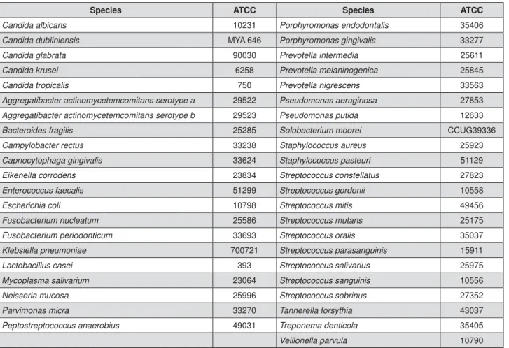
No significant differences were observed in
solutions yield the lowest percentage of biofilm,
followed by S3. S4 had the highest values (Figure 3).
scores at baseline, washout, and after treatments. A
change from score “1” (satisfactory) to “0” (absence)
and from score “3” (unsatisfactory) to “2” (regular) was found. Table 2 shows the score movement for each
solution. S3 and S1 had the highest percentages of
the “improved” and “cured”, being equal to 50% and
46%, respectively.
Multinomial logistic regression shows that a
observed with S3 and S1. The order and sequence of
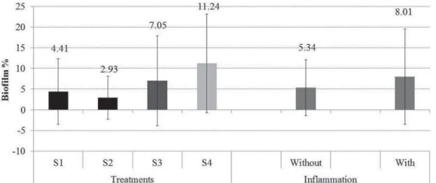
Patients’ satisfaction results are show in Table
4. In question 1, the effects of different solutions
Solution Baseline and Washout After treatment
0 1 2 3 Total 0 1 2 3 Total
S1 F 1 7 12 4 24 6 9 9 0 24
% 4.2 29.2 50.0 16.7 100 25.0 37.5 37.5 0.0 100
S2 F 3 6 11 4 24 6 7 8 3 24
% 12.5 25.0 45.8 16.7 100 25.0 29.2 33.3 12.5 100
S3 F 3 8 8 5 24 6 10 7 1 24
% 12.5 33.3 33.3 20.8 100 25.0 41.7 29.2 4.2 100
S4 F 1 9 11 3 24 0 9 11 4 24
% 4.2 37.5 45.8 12.5 100 0.0 37.5 45.8 16.7 100
Total F 8 30 42 16 96 18 35 35 8 96
% 8.3 31.3 43.8 16.7 100.0 18.8 36.5 36.5 8.3 100.0
Table 1-)UHTXHQF\RILQÀDPPDWLRQVFRUH)DQGSHUFHQWDJHDWEDVHOLQHZDVKRXWDQGDIWHUWUHDWPHQW
Worse Maintained Improved Cured Total
S1 0 13 6 5 24
% 0.0% 54.2% 25.0% 20.8% 100%
S2 2 15 3 4 24
% 8.3% 62.5% 12.5% 16.7% 100%
S3 1 11 9 3 24
% 4.2% 45.8% 37.5% 12.5% 100%
S4 6 13 5 0 24
% 25.0% 54.2% 20.8% 0.0% 100%
Total 9 52 23 12 96
% 9.4% 54.2% 24.0% 12.5% 100%
Table 2- ,QÀDPPDWLRQUDWHVDIWHUWUHDWPHQWV
solution interaction could not be assessed due to lack
of variability of the responses. Regarding questions 2
p=0.8; interaction: p=0.08), and 6 (solutions: p=0.6;
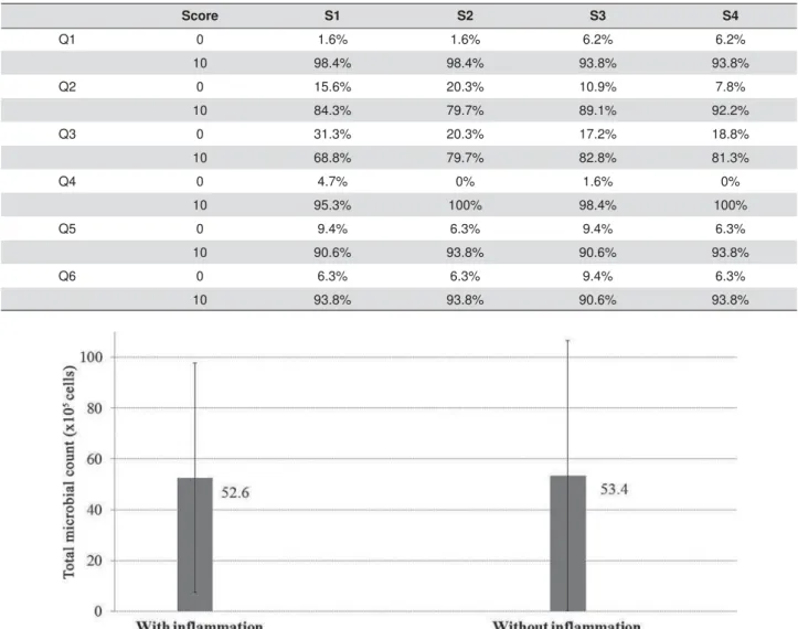
For DNA-Checkerboard hybridization results, no differences were found in the amount of total
microorganism count between groups with and without
candidiasis (p=0.75; Figure 4) or in the interaction
microorganisms counts were similar after use of S1,
S2, and S3 solutions and lower than S4 (Figure 5).
Num DF Den DF F Value Pr > F
Baseline and washout 3 61 4.51 0.0064
Treatment 3 61 4.44 0.0069
Order 3 61 0.52 0.6691
Sequence 3 20 0.74 0.5412
Num DF and Den DF: Degrees of freedom used in determining the F values.
3U!)SYDOXHDVVRFLDWHGZLWKWKH)YDOXHRIWKHVWDWLVWLFDOWHVW7KHQXOOK\SRWKHVLVWKHVSHFL¿HGFDQRQLFDOFRUUHODWLRQVDUHHTXDOWR]HUR LVHYDOXDWHGZLWKUHJDUGWRWKLVSYDOXH7KHQXOOK\SRWKHVLVLVUHMHFWHGLIWKHSYDOXHLVOHVVWKDQWKHVSHFL¿HGDOSKDOHYHO
F Value - F Value - Test the hypothesis that both canonical correlations are equal to zero in the sample.
Table 3- Effect of source factors on remission of candidiasis
Score S1 S2 S3 S4
Q1 0 1.6% 1.6% 6.2% 6.2%
10 98.4% 98.4% 93.8% 93.8%
Q2 0 15.6% 20.3% 10.9% 7.8%
10 84.3% 79.7% 89.1% 92.2%
Q3 0 31.3% 20.3% 17.2% 18.8%
10 68.8% 79.7% 82.8% 81.3%
Q4 0 4.7% 0% 1.6% 0%
10 95.3% 100% 98.4% 100%
Q5 0 9.4% 6.3% 9.4% 6.3%
10 90.6% 93.8% 90.6% 93.8%
Q6 0 6.3% 6.3% 9.4% 6.3%
10 93.8% 93.8% 90.6% 93.8%
Table 4- Percentage of patients for score 0 or 10 for each question and treatment
The effects of solutions on individual microorganism
count are shown in Table 5. S1 and S3 showed
C. t r opicalis; C. kr usei; S. sanguinis; S. or alis; S. m ut ans; P. int er m edia; L. casei; C. rect us; A. act inom ycet em com it ans serot ype b; S. m oorei; S. const ellat us; P. put ida; P. m icra; P. anaerobios; K. penum oniae
against C. dubliniensis and P. m elaninogenica. S1 and
F. nucleat um , S. p ast eu r i, P. en d od on t alis, N. m u cosa, and F. periodont icum. S3 was effective against P. aeruginosa.
S1 and S3 caused a mild reduction in the count of E. coli and A. act inom ycet em com it ans serotype a, against
Discussion
The association of mechanical and chemical methods have been recommended for the control of denture
7,14,16,22. The most commonly
used chemical solution is NaClO, however it can
cause deleterious effects to the denture when used
at 1% or 0.5% concentrations4,17,19,20. Therefore, the
assessment of NaClO at lower concentrations, as well as of new chemicals, is needed to help clinicians and
followed by S3. Results showed that S1, S2, and
individual microbial counts of target species. All
treatments were better than control (S4). Previous
studies have shown that the immersion of dentures
removal2 and in the reduction of microorganism
count22,23. These results demonstrate that lower
concentrations of sodium hypochlorite or the use of
R. com m unis
formation and for microorganism reduction and an
alternative for hypochlorite at 0.5%, which have been recommended from other studies8,22,23. Percentages
However, it is still necessary to evaluate the adverse effects of 0.25% NaClO and 10% R. com m unis (S3) on the acrylic resin of the denture. In the literature,
only one study evaluated the surface roughness with
the same solutions, which demonstrated clinically
4.
R. com m unis was used in this investigation once it shows antimicrobial properties similar to NaClO
when used in root canals with necrotic lesions11. In
addition, it is also biocompatible9 and has detergent
of R. com m unis solution in achieving complete denture hygiene, although experimental designs are diverse
and results are inconclusive2,10,19,20,22,23.
Andrade, et al.2 (2014) reported similar ratios between
2% R. com m unis and alkaline peroxide, but different ratios from 1% NaClO. Based on those previous
R. com m unis
concentration (10%), as an attempt to reach similarity
lower than with hypochlorite, S3 presented better
332
J
Appl Oral Sci.
S1 S2 S3 Control
Mean ±SD Lower Median Upper Mean ±SD Lower Median Upper Mean ±SD Lower Median Upper Mean ±SD Lower Median Upper p-value
quartile quartile quartile quartile quartile quartile quartile quartile
C. tropicalis 0.61 1.34 0 0ab 0 0.33 1.05 0 0b 0 0.68 1.44 0 0ab 0 1.37 1.77 0 0a 3.51 0.00
C. krusei 0.45 1.20 0 0ab 0 0.27 0.91 0 0b 0 0.36 1.03 0 0ab 0 1.03 1.66 0 0a 3.45 0.00
C. glabrata 0.42 1.20 0 0b 0 0.27 0.93 0 0b 0 0.44 1.19 0 0b 0 1.06 1.71 0 0a 3.26 0.02
C. dubliniensis 0.50 1.32 0 0ab 0 0.26 1.03 0 0b 0 0.42 1.21 0 0b 0 0.89 1.70 0 0a 0 0.02
C. albicans 0.36 1.13 0 0a 0 0.26 1.02 0 0a 0 0.21 0.83 0 0a 0 0.64 1.40 0 0a 0 0.12
V. parvula 0.27 1.07 0 0b 0 0.40 1.27 0 0b 0 0.65 1.63 0 0b 0 0.93 1.81 0 0a 0 0.02
T. denticola 1.36 2.11 0 0a 3.96 1.24 2.10 0 0a 3.36 1.59 2.21 0 0a 4.1 1.77 2.16 0 0a 39.3 0.43
T. forsythia 0.96 1.75 0 0a 2.54 0.45 1.29 0 0a 0 0.63 1.39 0 0a 0 1.13 1.89 0 0a 3.53 0.07
S. sobrinus 1.38 2.02 0 0a 3.98 1.04 1.92 0 0a 3.75 1.40 2.05 0 0a 4.1 2.32 2.18 0 0a 4.11 0.43
S. sanguinis 1.01 1.87 0 0ab 0 0.49 1.42 0 0b 0 1.23 2.19 0 0ab 4.1 1.45 2.18 0 0a 4.11 0.01
S. salivarius 1.69 2.25 0 0b 4.19 1.52 2.68 0 0b 3.63 2.51 2.24 0 3.52b 4.37 3.53 2.40 0 3.86a 5.26 0.00
S. pasteuri 0.87 1.75 0 0b 0 0.57 1.55 0 0b 0 1.15 1.86 0 0ab 3.38 2.03 2.42 0 0a 3.93 0.00
S. parasanguinis 1.82 2.51 0 0b 4.07 1.50 2.52 0 0b 3.58 2.02 2.39 0 0ab 4.16 3.32 2.01 3.13 3.84a 4.81 0.00
S. oralis 1.11 1.87 0 0ab 3.19 0.68 1.52 0 0b 0.00 1.18 1.90 0 0ab 3.31 1.89 2.14 0 0a 4.13 0.01
S. mutans 0.57 1.52 0 0b 0 0.35 1.10 0 0b 0 0.71 1.65 0 0ab 0 1.04 1.84 0 0a 2.61 0.04
S. moorei 1.82 1.71 0 0b 0 1.50 1.50 0 0b 0 2.02 1.50 0 0b 0 3.32 2.43 0 0a 2.82 0.00
S. mitis 2.02 2.62 0 0b 4.27 1.69 2.92 0 0b 3.47 2.39 2.24 0 3.46b 4.31 3.95 2.41 3.28 4.37a 5.47 0.00
S. gordonii 1.72 2.58 0 0b 3.89 1.29 2.71 0 0b 0 1.40 2.02 0 0b 3.42 2.89 2.51 0 3.56a 5.09 0.00
S. constellatus 0.72 1.70 0 0ab 0 0.50 1.36 0 0b 0 0.85 1.72 0 0ab 0 1.44 2.08 0 0a 3.88 0.00
S. aureus 2.11 2.77 0 0a 4.56 1.72 2.37 0 0a 4.28 1.72 2.27 0 0a 4.09 1.78 2.36 0 0a 4.29 0.35
P. putida 1.08 1.97 0 0ab 0 0.63 1.56 0 0b 0 1.06 1.79 0 0ab 3.17 1.62 2.26 0 0a 3.92 0.02
P. nigrescens 0.96 1.85 0 0b 0 0.82 1.73 0 0b 0 0.97 1.78 0 0b 0 2.00 2.09 0 0a 4.02 0.00
P. micra 1.19 2.08 0 0ab 3.6 0.86 1.82 0 0b 0 1.10 2.05 0 0ab 0 1.67 2.22 0 0a 3.67 0.01
P. melaninogenica
1.93 2.38 0 0ab 4.43 1.57 2.36 0 0b 4.89 1.59 2.15 0 0b 3.93 2.78 2.49 0 4.23a 4.74 0.02
P. intermedia 0.90 1.82 0 0ab 0 0.57 1.53 0 0b 0 0.85 1.63 0 0ab 0 1.66 2.03 0 0a 3.81 0.01
P. gingivalis 1.07 2.02 0 0a 0 0.54 1.43 0 0b 0 0.93 1.81 0 0ab 0 1.38 2.10 0 0a 3.6 0.01
P. endodontalis 0.98 2.10 0 0b 0 0.82 1.74 0 0b 0 1.29 2.01 0 0ab 3.05 2.08 2.16 0 2.82a 3.92 0.00
P. anaerobios 0.72 1.61 0 0ab 0 0.26 1.02 0 0b 0 0.52 1.40 0 0b 0 0.96 1.88 0 0a 0 0.02
P. aeruginosa 1.14 1.84 0 0b 3.39 1.36 3.81 0 0b 0 0.52 1.39 0 0c 0 1.91 2.14 0 0a 4.08 0.00
N. mucosa 1.11 1.97 0 0b 3.24 0.97 1.88 0 0b 0 1.47 2.09 0 0ab 3.75 2.30 2.20 0 3.41a 4.01 0.00
M. salivarium 2.66 2.46 0 4.01a 4.67 2.28 2.45 0 0a 4.81 2.54 2.44 0 3.8a 4.7 3.10 2.28 0 4.23a 4.83 0.17
L. casei 0.84 1.91 0 0ab 0 0.57 1.41 0 0b 0 1.30 1.99 0 0ab 3.18 1.58 2.15 0 0a 3.91 0.00
K. pneumoniae 1.37 2.20 0 0ab 3.78 0.86 1.81 0 0b 0 1.49 2.10 0 0ab 3.71 2.10 2.35 0 0a 4.27 0.00
F. periodonticum 0.87 1.66 0 0b 0.00 0.70 1.67 0 0b 0 1.25 1.90 0 0ab 3.27 1.79 2.13 0 0a 3.92 0.00
F. nucleatum 1.82 2.23 0 0b 4.12 1.97 2.54 0 0b 4.5 2.30 2.47 0 0ab 4.65 2.96 2.34 0 4.12a 4.75 0.00
E. faecalis 0.80 1.65 0 0b 0 0.77 1.71 0 0b 0 0.81 1.71 0 0b 0 1.79 2.10 0 0a 3.89 0.01
E. corrodens 1.34 2.01 0 0b 3.81 1.42 2.17 0 0b 3.6 1.20 1.96 0 0b 3.41 2.27 2.25 0 3.25a 4.25 0.03
E. coli 0.62 1.45 0 0b 0 0.82 1.75 0 0b 0 1.02 1.79 0 0ab 2.82 1.56 1.98 0 0a 3.83 0.03
C. rectus 0.19 0.87 0 0bc 0 0.14 0.78 0 0c 0 0.49 1.43 0 0ab 0 0.78 1.66 0 0a 0 0.00
C. gingivalis 1.19 1.94 0 0b 3.66 1.29 2.19 0 0ab 3.78 1.37 2.08 0 0ab 3.69 2.21 2.12 0 3.24a 4.04 0.04
B. fragilis 0.45 1.20 0 0a 0 0.54 1.34 0 0a 0 0.73 1.46 0 0a 0 1.05 1.70 0 0a 3.24 0.05
Aa serotype a 0.91 1.59 0 0b 2.92 1.14 1.88 0 0ab 3.34 1.36 1.83 0 0ab 3.38 1.82 1.96 0 0a 3.63 0.02
Aa serotype b 0.57 1.32 0 0ab 0 0.27 0.95 0 0b 0 0.61 1.36 0 0a 0 0.83 1.57 0 0a 0 0.04
Aa:Aggregatibacter actinomycetemcomitans
BADARÓ MM, SALLES MM, LEITE VMF
, ARRUDA
CNF
, OLIVEIRA
VC, NASCIMENT
O
C, SOUZA
RF
, P
ARANHOS HFO, SIL
V
A-LOV
A
T
O CH
patients, once it also presents biocompatibility with
living tissues6,9.
When the effects of the solutions on individual microorganisms were evaluated, S3 showed similar
results to hypochlorite (S1 and S2) against C. glabrat a, V. par v ula, S. saliv ar ius, S. m it is, S. gor donii, S. m oorei, P. nigrescens, E. faecalis, and E. corrodens. S3 had also the same effect as S2 against P. anaerobius
and C. du blin ien ses; S3 was more effective than S1 and S2 against P. aer u g in osa. Against other microorganisms such as C. t r opicalis, C. kr usei, E. coli, and S. m ut ans, S3 showed results that were mild, less effective than both concentrations of hypochlorite
but more effective than saline. It is noteworthy that no difference between treatments was found in the
count of C. albicans and S. aur eus, two important
reported that the detergent properties of R. com m unis
cause damage to the cell wall, resulting in loss of
the constituents of cytoplasm and subsequent cell
death12,30. These action mechanisms however need to
be further investigated.
The use of saline as a control substance resulted
in the highest percentage of biofilm among the
evaluated solutions. This result was expected and
2 (2014).
However, the act of brushing followed by immersion in
brushing found in previous studies16. However,
from solid particles, it is not enough for eliminating
microorganism from micro-irregularities of denture
surfaces. Thus, the association of mechanical and chemical methods is recommended for proper denture
hygiene7,22. This effective association explains the
dentures treated with antimicrobial solutions which,
matrix.
Regarding the remission of candidiasis, the immersion in 10% R. com m unis and 0.25% sodium
than 0.5% sodium hypochlorite. S3 solution had the
best results for remission of candidiasis in 50% of
18
(2013), in whose study a castor oil based solution
improved clinical symptoms of candidiasis in older
adult patients, similarly to the effect of Miconazole. In
than at 0.5% concentration. This result is contrary to
and antimicrobial action. Perhaps an allergic and/
or irritant action caused by residual waste solutions
and/or alveolar ridge3. A limitation of this study was
that residual effect of NaClO on the acrylic resin was
not evaluated. Moreover, clinical trials evaluating the irritating action of hypochlorite on the oral mucosa
and long-term evaluation are necessary.
Patients with and without denture stomatitis
participated in this study in order to determine whether the analyzed solutions can be used for cleaning of
dentures giving preventive and curative actions against
candidiasis.
Results of the questionnaire showed that S1, S2, and S3 had similar patient approval than
saline, rejecting the second null hypothesis. This
demonstrates that the use of these solutions did not
cause any inconvenience to participants, which would
prostheses home care. However, this is in contrast with
some studies that emphasize malodor and unpleasant
taste of NaClO as one of its disadvantages.
Finally, this study reinforces that 10% R. com m unis
and 0.25% NaClO solutions can be used as denture
cleanser replacing the 0.5% NaClO as auxiliary agent
for the mechanical method of brushing. Other studies should be used in addition, evaluating these solutions
to reinforce their viability of use such as research on
biomechanical analysis.
Conclusion
and were approved by the participants. R. com m unis
solution and 0.25% NaClO were effective in the remission of candidiasis. 0.25% sodium hypochlorite
and R. com m u n is can be indicated as a denture cleanser.
Acknowledgement
The authors would like to thank – São Paulo
Research Foundation (process number:
(ICQ, São Carlos, University of São Paulo).
References
1- Ambjornsen E, Rise J, Haugejorden O. A study of examiner errors associated with measurement of denture plaque. Acta Odontol Scand.
1984;42(3):183-91.
2- Andrade IM, Andrade KM, Pisani MX, Silva-Lovato CH, Souza RF,
Paranhos HF. Trial of an experimental castor oil solution for cleaning dentures. Braz Dent J. 2014;25(1):43-7.
3- Aparecida Guimarães M, Rocchetto Coelho L, Rodrigues Souza R, Ferreira-Carvalho BT, Marie Sá Figueiredo A. Impact of biocides
St aphylococcus aureus
(ST239-SCCmecIII) isolates. Microbiol Immunol. 2012;56(3):203-7.
4- Badaró MM, Salles MM, Arruda CN, Oliveira VC, Souza RF, Paranhos HF, et al. I n vit ro analysis of surface roughness of acrylic resin exposed
to the combined hygiene method of brushing and immersion in Ricinus
com m unis and sodium hypochlorite. J Prosthodont. In press 2016. doi: 10.1111/jopr.12436.
5- Bloem TJ, Razzoog ME. An index for assessment of oral health in the
edentulous population. Spec Care Dentist. 1982;2(3):121-4.
histometric evaluation of rat alveolar wound healing around polyurethane resin implants. Int J Oral Maxillofac Surg. 1997;26(2):149-52.
7- Cruz PC, Andrade IM, Peracini A, Souza-Gugelmin MC, Silva-Lovato CH, Souza RF, et al. The effectiveness of chemical denture cleansers
Appl Oral Sci. 2011;19(6):668-73.
8- Freitas Fernandes FS, Pereira-Cenci T, Silva WJ, Ricomini AP Filho,
Candida
J Prosthet Dent. 2011;105(1):51-8.
9- Huo L, Wang D, Liu H, Jia P, Gao J. Cytoxicity, dynamic and thermal
properties of bio-based rosin-epoxy resin/ castor oil polyurethane/ carbon nanotubes bio-nanocomposites. J Biomater Sci Polym Ed.
2016;27(11):1100-14.
10- Leite VM, Pinheiro JB, Pisani MX, Watanabe E, Souza RF, Paranhos
HF, et al. I n vit ro antimicrobial activity of an experimental dentifrice based on Ricinus com m unis. Braz Dent J. 2014;25(3):191-6.
11- Meneghin MP, Nomelini SM, Sousa-Neto MD, Marchesan MA, Franca SC, Santos HS. Morphologic and morphometric analysis of the root
canal apical third cleaning after biomechanical preparation using 3.3%
Ricinus com m unis detergent and 1% NaOCl as irrigating solutions. J Appl Oral Sci. 2006;14(3):178-82.
12- Messetti MA, Santos AM, Angelis DF, Chierice GO, Claro Neto S.
Estudo do derivado do óleo de Ricinus com m unis L. (mamona) como agente biocida e redutor da viscosidade produzida por Leuconostoc
mesenteroides em indústrias sucroalcooleiras. Arq Inst Biol. 2010;77(2):301-8.
13- Nascimento C, Albuquerque RF Jr, Monesi N, Candido-Silva JA. Alternative method for direct DNA probe labeling and detection
using the checkerboard hybridization format. J Clin Microbiol. 2010;48(8):3039-40.
14- Nishi Y, Seto K, Kamashita Y, Kaji A, Kurono A, Nagaoka E. Survival of microorganisms on complete dentures following ultrasonic
cleaning combined with immersion in peroxide-based cleanser solution. Gerodontology. 2014;31(3):202-9.
15- Panariello BH, Izumida FE, Moffa EB, Pavarina AC, Jorge JH,
Giampaolo ET. Effect of mechanical toothbrushing combined with different denture cleansers in reducing the viability of a multispecies
16- Paranhos HF, Peracini A, Pisani MX, Oliveira VC, Souza RF,
Silva-an acrylic resin submitted to simulated overnight immersion in denture
cleansers. Braz Dent J. 2013;24(2):152-6.
17- Paranhos HF, Silva-Lovato CH, Souza RF, Cruz PC, Freitas KM, Peracini A. Effects of mechanical and chemical methods on denture
18- Pinelli LA, Montandon AA, Corbi SC, Moraes TA, Fais LM. Ricinus
com m unis treatment of denture stomatitis in institutionalized elderly. J Oral Rehabil. 2013;40(5):375-80.
19- Pisani MX, Macedo AP, Paranhos HF, Silva CH. Effect of experimental
Ricinus com m unis solution for denture cleaning on the properties of acrylic resin teeth. Braz Dent J. 2012;23(1):15-21.
20- Pisani MX, Silva CH, Paranhos HF, Souza RF, Macedo AP. Evaluation
of experimental cleanser solution of Ricinus com m unis: effect on soft denture liner properties. Gerodontology. 2012;29(2):e179-85.
oral mucosal membrane in patients with chronic obstructive pulmonary disease. Adv Exp Med Biol. 2015;839:25-30.
22- Salles MM, Badaró MM, Arruda CN, Leite VM, Silva CH, Watanabe
based on sodium hypochlorite and Ricinus com m unis: a randomized clinical study. J Appl Oral Sci. 2015;23(6):637-42.
23- Salles MM, Oliveira VC, Souza RF, Silva-Lovato CH, Paranhos HFO. Antimicrobial action of sodium hypochlorite and castor oil solutions for
denture cleaning: in vit ro evaluation. Braz Oral Res. 2015;29(1):1-6. 24- Sanita PV, Machado AL, Pavarina AC, Massucato EM, Colombo AL,
Vergani CE. Microwave denture disinfection versus nystatin in treating patients with well-controlled type 2 diabetes and denture stomatitis:
a randomized clinical trial. Int J Prosthodont. 2012;25(3):232-44. 25- Sanitá PV, Pavarina AC, Giampaolo ET, Silva MM, Mima EG, Ribeiro
DG, et al. Candida spp. prevalence in well controlled type 2 diabetic patients with denture stomatitis. Oral Surg Oral Med Oral Pathol Oral
Radiol Endod. 2011;111(6):726-33.
three brushes in the control of complete denture cleansing. J Appl Oral Sci. 2006;14(6):454-9.
27- Silva-Lovato CH, Wever B, Adriaens E, Paranhos HdeF, Watanabe
based disinfecting cleaning tablets in complete denture wearers. J Appl Oral Sci. 2010;18(6):560-5.
28- Silva MM, Mima EG, Colombo AL, Sanitá PV, Jorge JH, Massucato EM, et al. Comparison of denture microwave disinfection and
conventional antifungal therapy in the treatment of denture stomatitis: a randomized clinical study. Oral Surg Oral Med Oral Pathol Oral Radiol.
2012;114(4):469-79.
29- Souza RF, Nascimento C, Regis RR, Silva-Lovato CH, Paranhos
HFO. Effects of the domestic use of a disclosing solution on the denture
30- Takano EH, Busso C, Gonçalves AL, Chierice GO, Guimarães-Catazarro SA, Prado-Castro MA. Inibição do desenvolvimento de fungos
Ricinus