Authors
Carmen Antonia Sanches Ito1
Roberto Pecoits-Filho2 Larissa Bail1
Mônica Arcoverde Wosiack1
Danieli Ainovicz1 Aline Borsato Hauser3
1Universidade Estadual de Ponta Grossa – UEPG. 2Centro de Ciências Bioló-gicas e da Saúde da Ponti-fícia Universidade Católica do Paraná – PUC/PR. 3Universidade Federal do Paraná – UFPR.
Submitted on: 05/17/2011 Approved on: 07/13/2011
Correspondence to: Aline Borsato Hauser Rua Prefeito Lothario Meissner, 632
Universidade Federal do Paraná – Sede Botânico – Jardim Botânico Curitiba – PR – Brazil Zip code 80210-170 E-mail: [email protected] Financial support: UEPG.
This study was undertaken at the Clinical Laboratory of the UEPG.
The authors report no conlict of interest.
R
ESUMOIntrodução: A presença de hemácias dis-mórficas na urina é um forte indicativo da origem glomerular do sangramento, sendo uma ferramenta importante no diagnóstico de glomerulonefrites. Os cilindros hemáti-cos geralmente acompanham as hemácias dismórficas, sendo também fortes indica-dores de hematúria glomerular, embora não sejam encontrados com frequência no exa-me parcial de urina. Objetivo: Comparar duas técnicas de concentração de amostras em uma série de exames de urina com he-matúria dismórfica. Material e Métodos: Foram selecionadas 249 amostras com hematúria dismórfica a partir de 4.277 amostras de urina de rotina. As amostras foram processadas utilizando-se duas téc-nicas: a convencional e a de concentração. O percentual de identificação dos cilindros hemáticos foi comparado de acordo com a metodologia utilizada. Resultados: A pre-sença de cilindros hemáticos pela técnica de concentração foi estatisticamente maior (52,6%) em comparação com a positivida-de pela metodologia convencional (8,4%) (p < 0,001). Discussão e Conclusão: Sugere-se que a técnica convencional não concentrou suficientemente a amostra de urina e os cilindros hemáticos ficaram no sobrenadante, sendo descartados. A utili-zação da técnica de concentração aumen-tou a sensibilidade técnica para a pesquisa dos cilindros hemáticos. Portanto, a técni-ca de concentração, associada à presença de hemácias dismórficas, mostrou-se útil para aumentar a concordância dos dois parâmetros laboratoriais para a detecção da hematúria de origem glomerular como auxílio diagnóstico das glomerulopatias, importante causa de doença renal crônica. Palavras-chave: Sedimento urinário. Dismorfismo hemático. Cilindros hemáticos.
A
BSTRACTIntroduction: Dysmorphic red blood cells (RBCs) in the urine are a strong in-dicator of a glomerular bleeding source. RBC casts, which while generally fol-lowing RBC dysmorphism are not fre-quently seen on routine urinalysis, are also important indicators of glomerular
hematuria. Objective: This study tested
the superiority of a urine concentra-tion technique (CT) over the standard method (SM) for RBC cast identifica-tion in a group of patients suspected of
glomerular hematuria. Material and
methods: Of a total of 4,227 routine urinary samples, 249 with dysmorphic hematuria were selected. The samples were processed according to two tech-niques: standard method (SM) and concentration technique (CT). The per-centages of RBC cast identification ac-cording to each method were compared. Results: The CT showed a higher rate of RBC casts (52.6%) compared to the SM (8.4%) (p < 0.001). Discussion and Conclusion: We suggest that the SM did not sufficiently concentrate the urine sample, the RBC casts remaining in the supernatant and being discarded. The CT increased the sensitivity of the RBC cast yield. The CT, associated with the presence of RBC dysmorphism, was use-ful to increase the agreement of the two parameters used for identification of glomerular-based bleeding and the diag-nosis of glomerular diseases, important causes of chronic kidney disease. Keywords: Urinary sediment.
Dysmorphic red blood cells. Red blood cell casts. Glomerular hematuria. Glomerulonephritis.
Comparative analysis of two methodologies for
the identiication of urinary red blood cell casts
I
NTRODUCTIONThe presence of dysmorphic red blood cells (RBCs) in the urine is a strong indicator of a glomerular source for the bleeding, being an important tool for the diagnosis of glomerulonephritides. In addition, the presence of RBC casts in the urine of patients with dysmorphic hematuria is directly associated with a glomerular source for the bleeding; therefore, the finding of a single RBC cast is enough to indicate
the glomerular nature of the hematuria.1,2 Because
glomerulopathies are potentially progressive renal diseases, tools to increase our diagnostic threshold are important for the prevention of chronic kidney
disease (CKD).3,4
An abnormal number of RBCs on urine micros-copy is a relatively common finding in laboratory practice, and definition of its origin makes it easier to identify its source in the urinary tract. Besides clinical history and physical examination, laboratory investi-gation of RBC morphology is the initial step towards diagnosis.
Birch & Farley,5 in the 1970s, developed a method
to determine the source of hematuria, which is now used in most clinical laboratories. In the 1990s, some
studies, such as those by Kohler et al. and Tomita
et al.,6,7 sought standardization of the criteria used to classify dysmorphic RBCs. In spite of these advances, complementary methods for identification of RBC casts may increase our sensitivity in the diagnosis of glomerulopathies, once urine, a colloidal suspension, may have some elements in the supernatant of a spun
sample which will not be seen in the sediment,8
justi-fying the low rate of RBC casts in urine samples from
patients with hematuria.2
The hypothesis of this study is that the low rate of RBC cast identification is due to the insufficient concentration reached with conventional urinalysis methods.
O
BJECTIVECompare the two techniques for sample analysis, con-ventional method (CM) and concentration technique (CT), in a series of urine exams presenting dysmor-phic hematuria.
M
ETHODSWe assessed urine samples from patients referred for several medical reasons, without a suspicion of glom-erulopathy, within the routine of the Clinical Analysis Laboratory of the State University of Ponta Grossa.
4,277 urine samples were collected, according to guidelines for routine urinalysis. Hematuria was de-fined as greater than 5,000 RBCs/mL of urine. After microscopy of the sediment, 991 samples with hema-turia were selected for investigation of RBC dysmor-phism and casts.
RBC dysmorphism was investigated in the 991 selected samples according to the characteristics
proposed by Birch & Farley,5 who considered RBC
dysmorphism to consist of the presence of more than three different cell populations which, when greater than 80%, indicates a glomerular source for the hematuria. The study was undertaken with
bright-field microscopy.9 Of the 991 samples
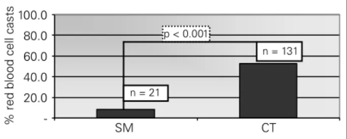
ana-lyzed, 249 had RBC dysmorphism, and were in-vestigated for RBC casts according to the standard method (SM) or concentration technique (CT), as shown in Chart 1.
Routinely, the SM uses a 10 mL aliquot of urine which has undergone 5-minute low-speed centrifu-gation (relative centrifugal force 400g). The urine is concentrated to 1:10, with removal of 9.0 ml of supernatant, the remaining 1.0 mL of sediment be-ing analyzed in a Neubauer chamber for investi-gation and count of the urinary elements, such as RBC casts. In the CT, after sample analysis with the SM, those samples with dysmorphic hematuria underwent a further step, which consisted of the ad-dition of 10 ml urine to the initial cone-shaped tube, which was centrifuged at a relative centrifugal force of 2,000 g for 10 minutes, that is, concentrating the urine to 1:100, taking into account that 0.2 mL of the sediment was used for a final volume of 20 mL.
Urine samples
n = 4,277
> 5,000 red blood cells/mL
n = 991
Red blood cell dysmorphism
n = 249
Search for red blood cell casts
Standard method
n = 21 (8.4%)
Concentration technique
n = 131 (52.6%)
After the supernatant was discarded, we searched for casts, including RBC casts, analyzing the sedi-ment between slide and coverslip, under light mi-croscopy, with 400X magnification. The result was expressed as type and number of casts per prepa-ration (p/p), after scanning the whole extension of the sediment. RBC casts were defined as casts with entrapped RBCs (RBC casts and mixed casts, com-posed of RBCs and other cells) and those with he-moglobin (hehe-moglobin casts), according to Figure 1. The latter were always associated with the presence of other RBC casts, once in isolation they may indi-cate hemoglobinuria, myoglobinuria or the presence of other similar pigments.
Statistical analysis was performed with the χ2 test.
R
ESULTSOf a total of 4,277 processed samples, 991 (23.2%) had hematuria on sedimentoscopy, with greater than 5,000 RBC/mL. Women had a higher prevalence rate (25.4% versus 16.7% in men; p < 0.001). Of the samples with hematuria, 249 (25.6%) had
dysmor-phic hematuria according to Birch & Farley`s criteria,5
there being no statistically significant difference in the prevalence of hematuria between the genders in this group (Table 1).
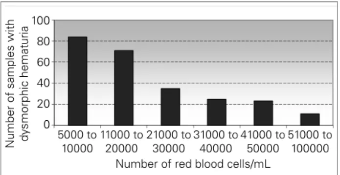
RBC count was divided in ranges from 5,000 to 100,000/mL. Most samples were in the 5,000 – 20,000 RBCs/mL range, only 4.4% of the samples having more than 100,000 RBCs/mL, being then clas-sified as gross hematuria (Figure 2). The rate of sam-ples with dysmorphic hematuria varied according to age range, with patients older than 60 years showing a statistically significant difference when compared to those younger than 40 years and those between 40 and 60 years (p = 0.04 and p = 0.02, respectively), whereas those older than 40 years and between 40 and 60 years had no statistically significant difference (p = 0.78) (Figure 3).
The 249 samples with dysmorphic hematuria were submitted to the two techniques for RBC cast investigation: SM and CT. The SM was positive in 8.4% (n = 21), while the CT was positive in 52.6% (n = 131) of the samples analyzed (p < 0.001) (Figure 4).
D
ISCUSSIONAND CONCLUSIONStarting in the mid 1970s, Birch & Farley5
devel-oped a method to determine the origin of hematuria. Since then, other studies have confirmed the impor-tance of urinary RBC morphometry. However, there has been disagreement concerning the cut-off values
1: dysmorphic red blood cells; 2: red blood cell cast.
Figure 1. Dysmorphic red blood cells and red blood cell casts found in the study samples.
1
2
100
80
60
40
20
Number of samples with dy
smorphic hemat
uria
0
11000 to 20000 5000 to
10000
21000 to 30000
Number of red blood cells/mL
31000 to 40000
41000 to 50000
51000 to 100000
Figure 2. Number of samples with dysmorphic hematuria in relation to the total number of red blood cells/mL.
0 10 20 30 40 50 60
0 to 10 11 to 20 21 to 30 31 to 40 41 to 50 51 to 60 > 61 Age Range
Number of samples with dy
smorphic hemat
uria
Figure 3. Number of samples with dysmorphic hematuria according to age range.
Number of
samples Men Women Total
Urinalysis 1,086 3,191 4,277 General
hematuria 180 (16.7%) 811 (25.4%) 991 (23.2%) Dysmorphic
hematuria 56 (5.2%) 193 (6.0%) 249 (5.8%)
Table 1 NUMBERANDPERCENTAGEOFURINALYSIS SAMPLESBYGENDER, GENERAL
-20.0 40.0 60.0
80.0 p < 0.001
n = 21
n = 131
100.0
SM CT
% red blood cell casts
Figure 4. Percentage of red blood cell casts found on the standard method (SM) and on the concentration technique (CT).
to be observed, once these varied among the stud-ies. In an attempt to overcome this obstacle, Hans
Kölher et al.,6 in 1991, classified urinary RBCs in
nine types, observing that acanthocytes and codo-cytes were frequently related to glomerulopathy-as-sociated hematuria, when present at concentrations of 5% or above. Yet, because the absence of these cells does not rule out the glomeruli as the source of hematuria, these markers have high specificity but low sensitivity.
In the following year, Tomita et al.7 proposed
another classification of urinary RBCs, with the aim of standardizing the dysmorphic forms and avoiding inter-observer variability. The RBCs were divided in five non-glomerular or urologic types (N1, N2, N3, N4 and N5) and five glomerular types (G1, G2, G3, G4 and G5), with their respec-tive ghost forms, that is, RBCs which have lost their hemoglobin. G1 cells are similar to acanthocytes, and considered very specific. Therefore, rates of only 1% of these cells already indicate glomerular hematuria. When G1 cells are not present (or their rate is lower than 1%), a total G-cell count can be made, with the cut-off point for glomerular hema-turia rising to 15%.
Even when these criteria are used, analysis is at times inconclusive, the association of other param-eters, such as the presence of RBC casts, being
nec-essary. 10 In addition, assessment of urinary RBC
dysmorphism, in almost all studies, is made with phase-contrast microscopy, a method which is not available in most clinical laboratories, as is the case
with automated urinalysis.8,11
In general, the prevalence of hematuria depends on the study population, and may range from 1 to 22% or more, males over the age of 50 years having higher rates of hematuria associated with severe
uro-logic diseases, such as neoplastic disorders.12-15 In our
study we found a 23.2% (n = 991) prevalence rate of
hematuria for the general population. When only the samples with dysmorphic hematuria (n = 249) were assessed, the rate was 5.8%.
As reported in other studies,16 women had a higher
general rate of hematuria. Yet, when only dysmor-phic hematuria was considered, there was no signifi-cant gender difference. This fact may be explained by the presence of several interfering factors that were not excluded in the first group, such as urinary tract infection and contamination by menstrual blood and vaginal secretions. These factors were excluded by the investigation and selection of samples with dys-morphic RBC.
As for age, our study showed that 59.8% of the samples (n = 149) belonged to patients aged 50 years or less, in disagreement with other studies which re-ported that microscopic hematuria was rare below
the age of 50 years.16 This fact points to the early
investigation of glomerular diseases in younger per-sons, as glomerulopathies are the second cause of
CKD in Brazil.17 Moreover, Schroder16 reported the
importance of investigating microscopic hematuria, present in 95.6% of his sample. Figure 3 includes the samples with hematuria (n = 991), with the statisti-cal analysis of dysmorphic hematuria in the follow-ing age ranges: < 40 years, between 40 and 60 years, and > 60 years. There was no statistically significant difference between the 40–60 years and > 60 years groups, while the difference was statistically signifi-cant between the > 60 years group and the others. This is probably due to an increased number of he-maturia-provoking conditions in persons > 60 years, such as prostatitis, neoplastic disorders and other urologic problems.
Hematuria may, therefore, be related to nephro-logic or uronephro-logic problems. Once hematuria is con-firmed, investigation of the bleeding source, under-taken with a search for RBC dysmorphism and RBC casts in the urine, is warranted. The additional pres-ence of proteinuria, glycosuria and other pathological casts is helpful.
Several authors have found it difficult to establish a cut-off point to characterize glomerular hematuria.
In an attempt to raise specificity, Kölher & Tomita6,7
strongly associated with glomerular hematuria, may
help with the diagnosis of the bleeding source.16
RBC casts are not frequently visualized in routine
urinalysis.18 This is believed to be due to the
insuffi-cient sample concentration afforded by the SM. We proposed to use a CT to investigate the presence of RBC casts in samples with dysmorphic hematuria.
We employed Birch & Farley`s5 criteria to classify
25.1% (n = 249) of the 991 orginal samples as dysmor-phic hematuria. The presence of specific cells, such as codocytes, acanthocytes and G1 cells was investigated, but. taking into account that these structures are not always present, the main criteria used were changes in the form and size of the RBCs. The presence of RBC casts was investigated in all samples with dysmorphic hematuria, in parallel, with the SM and the CT. The SM showed an 8.4% (n = 21) positivity. The 228 nega-tive samples on the SM were submitted to the CT, in which the sediment was re-processed under high speed rotation for a longer period, aiming to increase the concentration of the urinary elements. The CT was positive in 52.6% (n = 120), meaning a significant improvement of RBC-cast identification on the SM (r < 0.001), as can be seen in Figure 4.
The CT was effective in our study, raising the sensitivity of RBC-cast investigation. It must be highlighted that no lysis of the RBC casts happened during high speed rotation centrifugation (2,000 g), contrary to the literature, which recommends that sedimentoscopy should be performed on uncentri-fuged urine, or on urine which has been submitted
to a lower rotation speed (400 g).15,11 This result was
observed for other casts (hyaline, granular, epithe-lial, leukocyte, fatty and oval fat bodies), which ap-peared on CT microscopy.
These data point to the possibility that a myth surrounding cast degeneration by urine centrifuga-tion has been created. Although this concept of centrifugation-associated destruction of urinary ele-ments has been widely accepted by the laboratory community, our data show that this is not valid as far as casts are concerned. We cannot infer about the existence of glomerulopathies in the study pop-ulation, but we can certainly state that the CT in-creases the positivity for RBC casts.
Our population was composed of random samples not specified for glomerulopathy suspicion and with-out further investigation of a bleeding source. Indeed, this was a typical primary care population, for which hematuria assessment may avoid delay in the refer-ral to a nephrologist, that is, when end-stage CKD is
established and dialysis is necessary. In spite of the absence of epidemiologic studies on the occurrence
of glomerular diseases in Brazil, recent studies19 have
shown glomerular disorders to be the second cause of end-stage CKD in the country. These data show the
importance of referral of persons with RBC casts,18
there being consensus on the primary care responsi-bility for diagnosing and adequately referring patients with hematuria, aiming the early detection of urologic
and nephrologic conditions.20,21
Inter-laboratory methodological variation and inter-examiner variability may be obstacles to labo-ratory quality control. Therefore, greater attention should be paid to standardized urinalysis, while new techniques for identification of markers of renal dis-ease are sought. While other methods are not avail-able, we suggest the association of the CT-based RBC cast investigation and the search for RBC dysmor-phism for the assessment of renal diseases in the labo-ratory investigation of the bleeding origin, in patients with confirmed hematuria.
This study opens new perspectives towards the study of new ways for the urinalysis-based early de-tection of renal diseases. The CT-based search for RBC casts must be associated with the presence of dysmorphism on routine urinalysis to prove the glom-erular origin of the hematuria.
R
EFERENCES1. Abreu PF, Requião-Moura LR, Sesso R. Avaliação diagnóstica de hematúria. J Bras Nefrol 2007;29:158-63. 2. Rizzoni G, Braggion F, Zacchello G. Evaluation of
glomerular and nonglomerular hematuria by phase-contrast microscopy. J Pediatr 1983;103:370-4. 3. Margulis V, Sagalowsky AI. Assessment of hematuria.
Med Clin North Am 2011;95:153-9.
4. Barros E, Manfro RC, Thomé FS, Gonçalves LF. Nefrologia: rotinas, diagnóstico e tratamento. Porto Alegre: Artmed; 2006.
5. Birch DF, Fairley KF. Haematuria: glomerular or non-glomerular? Lancet 1979;2:845-6.
6. Kohler H, Wandel E, Brunck B. Acanthocyturia: a characteristic marker for glomerular bleeding. Kidney Int 1991;40:115-20.
7. Tomita M, Kitamoto Y, Nakayama M, Nakayama M, Sato T. A new morphological classification of urinary erythrocytes for differential diagnosis of glomerular hematuria. Clin Nephrol 1992;37:84-9.
8. Hannemann-Pohl K, Kampf SC. Automation of urine sediment examination: a comparison of the Sysmex UF-100 automated flow cytometer with routine manual diagnosis (microscopy, test strips, and bacterial culture). Clin Chem Lab Med 1999;37:753-64.
haematuria: role of dysmorphic red cell, G1 cell and bright-field microscopy. Scand J Clin Lab Invest 1997;57:203-8.
10. Rizzoni G. Evaluation of glomerular and nonglomerular hematuria by phase-contrast microscopy. J Ped 1983;103:370-4.
11. Venkat Raman G, Pead L, Lee HA, Maskell R. A blind controlled trial of phase-contrast microscopy by two observers for evaluating the source of hematuria. Nephron 1986;44:304-8.
12. Bastos MG, Martins GA, de Paula RB. Diagnóstico diferencial nas hematúrias. J Bras Nefrol 1998;20:425-40. 13. Grossfeld GD, Wolf JS Jr, Litwan MS, Hricak H, Shuler
CL, Agerter DC, et al. Asymptomatic microscopic hematuria in adults: summary of the AUA best practice policy recommendations. Am Fam Physician 2001;63:1145-54.
14. Messing EM, Young TB, Hunt VB, Emoto SE, Wehbie JM. The significance of asymptomatic microhematuria in men 50 or more years old: findings of a home screening study using urinary dipsticks. J Urol 1987;137:919-22.
15. Mohr DN, Offord KP, Owen RA, Melton LJ 3rd. Asymptomatic microhematuria and urologic disease. A population-based study. JAMA 1998;256:224-5. 16. Schroder FH. Microscopic haematuria. BMJ
1994;309:70-2.
17. Kirsztajn GM. A campanha nacional de prevenção de doenças renais. J Bras Nefrol 2006;28:2-3.
18. McDonald MM, Swagerty D, Wetzel L. Assessment of microscopic hematuria in adults. Am Fam Physician 2006;73:1748-54.
19. Alves MAR. Propedêutica das Glomerulopatias. In: Barros RT, Alves MAR, Kirztajn GM, Sens YAS, M Dantas. Glomerulopatias: Patogenia, Clínica e Tratamento. 2a ed. São Paulo: Editora Sarvier, 2006. v.
01. 480 p.
20. Patel JV, Chambers CV, Gomella LG. Hematuria: etiology and evaluation for the primary care physician. Can J Urol 2008;15:54-61.