Autores
Mariana Gascue de Alencastro1
Joana Raquel Nunes Lemos1 Nícia Maria Romano de Medeiros Bastos2 Alessandra Rosa Vicari2 Luiz Felipe Santos Gonçalves3
Roberto Ceratti Manfro3
1 Federal University of Rio Grande do Sul.
2 University Hospital of Porto Alegre.
3 University Hospital of Porto Alegre and Federal University of Rio Grande do Sul.
Data de submissão:12/11/2012. Data de aprovação: 14/05/2013.
Correspondência para: Roberto Ceratti Manfro. Nephrology Department of the University Hospital of Porto Alegre. Rua Ramiro Barcelos, nº 2350, sala 2030. Porto Alegre, RS, Brazil. CEP: 90035-003.
E-mail: [email protected] Fax: (51) 3359-8121.
Financial support from the Research Foster Funds of the University Hospital of Porto Alegre (FIPE-HCPA).
Avaliação da síndrome metabólica e suas associações com inflamação
e função do enxerto em pacientes receptores de transplante renal
Evaluation of metabolic syndrome and associations with
inflammation and graft function in renal transplant recipients
Introdução: A doença cardiovascular (DCV) é um dos principais determinantes da mortalidade em receptores de trans-plante renal (RTR). A síndrome metabó-lica (SM) e a inflamação crônica atual-mente são considerados fatores de risco não tradicionais para doença cardiovas-cular. Objetivo: Avaliar a frequência da SM e da inflamação e suas associações com a função do enxerto em receptores de transplante renal. Métodos: Foi rea-lizado um estudo transversal com 200 RTR. A SM foi definida pelos critérios do NCEP-ATP III. A inflamação foi avaliada por meio dos níveis de PCR. A função renal foi avaliada pela esti-mativa da TFG por meio da equação MDRD. Resultados: A SM ocorreu em 71 pacientes (35,5%). Pacientes com SM apresentaram maior PCR e diminuição dos níveis de TFG. A inflamação esteve presente em 99 pacientes (49,5%). A cir-cunferência abdominal, índice de massa corporal, triglicérides e colesterol total foram significativamente maiores em pacientes com inflamação. Foi demons-trada associação entre MS e inflamação, 48 (67,6%) pacientes com SM estavam inflamados e entre aqueles sem SM a taxa de inflamados foi de 39,5% (51 pa-cientes) (p < 0,001). Uma porcentagem significativamente maior de pacientes com SM foi observada no grupo de pa-cientes de doença renal crônica estágios III e IV. Conclusão: Em RTR há associa-ção significativa entre MS e inflamaassocia-ção. A SM está negativamente associada com a função do enxerto. As implicações clínicas destes achados devem ser avaliadas em estudos longitudinais.
R
ESUMOIntroduction: Cardiovascular disease (CVD) is a major determinant of mortality in renal transplant recipients (RTR). Met-abolic syndrome (MS) and chronic in-flammation are currently considered non traditional risk factors for cardiovascular disease. This study evaluates the frequency of these conditions their associations with graft function. Objective: To evaluate the prevalence of metabolic syndrome (MS) and inflammation and their associations with graft function in renal transplant recipients. Methods: A cross-sectional study was carried out with 200 RTR. MS was defined by the NCEP-ATP III criteria. Inflammation was assessed by CRP levels. Renal function was assessed by GFR estimation using the MDRD equation. Results: MS occurred in 71 patients (35.5%). Patients with MS had higher CPR and decreased GFR levels. Inflammation was present in 99 patients (49.5%). Mean waist perimeter, body mass index, triglycerides and serum total cholesterol were significantly higher in in-flamed patients. An association between MS and inflammation was demonstrat-ed, 48 (67.6%) patients with MS were inflamed and among those without MS the rate of inflamed patients was 39.5% (51 patients) (p < 0.001). A significantly higher percentage of patients with MS in the group of patients in chronic renal disease stages III and IV was observed.
Conclusion: In RTR there is a signifi-cant association among MS and inflam-mation. MS is negatively associated with graft function. The clinical implications of these findings must be evaluated in longitudinal studies.
ABSTRACT
Palavras-chave: inflamação; obesidade; proteína c-reativa; síndrome X metabólica; transplante de rim.
Keywords: C-reactive protein; inflamma-tion; kidney transplantainflamma-tion; metabolic syn-drome X; obesity.
Metabolic Syndrome and Inflammation in RTR
INTRODUCTION
Renal transplantation has become the treatment choice for a significant proportion of patients with terminal chronic renal disease (CRD). Over the last decades, the advances in the field lead to significant reduction of the acute rejection rates and improve-ments in short-term survival of patients and grafts. However in the long term the results still need to improve and most of the losses occur due to chro-nic allograft failure, mainly chrochro-nic rejection and death with functioning graft.1 Among the causalities,
cardiovascular disease (CVD) is the leading cause accounting for approximately half of the observed mortality,2 Many risk factors for CVD in the general
population are present in renal transplant recipients. The most prevalent are hypertension, diabetes melli-tus, hyperlipidemia, obesity, smoking and anemia.2 In
addition, other risk factors have been suggested in the pathogenesis of CVD in renal transplant recipients (RTR), among these factors proteinuria and inflam-mation have being described.3,4
The components of the metabolic syndrome (MS) namely, hypertension, diabetes mellitus, dyslipidemia and obesity, are independent risk factors for CVD. MS is an evolving concept, however its relevance in the re-nal transplant population has already being shown.5,6
Its prevalence has been evaluated and reported to be as high as 63% in one study.7 Recent studies report that
MS may be associated with impaired long-term graft function, cardiovascular events, new onset diabetes after transplantation, graft loss and patient’s death.7-10
Inflammation, as in the general population and uremic patients, is associated to cardiovascular events in RTR. In clinical practice it is diagnosed by the incre-ment of acute phase proteins, and the C-reactive pro-tein (CRP) is the clinically used parameter for such pur-pose.11 CRP is produced by hepatocytes in response to
infections, inflammation, injury and other stimuli. Its increment is well correlated to other inflammation ma-rkers, such as interleukin-1 (IL-1), interleukin-6 (IL-6) and tumor necrosis factor-alpha (TNF-α).12 It has been
identified as a predictor of cardiovascular events in the general population, patients undergoing dialysis, and in RTR.13-15 Moreover, there is evidence that increased
post-transplant CRP levels are associated with a higher risk of chronic graft disease.16
After transplantation, appetite restoration, end of alimentary restrictions and the side effects of the immunosuppressive agents commonly lead to weight
gain and obesity, a major problem after renal trans-plantation, occurring in up to 50% of the patients. The average weight gain is reported to be of 10 kg during the first post-transplant year.17 Previous studies
suggested that obesity is associated with an increased cardiovascular morbidity and mortality, and reduced survival of patients and grafts.18
The present study was undertaken to evaluate the prevalence and associations of metabolic syndrome and inflammation in a population of RTR in southern Brazil.
METHODS
A cross-sectional study was conducted including RTR followed at the kidney transplant clinic at Hospital de Clínicas de Porto Alegre. The study was approved by the Hospital de Clínicas de Porto Alegre - Federal University of Rio Grande do Sul Institutional Review Board (IRB) and Ethics Committee, in adherence with the Declaration of Helsinky.
Outpatient RTR that met the following criteria: (a) transplant time between one and ten years; (b) stable graft function in the last three months defined by the variation of serum creatinine ≤ 0.3 mg/dL and; (c) accepting to participate in the study by signing the written informed consent, were included in the study. Patients with clinical or laboratorial evidence of infection, inflammation, auto immune diseases and with estimated glomerular filtration rate (GFR)
≤ 15 mL/minute were excluded.
Demographic data including age, gender, ethnicity, post-transplant time, organ source (living/deceased), primary renal disease, immunosuppressive treatment and medication use were recorded. The comorbidities evaluated included hypertension, hyperlipidemia, obesity, pre transplant and posttransplant diabetes mellitus and smoking. Laboratory data including the CRP levels were obtained in a routine clinic appoint-ment in conjunction with measureappoint-ments of the blood pressure, body weight, height, and waist perimeter. Body weight was measured in a 0.1 kg precision scale and the height was measured by using a 0.5 cm precise stadiometer. Body mass index was calculated as bo-dy weight (kilograms) divided by the squared height (meters). Patients were classified, according to BMI: undernourished (BMI < 18.5 kg/m2), eutrophic (BMI
18.5 to 24.9 kg/m2), overweight (25 to 29 kg/m2),
obesity class I (30 to 34.9 kg/m2), obesity class II (35
to 39.9 kg/m²) and obesity class III (≥ 40 kg/m2).19 The
metric tape, positioning half way between the lower rib and the superior iliac crest.
METABOLICSYNDROME
The National Cholesterol Education Program’s Adults Treatment Panel III (NCEP-ATP III) definition crite-ria was used and include: central obesity, measured by waist circumference (WC), (> 102 cm for men and > 88 cm for women); triglycerides (TG) ≥ 150 mg/dL; HDL cholesterol (HDL-c), (< 40 mg/dL for men and < 50 mg/dL for women); systolic pressure (SP)
≥ 130 mmHg or diastolic pressure (DP) ≥ 85 mmHg and fasting glucose ≥ 100 mg/dL. Patients were diag-nosed with MS when presenting at least three of the components.20
INFLAMMATION
The inflammatory state was accessed by the measurement of CRP that was analysed by nephelometry, using the reagent CardioPhase hsCRP (Dade Behring, Germany). In the absence of validated values for the renal transplant recipients population, the median value observed in the sample of this study was used as a cutoff for defining inflammation.
RENALFUNCTION
Renal function was estimated through creatinine based GFR estimation, according to MDRD (Modification of Diet in Renal Disease) equation: GFR = 175 x (creatinine)-1.154 x (age)-0.023 x (0.742
woman) x (1.210 black race).21 After calculating
the GFR, patients were classified according to CRD stages: stage I: > 90 mL/min/1.73m²; stage II: 60-89 mL/min/1.73m², stage III: 30-59 mL/min/1.73m²; stage IV: 15-29 mL/min/1.73m²; and stage V: < 15 mL/min/1.73m² (excluded).22
STATISTICALANALYSIS
Statistical analyses were performed by using the SPSS (Statistical Package for the Social Sciences) software, for Windows 16 version. Normality was tested by using the Kolmogorov-Smirnov test. Normally dis-tributed data were expressed as mean ± standard deviation. Median and quartile interval were used for variables without normal distribution. Paired data were compared by Student´s t test, ANOVA was used for multiple comparisons and unpaired varia-bles through by Mann-Whitney U test. The categori-cal variables were associated according to chi square
test with Yates’ correction. Poisson’s regression with robust variance was used for the estimation of the prevalence ratios. The continuous variables were correlated by the Spearman’s test. Multiple compari-sons of continuous variables with asymmetric distri-bution were made by using the Kruskal-Wallis test. A rank transformation of the variables with asymmetric distribution was performed and used for comparison between groups through Tukey’s test. p values lower than 0.05 were considered statistically significant.
RESULTS
Two hundred renal transplant recipients were evaluated, 113 (56.5%) men, mean age 45.7 ± 11.5 years. The median of transplant time was 44 (19-71) months, and 135 patients (67.5%) received organs from deceased donors. Primary renal diseases were hypertension in 49 patients (24.5%), primary glome-rular disorders in 36 patients (18%), adult polycystic kidney disease in 27 patients (13.5%) diabetic nephopathy in 15 (7.5%) chronic pyelonephritis in 14 (7%), other causes in 14 (7%) and in 52 patients (26%) the etiology of the renal disease was unknown. The more frequent co-morbidities were: hypertension in 159 patients (79.5%), hyperlipidemia in 56 (28%), obesity in 35 (17.5%) and hepatitis C virus (HCV) infection in 33 (16.5%). Sixteen patients (8%) were diabetic before transplantation and new onset diabetes after transplantation occurred in 18 patients (9%). Eleven patients (5.5%) were current smokers.
All patients were using low dose of prednisone (5 mg/day), calcineurin inhibitors were used by 179 patients (89.5%) and mycophenolate sodium or mofetil in 168 patients (84%), azathioprine in 15 (7.5%) and rapamycin in 7 (6.0%). The main non-immunosuppressive medications used were: antihypertensive drugs in 158 patients (79%), pro-ton pump inhibitors in 123 (61.5%), diuretics in 73 (36.5%), statins in 56 (28%), low dose aspirin in 26 (13%), insulin in 25 (12.5%), and other diabetes controlling drugs in 6 (3%).
The nutritional assessment, according to BMI categories, revealed that 82 patients (41%) were eutrophic, 83 (41.5%) were overweight and 35 (17.5%) obese, being 24 (12%) classified as class I obesity, 9 (4.5%) class II obesity and 2 (1%) class III obesity. There were no undernourished patients.
Metabolic Syndrome and Inflammation in RTR
CRD stage I, 49 (24.5%) stage II, 116 (58%) stage III and 25 (12.5%) stage IV.
Demographic and laboratory data of patients with and without MS and inflammation are shown in Table 1. MS occurred in 71 patients (prevalence 35.5%). Apart from variables involved in the defini-tion of MS, which were expectedly higher in patients with MS, it was also found that patients with MS were older, presented significantly higher serum urea, CRP and BMI. They also presented significantly lower estimated GFR values (Table 1).
According to the criteria established for this analysis inflammation was defined by a serum CRP level higher than 1.6 mg/L. Ninety nine patients were considered inflamed (49.5% prevalence). Among the variables shown in Table 1, it was observed that the mean waist circumference, BMI, TG and serum total cholesterol were significantly higher in this group.
An association between MS and inflammation was observed. Forty-eight (67.6%) patients with MS were inflamed and among patients without MS the percentage of inflamed patients was 39.5% (51 patients) (p < 0.001). As shown in Figure 1 median and quartile CRP serum values were significantly higher in the group of patients with MS [3.2 (1.2-5.4)]
as compared to the group of patients without MS [1.2 (0.6-3.8)] (p < 0.001).
A significant association was found between increased BMI and inflammation. As shown in Table 2, firstly considering all patients, the prevalen-ce rate of inflammation significantly increased in the overweight group and further increased significantly in the obese group. In patients with CRD stages I + II (excellent and good graft function) a significant effect of weight in the prevalence rate of inflamma-tion was observed only in the obese group. However in the group of patients in CRD stages III + IV (fair and poor graft function) this prevalence rate increa-sed significantly in the overweight and in the obese subgroups uncovering a possible association between loss of graft function and inflammation.
The presence of inflammation was tested against MS individual components. Positive and significant correlations between CRP with waist circumference (rs = 0.270; p < 0.001), with fasting glucose (rs = 0.174; p = 0.014) and with serum triglycerides (rs = 0.229; p = 0.001) were found. No correlation was found between inflammation and blood pressure, either systolic or diastolic, or inflammation and HDL-cholesterol. To further investigate the
TABLE 1 COMPARISONOFTHEGROUPSWITHANDWITHOUTMETABOLICSYNDROMEANDINFLAMMATION
MS present (n = 71)
MS absent
(n = 129) p
Inflamed (n = 99)
Not Inflamed
(n = 101) p
Age (years)a 48.45 ± 10.08 44.13 ± 11.98 0.007 47.01 ± 11.11 44.35 ± 11.78 0.102 Ethnicity
(caucasian/non-caucasian) 57/14 107/22 0.702 84/15 80/21 0.400
Time post-renal
tranplantation (months)b 37 (16-75) 45 (19-71) 0.486 44 (19-88) 44 (18-61) 0.384
Donor (living/deceased) 20/51 45/84 0.349 28/71 37/64 0.207
Waist Circumference (cm)a 99.87 ± 13.21 87.16 ± 11.43 < 0,001 94.81 ± 12.97 88.59 ± 13.37 < 0.001 Serum triglycerides (mg/dL)a 236.01 ± 72.85 145.52 ± 61.33 < 0.001 195.26 ± 80 160.38 ± 73.37 0.002
HDL-c (mg/dL)b 41 (35-49) 53 (42-65) < 0.001 46 (37-57) 49 (40.5-62) 0.146
Hemoglobin (mg/dL)a 13.29 ± 1.98 12.91 ± 1.64 0.147 13.12 ± 1.74 12.97 ± 1.80 0.541
Hematocrit (mg/dL)a 40.72 ± 5.86 40.07 ± 4.85 0.403 40.54 ± 5.23 40.06 ± 5.23 0.520
Serum total cholesterol
(mg/dL)a 207.31 ± 43.50 193.73 ± 48.62 0.051 209.35 ± 48.53 187.96 ± 43.56 < 0.001
Glucose (mg/dL)b 109 (92-128) 91 (86-99.5) < 0.001 96 (89-109) 93 (86,5-99) 0.070
Creatinine (mg/dL)b 1.52 (1.19-2.06) 1.48 (1.16-1.76) 0.189 1.47 (1.11-1.91) 1.5 (1.23-1.90) 0.487 Serum urea (mg/dL)b 63 (46-90) 56 (43-69) 0.031 58 (45-79) 59 (43.5-72.5) 0.516
eGFR (mL/min/1.73m²)a 48.17 ± 18.88 54.17 ± 20.14 0.041 52.06 ± 21.78 52.03 ± 17.89 0.992
Body Mass Index (Kg/m²)a 28.93 ± 4.23 25.14 ± 3.99 < 0.001 27.77 ± 4.65 25.23 ± 3.88 < 0.001
C-reactive Protein (mg/L)b 3.2 (1.2-5.4) 1.2 (0.6-3.8) < 0.001 4,2 (3-6.7) 0.8 (0.4-1.15) < 0.001
a Values expressed as mean ± SD; b Values expressed as median and interquartile interval (p25-75); HDL-c: Serum HDL cholesterol;
An evaluation of the serum creatinine, BMI, estimated GFR and metabolic syndrome was made according to CRP quartiles and is shown in Table 4. BMI values where higher in the third and fourth quartiles and the percentages of patients with metabolic syndrome were higher in the third quartile as compared to the first quartile.
To explore a possible association between renal function and MS and renal function and inflamma-tion we grouped the patients at CRD stages I and II (59 patients), and the patients at CRD stages III and IV (141 patients). MS was present in 14 patients (23.7%) and in 57 patients (40.4%) respectively in the first and second groups (p = 0.037). However, the prevalences of inflammation were 49.1% (29 patients) and 49.6% (70 patients) in the respective groups (p = 0.949).
DISCUSSION
Several factors contribute to the elevated prevalence of MS observed in RTR. Among them factors related to the use of immunosuppressive drugs including weight gain, altered lipid profiles, effects on blood pressure, glucose metabolism and possibly the renal graft function have being described.8
Immunosuppressive therapy with corticosteroids, calcineurin inhibitors and rapamycin is associated important modifications in lipid and glucose metabolism and may impact on de novo MS.23,24
Furthermore correction of uremia and the use of cor-ticosteroids lead to increased appetite and to develo-pment of post-transplant overweight and obesity.25
An elevated prevalence of MS was previously reported in studies with RTR.7,9,26 Studies adopting the
NCEP-ATP III diagnostic criteria reported a prevalence
Figure 1. CRP (mg/L): Values distribution among the group of patients with and without metabolic syndrome. Box-plot graphs presenting the median values 25-75. percentiles 10-90 and outliers.
TABLE 2 ASSOCIATIONBETWEENTHEBODYMASSINDEXCLASSIFICATIONSANDTHEPREVALENCERATIOOFINFLAMMATIONIN
ALLPATIENTSANDINTHECHRONICRENALDISEASE (CRD) STAGEIII + IVGROUP
All patients (n = 200)
Patients with CRD Stage I+II (n = 59)
Patients with CRD Stage III+IV (n = 141)
Inflammation
n (%) PR - CI 95% p*
Inflammation
n (%) PR - CI 95% p*
Inflammation
n (%) PR - CI 95% p* Eutrophic
(n = 82/25/57) 30 (36.6) 1 - 10 (40) 1 - 20 (35.1) 1
-Overweight
(n = 83/26/57) 44 (53.0)
1.45
(1.02-2.05) 0.038 12 (46.2)
1.1
(0.61-2.18) 0.659 32 (56.1)
1.6
(1.05-2.44) 0.029
Obese
(n = 35/8/27) 25 (71.4)
1.95
(1.37-2.78) 0.000 7 (87.5)
2.187
(1.27-3.78) 0.005 18 (66.7)
1.9
(1.22-2.96) 0.004
* Prevalence ratio (Poisson’s regression); PR: Prevalence ratio; CI: Confidence interval; CRD: Chronic renal disease. The first number in each category corresponds to the general sample, the second corresponds to the sample of patients with I+ II CRD and the third corresponds to the sample of patients with III + IV CRD. The statistical comparisons were made against the group of eutrophic patients.
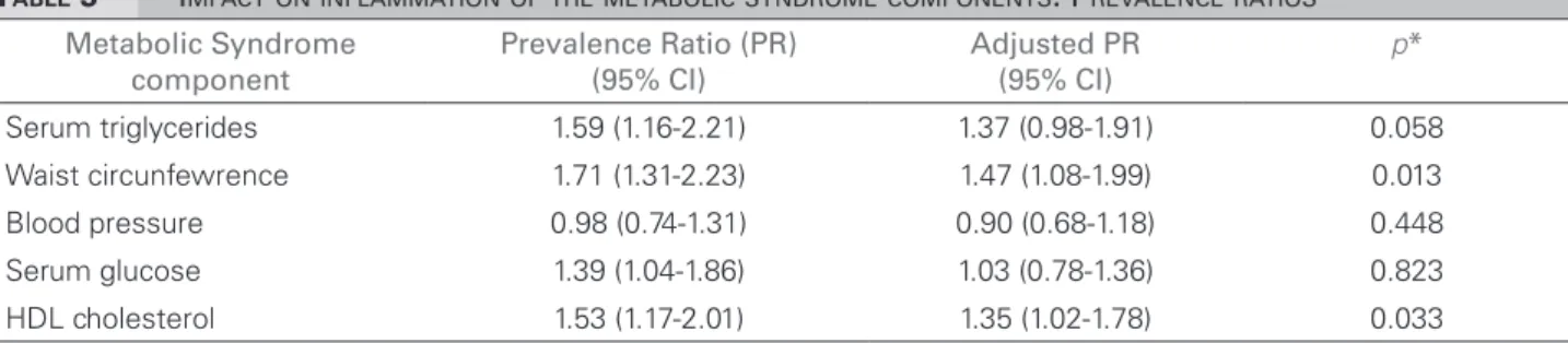
association between inflammation and the individual components of MS Poisson´s regression was used to analyze the prevalence ratios of each component against the presence of inflammation (Table 3). Here we found that the waist circunference, and HDL cho-lesterol are the components that significantly impact in the association. Further analyses showed that CRP positively correlated with BMI (rs = 0.315; p < 0.001) and with total cholesterol (rs = 0.173; p = 0.015).
Metabolic Syndrome and Inflammation in RTR
TABLE 3 IMPACTONINFLAMMATIONOFTHEMETABOLICSYNDROMECOMPONENTS. PREVALENCERATIOS
Metabolic Syndrome component
Prevalence Ratio (PR) (95% CI)
Adjusted PR (95% CI)
p*
Serum triglycerides 1.59 (1.16-2.21) 1.37 (0.98-1.91) 0.058
Waist circunfewrence 1.71 (1.31-2.23) 1.47 (1.08-1.99) 0.013 Blood pressure 0.98 (0.74-1.31) 0.90 (0.68-1.18) 0.448
Serum glucose 1.39 (1.04-1.86) 1.03 (0.78-1.36) 0.823
HDL cholesterol 1.53 (1.17-2.01) 1.35 (1.02-1.78) 0.033
* Poisson’s regression; CI: Confidence interval.
Figure 2. CRP (mg/L) values distribution among the BMI classification categories. Box-plot graphs presenting median values. percentiles 25-75. percentiles 10-90 and outliers. Eutrophic: BMI (18.5-24.9kg/m²); Overweight: BMI (25-29.9 kg/m²), Obese: BMI (≥ 30 kg/m²).
TABLE 4 EVALUATIONOFBODYMASSINDEX, SERUMCREATININE, ESTIMATEDGFRANDMETABOLICSYNDROMEACCORDING
TO C-REACTIVEPROTEINQUARTILES
Quartile (CPR levels)
1 (< 0.8 mg/L) (n = 49)
2 (0.8-1.6 mg/L) (n = 52)
3 (> 1.6-4.17 mg/L) (n = 49)
4 (> 4.17 mg/L)
(n = 50) p
Body Mass Index 24.51 ± 3.83ª 25.92 ± 3.84b 27.30 ± 4.60c 28.22 ± 4.69d < 0.001*
serum creatinine 1.66 ± 0.57 1.55 ± 0.49 1.74 ± 0.83 1.47± 0.50 0.133*
eGFR 51.31 ± 18.68 52.70 ± 17.23 51.80 ± 26.96 52.31 ± 15.38 0.987* Metabolic Syndrome 5 (10.2%)e 18 (34.6%) 25 (51%)f 23 (46%) < 0.001**
eGFR: Estimated glomerular filtration rate); CRP: C-Reactive Protein; * ANOVA; ** Chi square test; n: Number of patients. a versusc (p = 0.007); aversusd: (p < = 0.001); bversusd: (p = 0.035) and e versusf (p < 0.001).
of around 60%.7,27 Other reports with the same
criteria, but using BMI instead of waist circumference, reported prevalence between 22.6% and 32.0% one to six years after renal transplantation.9,28 In the
present study the prevalence of 35.5% was found. The observed variation is most possibly explained by the design of the studies, time of evaluation after transplantation and perhaps by population differen-ces in the frequency of the MS components in each study. The time of transplant is also an important variable to be taken into consideration.7,9 In addition,
other variables in the composition of study popula-tions including previous time of dialysis therapy, rate of preemptive transplantation, donor type (deceased or living) and the immunosuppressive drug regimen can potentially influence the MS prevalence.7,9,23
Obesity is a frequent post-transplant complication and a well established risk factor for atherosclerotic disease. Besides, it is associated to an increased risk for diabetes, dyslipidemia and hypertension.29 In this
study, 41.5% of patients were overweight and 17.5% were obese. These frequencies are similar to those reported in another studies.15
Inflammation is currently considered a risk factor for cardiovascular disease in RTR.4 In the clinical
practice it is detected by the increased CRP levels. However, the values correlated to cardiovascular outcomes are different in the general population and in uremic patients, and there are no validated cutoffs for renal transplant recipients. Cueto-Manzano et al.
measured CRP before and at different moments after renal transplantation, and found a significant decrease until one year after the transplant, leveling off around 3.2 mg/L afterwards.30 Another study found a similar
mean for the CRP levels.31 Besides, CRP and other
used in the analyses was the median value of the CRP (1.6 mg/dL) found in our study population. In sup-port to this approach a previous robust study found that CRP levels higher than 1.54 mg/L are associated with increased mortality in RTR.32 Using this cut off
to categorize inflammation resulted in half of the pa-tients being considered inflamed and higher levels of CRP were associated to increased weight, abdominal circumference and serum triglycerides. Also, in the evaluations of the BMI and MS according to the CRP quartiles, it was found that the groups of patients with CRP higher than 1.6 presented significantly hi-gher BMI values and percentages of patients with MS. The pro-inflammatory state has been considered one component of MS.33 Inflammation markers, such
as CRP, tumor necrosis factor, fibrinogen, interleukin-6, among others, are associated to MS.34,35 In the present
study significantly increased levels of CRP were found in patients with MS. These finding supports the asso-ciation, possibly clinically relevant, between metabolic syndrome and inflammation in the population of RTR.
A significant correlation between CRP and BMI was found. Also, as the CRP levels were analyzed according to the BMI classification (Figure 2) significant differen-ces were observed between the eutrophic and overweight and between the eutrophic and obese groups of patients. These data suggest that as the BMI increment after trans-plantation is paralleled by the increment of CRP levels.
In the regression analysis we found that waist circumference is the MS component with the stron-gest association to the inflammatory state. Previously Van Ree et al. reported the association between waist circumference and CRP.36 From these findings, it is
possible to suggest that in RTR the MS component most importantly associated with inflammation is obesity. The implication of this finding is perhaps relevant to the prevention and management of MS.
Similarly the evaluation of the BMI categories and the presence of MS and inflammation disclosed signi-ficant associations (Table 2). The overweight group of patients presented a higher prevalence of MS as com-pared to the eutrophic and obese group. Somehow these results are expected since obesity is one of the components of the MS. However, due to its relevance, different weights for the metabolic syndrome compo-nents should perhaps be established, especially obesity, should probably have a higher value in the definition. As for inflammation, overweight patients presented 1.4 times the prevalence of inflammation when
compared to the eutrophic group, in the obese group the increase in the prevalence was of 2.1 times. Again, these data support the notion that inflammation is significantly associated with obesity.37
Decreased GFR is an independent risk factor for cardiovascular events.38 In keeping with our findings,
previous studies in the unselected prevalent population of RTR showed that half of these patients were at CKD stage III.39 Additionally we also found that GFR
is significantly decreased in patients with metabolic syndrome, possibly due to the impact of conditions present in the syndrome that may contribute to the loss of renal function. Also the prevalence of patients with MS is significantly higher in the group of patients at CKD stages III and IV, supporting the hypothesis that MS and inflammation may be involved in the deterioration of renal function in these patients.
The study data allows the conclusion that in renal transplant recipients there are associations among MS, inflammation and graft function. In the late post-transplant period, complications such as hypertension, dyslipidemia, diabetes and obesity and even graft loss are frequent and toxicities of the immunosuppressive therapy, sedentary life style and unhealthy diet may contribute to these outcomes.40
MS may represent the sum of these factors that lead to increased mortality risk due to cardiovascular events.
CONCLUSION
In conclusion we believe that a more precise defi-nition of the inflammatory state in RTR is clearly needed. Longitudinal studies that correlate CRP levels, and perhaps other inflammation markers, to outcomes such as mortality and cardiovascular events are necessary to establish adequate prognostic indexes in this population.
ACKNOWLEDGMENTS
The present study received financial support from the Research Incentive Fund from Hospital de Clínicas de Porto Alegre.
MGA received a scholarship from Coordenação de Aperfeiçoamento de Pessoal de Nível Superior (CAPES).
REFERENCES
Metabolic Syndrome and Inflammation in RTR
2. Foley RN, Parfrey PS, Sarnak MJ. Epidemiology of cardio-vascular disease in chronic renal disease. J Am Soc Nephrol 1998;9:S16-23.
3. Fernández-Fresnedo G, Escallada R, Rodrigo E, Piñera C, de Francisco AL, Cotorruelo JG, et al. Proteinuria is an indepen-dent risk factor of cardiovascular disease in renal transplant patient. Transplant Proc 2002;34:367. PMID: 11959330 4. Varagunam M, Finney H, Trevitt R, Sharples E, McCloskey
DJ, Sinnott PJ, et al. Pretransplantation levels of C-reactive protein predict all-cause and cardiovascular mortality, but not graft outcome, in kidney transplant recipients. Am J Kidney Dis 2004;43:502-7. PMID: 14981609
5. Sharif A, Baboolal K. Metabolic syndrome and solid-organ transplantation. Am J Transplant 2010;10:12-7. PMID: 19958337
6. Goldsmith D, Pietrangeli CE. The metabolic syndrome following kidney transplantation. Kidney Int Suppl 2010;78:S8-14. PMID: 20706225
7. de Vries AP, Bakker SJ, van Son WJ, van der Heide JJ, Ploeg RJ, The HT, et al. Metabolic syndrome is associated with im-paired long-term renal allograft function; not all component criteria contribute equally. Am J Transplant 2004;4:1675-83. PMID: 15367224
8. Courivaud C, Kazory A, Simula-Faivre D, Chalopin JM, Du-cloux D. Metabolic syndrome and atherosclerotic events in renal transplant recipients. Transplantation 2007;83:1577-81. PMID: 17589340
9. Porrini E, Delgado P, Bigo C, Alvarez A, Cobo M, Checa MD, et al. Impact of metabolic syndrome on graft function and survival after cadaveric renal transplantation. Am J Kidney Dis 2006;48:134-42. PMID: 16797396
10. Soveri I, Abedini S, Holdaas H, Jardine A, Eriksson N, Fe-llström B. Graft loss risk in renal transplant recipients with metabolic syndrome: subgroup analyses of the ALERT trial. J Nephrol 2012;25:245-54.
11. Pepys MB, Hirschfield GM. C-reactive protein: a critical upda-te. J Clin Invest 2003;111:1805-12. PMID: 12813013 12. Stenvinkel P, Ketteler M, Johnson RJ, Lindholm B, Pecoits-Filho
R, Riella M, et al. IL-10, IL-6, and TNF-alpha: central factors in the altered cytokine network of uremia--the good, the bad, and the ugly. Kidney Int 2005;67:1216-33. PMID: 15780075 13. Jalal D, Chonchol M, Etgen T, Sander D. C-reactive protein
as a predictor of cardiovascular events in elderly patients with chronic kidney disease. J Nephrol 2012;25:719-25.
14. Ducloux D, Kazory A, Chalopin JM. Predicting coronary heart disease in renal transplant recipients: a prospective study. Kidney Int 2004;66:441-7. PMID: 15200454
15. Winkelmayer WC, Lorenz M, Kramar R, Födinger M, Hörl WH, Sunder-Plassmann G. C-reactive protein and body mass index independently predict mortality in kidney transplant recipients. Am J Transplant 2004;4:1148-54.
16. Teppo AM, Törnroth T, Honkanen E, Grönhagen-Riska C. Elevated serum C-reactive protein associates with deterio-ration of renal function in transplant recipients. Clin Nephrol 2003;60:248-56. PMID: 14579939
17. Baum CL. Weight gain and cardiovascular risk after organ transplantation. JPEN J Parenter Enteral Nutr 2001;25:114-9. 18. el-Agroudy AE, Wafa EW, Gheith OE, Shehab el-Dein AB,
Ghoneim MA. Weight gain after renal transplantation is a risk factor for patient and graft outcome. Transplantation 2004;77:1381-5.
19. World Health Organization. Physical Status: the use and in-terpretation of anthropometry. Report of a WHO Expert Committee. Technical Report Series No 854.Geneva: World Health Organization; 1995.
20. Grundy SM, Brewer HB Jr, Cleeman JI, Smith SC Jr, Lenfant C.; American Heart Association; National Heart, Lung, and Blood Institute. Definition of metabolic syndrome: Report of the National Heart, Lung, and Blood Institute/American Heart Association conference on scientific issues related to definition. Circulation 2004;109:433-8. PMID: 14744958
21. Levey AS, Coresh J, Greene T, Marsh J, Stevens LA, Kusek JW, et al.; Chronic Kidney Disease Epidemiology Collaboration. Expressing the Modification of Diet in Renal Disease Study equation for estimating glomerular filtration rate with standardized serum creatinine values. Clin Chem 2007;53:766-72. PMID: 17332152
22. K/DOQI clinical practice guidelines for chronic kidney disease: evaluation, classification, and stratification. Am J Kidney Dis 2002;39:S1-266.
23. Rike AH, Mogilishetty G, Alloway RR, Succop P, Roy-Chaudhury P, Cardi M, et al. Cardiovascular risk, cardiovascular events, and metabolic syndrome in renal transplantation: comparison of early steroid withdrawal and chronic steroids. Clin Transplant 2008;22:229-35.
24. Legendre C, Campistol JM, Squifflet JP, Burke JT.; Sirolimus European Renal Transplant Study Group. Cardiovascular risk factors of sirolimus compared with cyclosporine: early expe-rience from two randomized trials in renal transplantation. Transplant Proc 2003;35:151S-153S.
25. van den Ham EC, Kooman JP, Christiaans MH, Nieman FH, van Hooff JP. Weight changes after renal transplantation: a comparison between patients on 5-mg maintenance steroid therapy and those on steroid-free immunosuppressive therapy. Transpl Int 2003;16:300-6.
26. Faenza A, Fuga G, Nardo B, Donati G, Cianciolo G, Scolari MP, et al. Metabolic syndrome after kidney transplantation. Transplant Proc 2007;39:1843-6.
27. Sharif A, Ravindran V, Dunseath G, Luzio S, Owens DR, Baboolal K. Comparison of rival metabolic syndrome classifications against pathophysiological markers in re-nal transplant recipients. Transplantation 2010;89:347-52. PMID: 20145527
28. Soveri I, Abedini S, Holdaas H, Jardine A, Eriksson N, Fells-tröm B. Metabolic syndrome and cardiovascular risk in renal transplant recipients: effects of statin treatment. Clin Trans-plant 2009;23:914-20.
29. Friedman AN, Miskulin DC, Rosenberg IH, Levey AS. Demo-graphics and trends in overweight and obesity in patients at time of kidney transplantation. Am J Kidney Dis 2003;41:480-7. PMID: 12552513
30. Cueto-Manzano AM, Morales-Buenrosto LE, González-Espinoza L, González-Tableros N, Martín-del-Campo F, Correa-Rotter R, et al. Markers of inflamation before and after renal trans-plantation. Transplantation 2005;80:47-51.
31. Simmons EM, Langone A, Sezer MT, Vella JP, Recupero P, Morrow JD, et al. Effect of renal transplantation on biomarkers of inflammation and oxidative stress in end-stage renal disease patients. Transplantation 2005;79:914-9. PMID: 15849543 32. Abedini S, Holme I, März W, Weihrauch G, Fellström B,
Jardine A, et al. Inflammation in renal transplantation. Clin J Am Soc Nephrol 2009;4:1246-54.
33. Festa A, D’Agostino R Jr, Howard G, Mykkänen L, Tracy RP, Haffner SM. Chronic subclinical inflammation as part of the in-sulin resistance syndrome: the Inin-sulin Resistance Atherosclero-sis Study (IRAS). Circulation 2000;102:42-7. PMID: 10880413 34. Tamakoshi K, Yatsuya H, Kondo T, Hori Y, Ishikawa M,
35. Ford ES. The metabolic syndrome and C-reactive protein, fibrinogen, and leukocyte count: findings from the Third National Health and Nutrition Examination Survey. Atherosclerosis 2003;168:351-8.
36. van Ree RM, de Vries AP, Oterdoom LH, The TH, Ganse-voort RT, Homan van der Heide JJ, et al. Abdominal obesity and smoking are important determinants of C-reactive pro-tein in renal transplant recipients. Nephrol Dial Transplant 2005;20:2524-31.
37. Festa A, D’Agostino R Jr, Williams K, Karter AJ, Mayer-Davis EJ, Tracy RP, et al. The relation of body fat mass and distri-bution to markers of chronic inflammation. Int J Obes Relat Metab Disord 2001;25:1407-15.
38. Manjunath G, Tighiouart H, Coresh J, Macleod B, Salem DN, Griffith JL, et al. Level of kidney function as a risk fac-tor for cardiovascular outcomes in the elderly. Kidney Int 2003;63:1121-9. PMID: 12631096
39. Fernandez-Fresnedo G, de Francisco A, Ruiz JC, Cotorruelo JG, Alamillo CG, Valero R, et al. Relevance of chronic kidney disease classification (K/DOQI) in renal transplant patients. Transplant Proc 2006;38:2402-3. PMID: 17097948