ORIGIN
AL RESEAR
CH
Coactivation and peak torque of extensors and
lexors knee muscle in chronic hemiparetics in sitting
and supine positions
Coativação e pico de torque dos músculos extensores e lexores do joelho em indivíduos
hemiparéticos crônicos nas posições sentada e supina
Coactivación y pico de torque de los músculos extensores y lexores de rodilla en sujetos
hemiparéticos crónicos en las posiciones sentada y supina
Moisés Costa do Couto¹, Rafael Moreira Sales¹, Marco Aurélio Benedetti Rodrigues², Glória Elizabeth Carneiro Laurentino³, Alberto Galvão de Moura Filho³
Mailing address: Laboratório de Cinesiologia e Avaliação Funcional, Centro de Ciências da Saúde. Departamento de Fisioterapia. Federal University of Pernambuco – Av. Jornalista Aníbal Fernandes, s/n – Cidade Universitária. Recife, Pernambuco, Brazil – CEP: 50740-560 – E-mail: [email protected] – Presentation: Feb. 2015 – Accepted for publication: Dec. 2015 – This project was approved by Federal University of Pernambuco’s Research Ethics Committee – CAAE no. 05479912.9.0000.5208.
1Master in Physiotherapy, Physiotherapy Graduation Program, Universidade Federal de Pernambuco – Recife (PE), Brazil
2PhD and Professor of the Department of Electronics and Systems of the Universidade Federal de Pernambuco – Recife (PE), Brazil 3PhD and Professor of the Department of Physiotherapy of the Universidade Federal de Pernambuco – Recife (PE), Brazil
ABSTRACT | Disorders in co-activation and decreased muscle strength are often described in hemiparetic subjects. Changes in muscle length resulting from postural change may result in diferent responses of co-activation and strength of these individuals. The objective of this study was to evaluate the inluence of sitting and supine positions in Co-activation Index (CI) and peak torque (PT) of chronic hemiparetic subjects after stroke. The participants were twenty individuals with mean age of 54±12.14 years; mean body mass index of 26.93±3.34 kg/ m²; average stroke time of 55.85±49.4 months; Mini-Mental State Examination score between 27-30; and Fugl-Meyer of lower limb between 15-30. The electromyographic record was obtained while the volunteers performed ive isokinetic contractions (60º/s) of knee extension and lexion in the sitting and supine positions. The semitendinosus muscle of paretic limb exhibited lower CI in the supine position compared to sitting: 0.36±0.33; 0.44±0.33 (p=0.048). There was no diference in CI of the rectus femoris between positions: 0.28±0.25 sitting and 0.23±0.21 supine. The PT of extensor and lexor muscles of the paretic limb did not vary between positions (PT extensor: sitting = 56.48±37.62 Nm, supine = 52.29±32.37 Nm; PT lexor: sitting = 12±11.1 Nm, supine = 10.95±6.4 Nm). The supine position showed lower CI in the semitendinosus muscle of paretic limb. The change of position did not inluence the CI of rectus femoris muscle neither the PT of both muscle groups of the paretic limb. Thus, the supine position appears to be indicated during movement and
411
strength training of these muscles in chronic hemiparetic patients after stroke.
Keywords | Stroke; Muscle Strength; Electromyography.
supina mostrou menor ICa no músculo semitendíneo do membro parético. A mudança de posição não inluenciou o ICa do músculo reto femoral nem o PT de ambos os grupos musculares do membro parético. Assim, a posição supina parece ser indicada durante mobilizações e treinamento de força desses músculos em pacientes hemiparéticos crônicos pós-AVE.
Descritores | Acidente Vascular Cerebral; Força Muscular;
Eletromiograia.
RESUMEN | Los trastornos en la coactivación y disminución de la fuerza muscular están frecuentemente descriptos en sujetos hemiparéticos. Las alteraciones del tamaño muscular debido al cambio postural a los sujetos les pueden resultar diferentes respuestas de coactivación y de fuerza. En este estudio se pretende evaluar la inluencia de las posiciones sentada y supina en el Índice de Coactivación (ICa) y en el pico de torque (PT) de sujetos hemiparéticos crónicos pos-ACV. Veinte personas han participado del estudio con promedio de 54±12,14 años; Índice de Masa Corporal promedio de 26,93±3,34kg/ m²; promedio de tiempo del ACV de 55,85±49,4 meses;
puntuación del Mini-Examen Cognoscitivo entre 27-30 y Fugl-Meyer del miembro inferior entre 15-30. Se obtuvo el registro electomiográico mientras los participantes ejercían cinco contracciones isocinéticas (60º/s) de extensión y de lexión de la rodilla, en las posiciones sentada y supina. El músculo semitendinoso del miembro parético presentó ICa menor en la posición supina que en la sentada: 0,36±0,33; 0,44±0,33 (p=0,048). No hubo diferencias en el ICa del recto femoral entre las posiciones sentada (0,28±0,25) y supina (0,23±0,21). El PT de los músculos extensores y lexores del miembro parético no presentó variación entre las posiciones (PT extensor: sentada = 56,48±37,62Nm, supina = 52,29±32,37Nm; PT lexor: sentada = 12±11,1Nm; supina = 10,95±6,4Nm). La posición supina mostró menor ICa en el músculo semitendinoso del miembro parético. El cambio de posición no inluyó el ICa del músculo recto femoral tampoco el PT de ambos grupos musculares del miembro parético. De esta manera, la posición supina parece ser la indicada durante movilizaciones y entrenamientos de fuerza de dichos músculos de sujetos hemiparéticos crónicos pos-ACV.
Palabras clave | Accidente Cerebrovascular; Fuerza Muscular;
Electromiografía.
INTRODUCTION
Hemiparesis is the main sequela from cerebrovascular accidents (CVA), and it is characterized by the presence of spasticity and disorders in the reciprocity and inhibition of antagonist muscles, which generates excess muscle coactivation1. hese disorders lead to muscle weakness and wasting of energy2. Changes in muscle coactivation or cocontraction have been the targets of several studies that aim to understand and minimize their efects, such as the positioning a certain individual adopts3,4.
Sitting and supine positions may exert diferent inluences in the motor activity of hemiparetic patients, as they cause the length of rectus femoris and hamstring muscles to be changed, which results in diferent activation and muscle strength responses3,5. hus, understanding such inluences may help professionals dealing with this population’s rehabilitation choosing how to position patients, especially in tasks requiring action from these muscles.
Electromyography is the main technique used to evaluate muscle coactivation, although some studies only use activation intensities of antagonist muscles
to analyze it, which generates controversial results, as seen by Clark et al.6, who reported higher antagonist activation in their control group as compared to a group of spastic hemiparetic patients during knee extension; and Fleuren et al.7, who showed statistically higher antagonist activation of paretic limbs as compared to non-paretic limbs during the same movement. hus, the Coactivation Index (CaI) is an option for standardizing the evaluation of muscle cocontraction. his method expresses coactivation by comparing the electromyographic activity of a muscle while it is acting as an antagonist and agonist8.
found to evaluate torque during isotonic contractions or hemiparetic patients in such conditions.
Considering that, this study intended to evaluate the inluence from sitting and supine positions in the Coactivation Index (CaI) of rectus femoris and semitendinosus muscles and in the isokinetic concentric Peak of Torque (PT) of knee extensor and lexor muscle groups of post-CVA chronic, spastic hemiparetic subjects.
METHODOLOGY
his is an observational, cross-sectional study involving comparisons between muscle coactivation and peak of torque levels between sitting and supine positions and between paretic and non-paretic hemibodies of subjects with history of CVA. his research was conducted in the Federal University of Pernambuco’s Kinesiology and Functional Evaluation Laboratory, according to Resolution 466/2012 of the Brazilian Ministry of Health’s National Health Council, and it was approved by the Research Ethics Committee of the Federal University of Pernambuco (CAAE no. 05479912.9.0000.5208).
It included subjects who had sufered ischemic or hemorrhagic CVAs at least six months before, with no relapses of associated pathologies (orthopedic, rheumatic, or metabolic ones), who were capable of executing the proposed experimental protocol, with normal blood pressure levels, who did not make use of medications that could inluence motor performance (muscle relaxants or botulinum toxin), and who mostly had arm, thigh, or both sequelae, as long as they had scores between 10 and 30 in the lower extremity domain of the Brazilian version of the Fugl-Meyer Assessment12,13.
he excluded subjects were the ones who had scores lower than 19 (if illiterate) or 25 (if literate) in the Brazilian version of the Mini Mental State Examination (MMSE)14,15; who had hypertensive peak episodes (Systolic pressure greater than 140 mmHg or diastolic pressure greater than 90 mmHg) veriied before the evaluation; and those who were not capable of fully extending their knees (0°).
Volunteers were selected by convenience from a ile in the laboratory. he ones considered apt signed informed consent forms.
Twenty subjects took part in the study. hey were 54±12.14 years old in average; average Body Mass Index: 26.93±3.34 kg/m²; and Average time of CVA of 55.85±49.4 months. Subjects had Mini Mental State Examination scores between 27 and 30, and Fugl-Meyer Assessment of lower limb scores between 15 and 30.
Miotool 400 (Miotec®), electromyograph was used to record EMG values. It had four channels, gain of 1000, and common-mode rejection ratio of 110dB to 60Hz. Butterworth analog ilters (4th order), high pass at 20Hz, low pass at 500Hz, and a notch ilter at 60Hz. 14-bit analog/digital converter, and 2 KHz sampling frequency.
Surface electrodes of 3 cm in diameter (Meditrace®) were used to capture electric signals from Rectus Femoris (RF) and Semitendinosus (ST) muscles. Before the electrodes were placed, the patients’ skin was sanitized through local trichotomy, cleaning with 70% ethyl alcohol, and abrasion with a rugged sponge. he electrodes were placed pursuant to the recommendations from SENIAM – Surface Electromyography for the Non-Invasive Assessment of Muscle16. hey were 20 mm distant from each other, and the reference electrode was ixed on the head of the ibula in the evaluated limb. he captured data were exposed in theMiotecSuite® software. he electromyographic data were recorded at the same time patients performed an activity in the isokinetic dynamometer.
A Humac2009/Norm dynamometer (CSMi®) was used to record muscle strength - subjects were asked to perform ten knee extension and lexion repetitions in the concentric mode, the irst ive for warm-up purposes and the last ive after 30 seconds, for analysis purposes, using a speed of 60°/s. After a three-minute break, the same task was performed in the supine position.
he signals recorded by the dynamometer were provided by the Humac software and displayed at a sampling rate of 100Hz.
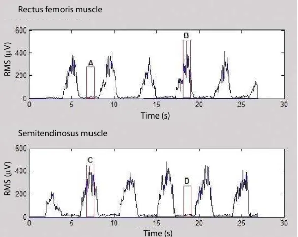
To evaluate cocontraction, Coactivation Index (CaI) was used. his index is expressed by dividing the average RMS value of the antagonist muscle and the average RMS value of this same muscle acting as an agonist. To do so, the crude signal was reiltered (pass band of 20-500Hz and notch 60Hz),
rectiied, and it in a moving window at each 50 ms. When the contraction in which subjects performed their highest extensor and lexor peak torques was identiied, the average RMS value was calculated in the 500 ms period, 250 ms before and 250 ms after the electromyographic signal peak from the agonist muscle. he average RMS value of the antagonist muscle was calculated within this same interval (Figure 1).
Rectus femoris muscle
Semitendinosus muscle
Time (s)
Time (s)
RMS (
µ
V
)
RMS (
µ
V
)
Figure 1. Title: RMS signal of rectus femoris and semitendinosus muscles. Caption: (A) Selection of rectus femoris muscle acting as an antagonist; (B) Selection of rectus femoris muscle acting as an agonist; (C) Selection of semitendinosus muscle acting as an agonist; (D) Selection of semitendinosus muscle acting as an antagonist
he following equations were used:
In which A is the time interval the rectus femoris muscle acted as an antagonist during the peak torque of knee lexors; and B is the time interval when this same muscle acted as an agonist during the peak of torque of knee extensors.
Peak of torque (PT) was used to evaluate muscle strength. PT represents the highest torque value recorded within the ive extension and lexion contractions performed, and it is expressed in Newton meters (Nm). he Matlab software (Mathworks® 7.6.0) was used to analyze the data.
he variables were compared between sitting and supine positions and between paretic and non-paretic hemibodies. To do so, the data were organized and the Kolmogorov-Smirnov test was performed. Upon verifying sample normality, student’s t test for paired samples was applied in the comparisons between positions and between hemibodies. In all cases, a signiicance level of p<0.05 was adopted. he SPSS statistical software, version 18.0, was used. Results were expressed as average ± standard deviation.
RESULTS
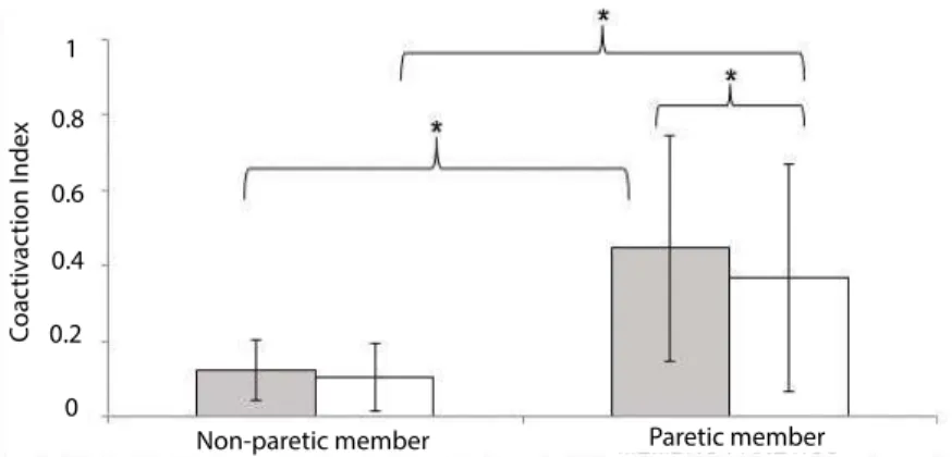
he CaI of the rectus femoris muscle was not shown to difer between positions for both limbs.
In the comparison between limbs in the sitting position, the paretic one was found to have a higher CaI value: 0.10±0.05; 0.28±0.25 (p=0.007), and there was a trend for the same result to be observed in the supine position: 0.11±0.10; 0.23±0.21 (p=0.05), (Figure 2).
he semitendinosus muscle in the paretic limb was found to have a lower CaI in the supine position as compared to the sitting one: 0.36±0.33; 0.44±0.33 (p=0.048). his muscle was also shown to have a higher CaI in the paretic limb as compared to the non-paretic limb for both positions: sitting 0.12±0.08; 0.44±0.33 (p=0.001); supine 0.10±0.09; 0.36±0.33 (p=0.005), as shown in Figure 3.
Extensor and lexor PT was shown to be statistically higher in the non-paretic limb as compared to the paretic one for both positions. In the comparison between positions, the only diference was observed in the lexor PT of the non-paretic limb, in which the sitting position was observed to result in a higher PT as compared to the supine position: 29.29±12.9 Nm; 21.38±8.5Nm (p=0.005), (Figure 4 and 5).
C
oac
tiv
ac
tion I
nde
x
1
0.8
0.6
0.4
0.2
0
Non-paretic member Paretic member
Figure 2. Coactivation Index of rectus femoris muscle. Caption: Grey bar = sitting position; White bar = supine position. *A diference was observed upon comparing paretic and nonparetic limbs in the sitting position (p<0.05)
C
oac
tiv
ac
tion I
nde
x
1
0.8
0.6
0.4
0.2
0
Non-paretic member Paretic member
Non-paretic member Paretic member
P
eak of t
or
que (N.m)
150
120
90
60
30
0
Figure 4. Title: Peak of extensor torque. Caption: Grey bar = sitting position; White bar = supine position. *A diference was observed upon comparing paretic and nonparetic limbs in both positions (p<0.05
P
eak of t
or
que (N.m)
150
120
90
60
30
0
Non-paretic member Paretic member
Figure 5. Title: Peak of lexor torque. Caption: Grey bar = sitting position; White bar = supine position. *A diference was observed upon comparing paretic and nonparetic limbs in both positions, as well as a diference in the comparison between positions for the nonparetic limb (p<0.05)
DISCUSSION
According to the results, the shift from a sitting to a supine position was clearly shown to imply reduced CaI of the semitendinosus muscle in the paretic limb and reduced lexor PT in the non-paretic limb. In the comparison between limbs, higher CaI and lower PT values were observed in the afected limb as compared to the opposite one.
he CaI results from the semitendinosus muscle showed that situations with more intense stretching of this muscle, i.e., in the sitting position, result in higher coactivation. he same was observed by Fleuren et al.3,
who evaluated the electromyographic activity of 19 post-CVA hemiparetic subjects in sitting and supine positions. hey found a higher level of antagonist activation in semitendinosus and rectus femoris muscles when these were further stretched. hat is due to muscle spindles being more sensitive to stretching, because once they are shortened, stretching is faster and more intensely transmitted in spastic muscles8.
stating that the increased antagonist activity of the rectus femoris muscle is more frequently seen in severely impaired patients. However, the subjects in this study were observed to have a moderate level of motor activity according to the Fugl-Meyer Assessment, which might have softened the coactivation of this muscle.
Higher CaI values of knee extensor and lexor muscles are commonly seen in spastic individuals as compared to healthy subjects, as seen by Neckel et al.19 and Hidler et al.20 through EMG and load cell. Regarding the comparison between paretic and non-paretic limbs, Newham and Hsiao21 found no signiicant diferences in twelve post-CVA subjects of both genders evaluated through an isokinetic dynamometer, which difers from the result of this study. his disagreement may be justiied by the fact that the authors recorded the myoelectric activity during isometric contractions, a mode in which the antagonist coactivation is lower in spastic muscles due to the absence of speed6. Another way to justify that is through the fact that the patients studied were in the acute phase (≤ 6 months), a period when CVA sequelae difer from the ones in the chronic phase. In the acute phase there is a predominance of paresis and lack of use of the afected limb, whereas in the chronic period neural adaptations also take place, and these result in muscle hyperactivity and consequent spasticity and cocontraction8.
he PT of the paretic limb was lower than the one in the non-paretic limb, and this result corroborates other studies11,21,22. his is justiied by alterations that take place in the afected hemibody, such as the diminished eferent commands from the corticospinal pathway, failed reciprocal inhibition that leads to excess coactivation of the antagonist muscle, and secondary muscle alterations, such as reduced cross-sectional area coupled with increased noncontractile tissue23,24.
In the comparison between positions, the sitting position was observed to favor higher PT of knee lexor muscles in the unafected limb, such as in other studies that evaluated healthy individuals10,25–27. his fact is justiied by the stretch-shortening principle, once the sitting position allows insertions to be more distant from each other and hamstring muscles to be consequently stretched. his principle states that a concentric muscle action is intensiied when it follows an eccentric action, and that is due to the storage of elastic energy in passive elements, increased cross-bridges, and the muscle spindle strain relex28,29.
Regarding the paretic limb, no diferences were found between positions. Unlike Bohannon11, who
reported higher isometric PT in the sitting position for both hemibodies of twelve acute post-CVA hemiparetic subjects; and Michaelsen et al.5, who reported the same thing for ten hemiparetic subjects in the chronic phase. Such disagreement may be justiied by the fact that these studies conducted their evaluations during isometrics, in which hemiparetic patients achieve a higher PT as compared to the concentric mode6.
In the case of extensor muscles, no PT variations were observed between positions, as there was no inluence from the stretch-shortening cycle, once vastus medialis, vastus lateralis, and vastus intermedius muscles 30, which account for up to 80% of the knee extensor torque, are monoarticular, and therefore sufer less inluence from the hip position10,25,26.
he indings of this study are important, as they provide information to physical therapists and other health care professionals who treat hemiparetic individuals, and also to the very patients with CVA sequelae. As, for example, the semitendinosus muscle in the paretic limb was observed to have reduced coactivation in the supine position and similar peaks of torque among positions, the supine position becomes the most indicated one during strength and motion training of hamstring muscles in this limb. And regarding the extensor muscles, as not diferences were observed in the coactivation intensity or either in the peak of torque among evaluated positions, the adoption of the supine position is also recommended when one performs mobilizations or strength training for this muscle group in hemiparetic patients who have respiratory comorbidities, due to the smaller efort this posture requires.
It is important to point out that these interpretations serve for tasks that speciically involve the discussed muscles, such as passive, assisted, active, or resisted mobilizations of knee lexion and extension. Other rehabilitation techniques are required to treat these patients. he adopted positions during the intervention should consequently vary. Besides that, all the interpretations in this study were obtained through evaluation in isokinetic mode, and that is why caution must be observed when these are used in activities involving isotonic or isometric contractions.
not controlled during the recruitment of the sample. However, the authors established as an inclusion criterion a predetermined range of scores in the bottom extremity domain of Fugl-Meyer Assessment, which represents a function level that is similar among subjects, regardless of physical activity levels or whether subjects were undergoing physiotherapy treatment.
CONCLUSION
he supine position was found to result in reduced Coactivation Index of the semitendinosus muscle in the paretic limb. In turn, the sitting position favored a higher lexor Peak of Torque in the non-paretic limb. Shifting positions has not inluenced the Coactivation Index of rectus femoris or the Peak of Torque of extensor muscles in both limbs. Considering that, it is clear that the supine position seems to be recommended when moving and training the strength of knee extensor and lexor muscles in chronic hemiparetic patients after cerebrovascular accidents.
REFERENCES
1. Rosa MCN, Marques A, Demain S, Metcalf CD. Lower limb co-contraction during walking in subjects with stroke: A systematic review. J Electromyogr Kinesiol. 2014;24(1):1-10. 2. Busse ME, Wiles CM, van Deursen RWM. Co-activation:
its association with weakness and speciic neurological pathology. J Neuroeng Rehabil. 2006;3:26.
3. Fleuren JF, Nederhand MJ, Hermens HJ. Inluence of posture and muscle length on stretch relex activity in poststroke patients with spasticity. Arch Phys Med Rehabil. 2006;87(7):981-8.
4. Daly JJ, Roenigk K, Cheng R, Ruf RL. Abnormal leg muscle latencies and relationship to dyscoordination and walking disability after stroke. Rehabil Res Pract. 2011;2011:313980. doi.org/10.1155/2011/313980
5. Michaelsen S, Ovando A, Bortolotti A, Bandini B. Strength deficit of knee flexors is dependent on hip position in adults with chronic hemiparesis. Braz J Phys Ther. 2013;17(1):86-91.
6. Clark DJ, Condlife EG, Patten C. Activation impairment alters muscle torque-velocity in the knee extensors of persons with post-stroke hemiparesis. Clin Neurophysiol. 2006;117(10):2328-37.
7. Fleuren JFM, Snoek GJ, Voerman GE, Hermens HJ. Muscle activation patterns of knee lexors and extensors during passive and active movement of the spastic lower limb in chronic stroke patients. J Electromyogr Kinesiol. 2009;19(5):e301-e10.
8. Gracies J-M. Pathophysiology of spastic paresis. II: Emergence of muscle overactivity. Muscle Nerve. 2005;31(5):552-71. 9. Dvir Z. Isokinetics: Muscle testing, interpretation, and clinical
applications. Churchill Livingstone; 2004.
10. Guex K, Gojanovic B, Millet GP. Inluence of hip-lexion angle on hamstrings isokinetic activity in sprinters. J Athl Train. 2012;47(4):390-5.
11. Bohannon RW. Decreased isometric knee lexion torque with hip extension in hemiparetic patients. Phys Ther. 1986;66(4):521-3.
12. Maki T, Quagliato E, Cacho E, Paz L, Nascimento N, M I. Estudo de coniabilidade da aplicação da escala de Fugl-Meyer no Brasil. Rev Bras Fisioter. 2006;10(2):177-83. 13. Fugl-Meyer A, Jääskö L, Leyman I, Steglind S. The post-stroke
hemiplegic patient: a method for evaluation of physical performance. Scand J Rehabil Med. 1974;7:13-37.
14. Lourenço RA, Veras RP. Miniexame do Estado Mental: características psicométricas em idosos ambulatoriais. Rev Saúde Pública. 2006;40(4):712-9.
15. Folstein M, Folstein S, McHugh P. “Mini-Mental State”: A practical method for grading the cognitive state of patients for the clinician. J Psychiat Res. 1975;12:189-98.
16. SENIAM - Surface Electromyography for the Non-Invasive Assessment of Muscle. http://www.seniam.org/. Accesso em 23/10/2013.
17. Horstman A, Gerrits K, Beltman M, Janssen T, Konijnenbelt M, de Haan A. Muscle function of knee extensors and lexors after stroke is selectively impaired at shorter muscle lengths. J Rehabil Med. 2009;41(5):317-21.
18. He J. Stretch relex sensitivity: efects of postural and muscle length changes. IEEE Trans Rehabil Eng. 1998;6(2):182-9. 19. Neckel N, Pelliccio M, Nichols D, Hidler J. Quantiication of
functional weakness and abnormal synergy patterns in the lower limb of individuals with chronic stroke. J Neuroeng Rehabil. 2006;3(17):1-11.
20. Hidler JM, Carroll M, Federovich EH. Strength and coordination in the paretic leg of individuals following acute stroke. IEEE Trans Neural Syst Rehabil Eng. 2007;15(4):526-34.
21. Newham DJ, Hsiao SF. Knee muscle isometric strength, voluntary activation and antagonist co-contraction in the irst six months after stroke. Disabil Rehabil. 2001;23(9):379-86. 22. Chow JW, Stokic DS. Force control of quadriceps muscle
is bilaterally impaired in subacute stroke. J Appl Physiol. 2011;111(5):1290-5.
23. Knutsson E, Mårtensson A, Gransberg L. Inluences of muscle stretch relexes on voluntary, velocity-controlled movements in spastic paraparesis. Brain. 1997;120(Pt 9):1621-33.
24. Ramsay JW, Barrance PJ, Buchanan TS, Higginson JS. Paretic muscle atrophy and non-contractile tissue content in individual muscles of the post-stroke lower extremity. J Biomech. 2011;44(16):2741-6.
25. Bohannon RW, Gajdosik RL, LeVeau BF. Isokinetic knee lexion and extension torque in the upright sitting and semireclined sitting positions. Phys Ther. 1986;66(7):1083-6. 26. Worrell TW, Perrin DH, Denegar CR. The inluence of hip
reciprocal muscle group ratio values. J Orthop Sports Phys Ther. 1989;11(3):104-7.
27. Deighan M, Serpell B, Bitcon M, Croix M. Knee joint strength ratios and efects of hip position in rugby players. J Strength Cond Res. 2012;26(7):1959-66.
28. Komi P V. Stretch-shortening cycle: A powerful model to study normal and fatigued muscle. J Biomech. 2000;33(10):1197-206.
29. Minozzo F, Lira C. Muscle residual force enhancement: A brief review. Clinics. 2013;68(2):269-74.