Copyright © by Sociedade Brasileira de Pediatria
Abstract
Objective: To assess the frequency and types of limb abnormalities observed among patients with trisomy 18, or Edwards syndrome (ES).
Method: The sample consisted of consecutive patients evaluated by a clinical genetics service in the period from 1975 to 2008. The results of the cytogenetic analysis, as well as the clinical data were retrieved from the medical records, with special attention to limb abnormalities indings. All the karyotype analysis was performed at the same laboratory.
Results: During the study period, 50 patients were identiied, 33 (66%) of them females, with ages at the irst evaluation ranging from 1 day to 16 years (median 14 days). The single lineage with free trisomy 18 was the most frequent chromosomal disorder (90%). Mosaicism was observed in 10% of the cases. Clenched ist with overlapping ingers was the predominant anomaly of the upper limbs (70%). Other common disorders included the single palmar crease (42%) and hypoplastic nails (36%). Radial abnormalities were found in 11 patients (22%). As for the lower limbs, hypoplastic nails were the most common abnormality (58%), followed by the rocker bottom foot with prominent calcaneus (50%). One patient had unilateral ectrodactyly as well.
Conclusions: Despite the classical description, limb anomalies can be much variable in ES. Some patients may show unusual abnormalities, such as radial defects and ectrodactyly. These indings are extremely important for the clinical suspicion and early identiication of patients with ES.
J Pediatr (Rio J). 2012;88(5):401-5: Chromosomes, human, pair 18, trisomy, chromosome aberrations, extremities.
401
O
riginala
rticleLimb abnormalities on trisomy 18:
evidence for early diagnosis
Rafael F. M. Rosa,1 Rosana Cardoso Manique Rosa,2 Marina Boff Lorenzen,3 Paulo R. G. Zen,4 Ceres A. V. de Oliveira,5 Carla Graziadio,6 Giorgio A. Paskulin7
1. PhD. Programa de Pós-Graduação em Patologia, Universidade Federal de Ciências da Saúde de Porto Alegre (UFCSPA), Porto Alegre, RS, Brazil. Invited professor, Programa de Pós-Graduação em Patologia, UFCSPA, Porto Alegre, RS, Brazil.
2. MSc. Programa de Pós-Graduação em Patologia, UFCSPA, Porto Alegre, RS, Brazil. 3. Medical student, UFCSPA, Porto Alegre, RS, Brazil.
4. PhD. Programa de Pós-Graduação em Patologia, UFCSPA, Porto Alegre, RS, Brazil. Professor, Disciplina de Genética Clínica, Programa de Pós-Graduação em Patologia, UFCSPA, Porto Alegre, RS, Brazil.
5. MSc. Engenharia de Produção, Universidade Federal do Rio Grande do Sul (UFRGS), Porto Alegre, RS, Brazil.
6. PhD candidate. Programa de Pós-Graduação em Patologia, UFCSPA, Porto Alegre, RS, Brazil. Professor, Disciplina de Genética Clínica, UFCSPA, Porto Alegre, RS, Brazil.
7. PhD. Genética Molecular, Programa de Pós-Graduação, UFRGS, Porto Alegre, RS, Brazil. Professor, Disciplina de Genética Clínica, UFCSPA, Porto Alegre, RS, Brazil. Coordinator, Programa de Pós-Graduação em Patologia, UFCSPA, Porto Alegre, RS, Brazil.
No conflicts of interest declared concerning the publication of this article.
Financial support: Coordenação de Aperfeiçoamento de Pessoal de Nível Superior (CAPES) and Programa de Iniciação Científica da Universidade Federal de Ciências da Saúde de Porto Alegre (PIC-UFCSPA).
Suggested citation: Rosa RF, Rosa RC, Lorenzen MB, Zen PR, de Oliveira CA, Graziadio C, et al. Limb abnormalities on trisomy 18: evidence for early diagnosis. J Pediatr (Rio J). 2012;88(5):401-5.
Manuscript submitted Apr 21 2012, accepted for publication May 28 2012. http://dx.doi.org/10.2223/JPED.2212
Introduction
Trisomy 18, or Edwards syndrome (ES), is considered a relatively common chromosomal disorder, which is observed
in one of every 3,600-8,500 live births. It is characterized
by a broad clinical picture that frequently includes multiple
malformations, and an ominous prognosis.1 To date, more
than 130 abnormalities have already been described, but none of them is present in 100% of the patients. Limb
Thus, the present study aimed to assess the frequency and types of limb abnormalities observed among patients with ES.
Methods
The sample was constituted of consecutive patients evaluated in genetic clinic of a reference hospital in the south of the country, in the period from 1975 to 2008. The results of the cytogenetic analysis, as well as clinical data, were retrieved from the medical charts, with special focus on limb abnormalities. The study was approved by
the Hospital Research Ethics Committee.
All the patients had the karyotype analysis performed at the Cytogenetic Laboratory of the Universidade Federal de Ciências da Saúde de Porto Alegre. Briely, this test
includes the performance of a cell culture with lymphocytes
stimulation using phytohemagglutinin, hypotonic shock, cell ixation with Carnoy solution, slides preparation and G banding staining with trypsin and Giemsa. All the slides analysis was done using an Axioskop Zeiss microscope, by counting a mean of 26 metaphasic plaques in each case (the number ranging from 6 to 40).
Data analysis was performed with the PEPI (Programs for EPIdemiologists) software (version 4.0) using the two-tailed Fisher’s exact test for comparison of frequencies. P values < 0.05 were considered signiicant.
Results
In the evaluation period, we identiied 50 patients with ES, 33 (66%) females. The patients’ ages at the irst evaluation ranged from 1 day to 16 years (74% under one month of age, median 14 days). Regarding the karyotype indings, the most common disorder was the single lineage with free trisomy 18, which was observed in 45 patients (90%). Two (4.4%) of these 45 patients also had trisomy X, and one (2.2%) an associated der (13;14). Mosaicism was identiied in ive patients (10%), with almost all of them
presenting one lineage with free trisomy 18 associated with
a normal one. In one case only there was a third lineage
with double aneuploidy: free trisomy 18 associated with the XXY constitution.
The most frequent abnormality observed in the upper
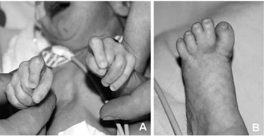
limbs in our sample (n = 50) was the clenched ists with overlapping ingers (campodactyly or contractures) (70%). Radial abnormalities were found in 11 patients (22%). As for the lower limbs, hypoplastic nails were the most common inding (58%), followed by the rocker-bottom foot with prominent calcaneus (50%). One of the patients had unilateral ectrodactyly (lobster claw foot) (Table 1). Figure 1 shows the clenched ists with predominant overlapping of the second over the third inger (in A), and foot with short hallux, hypoplastic nails and partial syndactyly of the second and third toes (in B).
To assess whether the presence of mosaicism could
inluence the frequency of limb abnormalities, we compared the main indings between patients with and without mosaicism. The most frequent indings were the clenched ists with overlapping ingers, hypoplasia of the toenails, and rocker-bottom foot with prominent calcaneus. There were no signiicant statistical differences between the groups, but a trend towards signiicance (p = 0.0502) was observed for the last inding.
Discussion
In spite of the classical description, limb abnormalities found in ES are highly variable. In our sample, only about one fourth (28%) of the patients had more than one of the
major limb abnormalities simultaneously, seen in more than
50% of the cases (Table 1). Clenched ists with overlapping ingers (usually the second over the third and the ifth over the fourth inger) are the main abnormality described, and
the one that usually leads to the clinical suspicion of ES (Table 1).3-10 This inding gets the maximum number of
points (out of 5) in the score of Marion et al.,3 a tool that
was developed with the aim of improving the identiication
of patients with ES in the neonatal period.
In our study, only 70% of the patients had clenched ists with overlapping ingers. This abnormality has also been reported in the literature as occurring from 58 to 96% of
the cases (usually over 90%) (Table 1).4-10 Our analysis,
however, shows that a considerable number of patients with
ES do not present this inding.4-10 Furthermore, this feature
is neither characteristic nor pathognomonic, as it has been reported in other conditions that comprise the differential
diagnosis of ES, such as trisomy 13 (Patau syndrome).2
Other common indings in this study, which have also been reported in the literature, are the single palmar crease (18-61%) and hypoplastic nails (13-96%). Nevertheless, the high variability in the frequency of these indings reported in
the studies should be highlighted.4-10 The lack of the distal
digital crease also seems to be frequent (observed in 28%
of our patients), but given little importance in the evaluation of the patients. The same applies to the presence of an
arches pattern on the digital pulps of six or more ingers
(with reported frequencies usually higher than 90%).4-10
To evaluate this feature, the use of stamp ink and paper, or even a magnifying glass may be necessary. In our study, this feature could not be evaluated. Both changes, the lack
of the distal phalangeal crease and the arch pattern on the digital pulps, represent dermatoglyphic disturbances (which
also include the sole palmar crease) and get the maximum
number of points in the score of Marion et al.3
Some patients can also present unusual abnormalities,
such as radial defects and ectrodactyly. Radial alterations
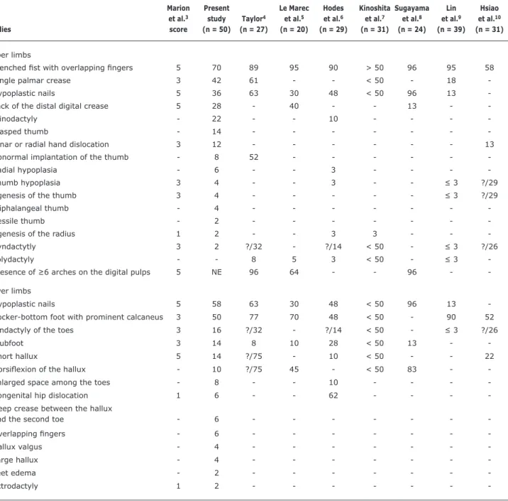
Table 1 - Abnormalities of the upper and lower limbs observed in the sample in comparison with the literature (%)
? = not determined; NE = not evaluated.
Marion Present Le Marec Hodes Kinoshita Sugayama Lin Hsiao
et al.3 study Taylor4 et al.5 et al.6 et al.7 et al.8 et al.9 et al.10 Studies score (n = 50) (n = 27) (n = 20) (n = 29) (n = 31) (n = 24) (n = 39) (n = 31)
Upper limbs
Clenched ist with overlapping ingers 5 70 89 95 90 > 50 96 95 58
Single palmar crease 3 42 61 - - < 50 - 18
Hypoplastic nails 5 36 63 30 48 < 50 96 13
Lack of the distal digital crease 5 28 - 40 - - 13 -
Clinodactyly - 22 - - 10 - - -
Clasped thumb - 14 - - -
Ulnar or radial hand dislocation 3 12 - - - 13
Abnormal implantation of the thumb - 8 52 - - -
Radial hypoplasia - 6 - - 3 - - -
Thumb hypoplasia 3 4 - - 3 - - ≤ 3 ?/29
Agenesis of the thumb 3 4 - - - ≤ 3 ?/29
Triphalangeal thumb - 4 - - -
Sessile thumb - 2 - - -
Agenesis of the radius 1 2 - - 3 3 - -
Syndactytly 3 2 ?/32 - ?/14 < 50 - ≤ 3 ?/26
Polydactyly - - 8 5 3 < 50 - ≤ 3
Presence of ≥6 arches on the digital pulps 5 NE 96 64 - - 96 -
-Lower limbs
Hypoplastic nails 5 58 63 30 48 < 50 96 13
Rocker-bottom foot with prominent calcaneus 3 50 77 70 48 < 50 - 90 52
Sindactyly of the toes 3 16 ?/32 - ?/14 < 50 - ≤ 3 ?/26
Clubfoot 3 14 8 10 28 < 50 13 -
Short hallux 5 14 ?/75 - 10 < 50 - - 22
Dorsilexion of the hallux - 10 ?/75 45 - < 50 83 -
Enlarged space among the toes - 8 - - 10 - - -
Congenital hip dislocation 1 6 - - 62 - - -
Deep crease between the hallux
and the second toe - 6 - - -
Overlapping ingers - 6 - - -
Hallux valgus - 4 - - -
Large hallux - 4 - - -
Feet edema - 2 - - -
Ectrodactyly 1 2 - - -
-Other indings in our study, which have also been reported
in other case series, were the hypoplasia or agenesis of the thumb and the radius (Table 1).4-11 One of our patients with radial abnormalities has been described in detail by
Zen et al.12 Polydactitly of the ingers, an unusual inding,
was not observed in our sample (Table 1).4-10
For the lower limbs, nails hypoplasia and rocker
bottom feet with prominent calcaneus represent important
indings.4-10 They get 5 and 3 points, respectively, in the
score of Marion et al.3 Other relatively common indings are
the syndactyly of the toes (particularly of the second and
third toes), clubfoot, and hallux abnormalities (short and
dorsilexed hallux). However, as shown in Table 1, there is a high variability in the frequency of these indings in the
literature.4-10 Congenital hip dislocation was observed in 6% of our patients, but it may have been underdiagnosed, as
Hodes at al. have found it in 62% of individuals with ES.6 In
those reports, ectrodactyly is considered a very rare inding,
which has been described in a few case reports only (our
study is the irst case series to report such inding) (Table 1). In those reports, as in our patient, ectrodactyly was observed
Figure 1 - Limb anomalies observed among the patients
In our review of the literature, we did not ind any comparison in the limb indings between patients with ES with and without mosaicism. Although our sample is the
largest reported in the literature (Table 1), we believe that the small number of patients with mosaicism (n = 5) may
have limited our analysis. Anyway, we did not observe signiicant differences in the frequency of the main limb
abnormalities observed.
In a clinical pathological study, Ramirez-Castro &
Bersu15 reported the limb anatomic variations observed
in 8 children with ES. The authors observed an inluence on the development of the pre-axial component of the upper limbs and a trend to longitudinal deiciency defects.
The anomalies reported included muscle defects along the radial margin of the forearm and hand, agenesis
of a deinitive musculocutaneous nerve in all the limbs,
and reduction of the radial artery. They suggested that
the abnormalities observed could be explained by certain
pathogenic mechanisms, such as a defect in the development of the peripheral nerve and tissue necrosis.15 Therefore, the recognition and understanding of the variability of limb
abnormalities are essential for the early identiication of
the patients with ES. This is of fundamental importance for the pediatricians (particularly, the neonatologists) who
are usually the irst health professionals to evaluate these patients. The deinition of the diagnosis has important
implications in the management and genetic counseling of
the patients and their families. Recurrence of the trisomy 18 is considered to be rare. Nevertheless, when the
trisomy 18 occurs as a consequence of translocations, a chromosomal study of the parents is indicated, as one of them can carry a balanced chromosome rearrangement,
which poses a higher risk for the future offspring.
Acknowledgements
We thank the Brazilian Federal Agency for Support and Evaluation of Graduate Education (Coordenação de Aperfeiçoamento de Pessoal de Nível Superior-CAPES) for
the scholarship received.
References
1. Rosa RF, Rosa RC, Lorenzen MB, de Moraes FN, Graziadio C, Zen PR, et al. Trisomy 18: experience of a reference hospital from the south of Brazil. Am J Med Genet A. 2011;155A:1529-35. 2. Jones KL. Smith’s recognizable patterns of human malformation.
6th ed. Philadelphia, PA: Elsevier Saunders; 2006.
3. Marion RW, Chitayat D, Hutcheon RG, Neidich JA, Zackai EH, Singer LP, et al. Trisomy 18 score: a rapid, reliable diagnostic test for trisomy 18. J Pediatr. 1988;113:45-8.
4. Taylor AI. Autosomal trisomy syndromes: a detailed study of 27 cases of Edwards’ syndrome and 27 cases of Patau’s syndrome.
J Med Genet. 1968;5:227-52.
5. Le Marec BM, Lair JC, Kérisit J, Le Mée F, Sénécal J. 20 cases of trisomy 18. Sex-ratio in relation to age of the mother. Ann Pediatr (Paris). 1977;24:125-36.
Correspondence: Giorgio Adriano Paskulin
Rua Sarmento Leite, 245/403, Centro CEP 90050-170 - Porto Alegre, RS - Brazil Tel.: +55 (51) 3303.8771
Fax: +55 (51) 3303.8810 E-mail: [email protected] 7. Kinoshita M, Nakamura Y, Nakano R, Morimatsu M, Fukuda
S, Nishimi Y, et al. Thirty-one autopsy cases of trisomy 18: clinical features and pathological indings. Pediatr Pathol. 1989;9:445-57.
8. Sugayama SM, Kim CA, Utagawa CY, Albano LM, Bertola DR, Koiffmann CP, et al. Estudo genético-clínico de 24 pacientes com trissomia 18 (síndrome de Edwards). Pediatria (São Paulo). 1999;21:133-43.
9. Lin HY, Lin SP, Chen YJ, Hung HY, Kao HA, Hsu CH, et al. Clinical
characteristics and survival of trisomy 18 in a medical center in
Taipei, 1988-2004. Am J Med Genet A. 2006;140:945-51.
10. Hsiao CC, Tsao LY, Chen HN, Chiu HY, Chang WC. Changing clinical presentations and survival pattern in trisomy 18. Pediatr Neonatol. 2009;50:147-51.
11. Sugayama SM, Kim CA, Barba MF, Albano LM, Bertola DR, Utagawa CY, et al. Síndrome de Edwards com aplasia radial – relato de dois casos e revisão das anomalias esqueléticas na síndrome. Radiol Bras. 2000;33:241-7.
12. Zen PR, Rosa RF, Rosa RC, Dale Mulle L, Graziadio C, Paskulin GA. Unusual clinical presentations of patients with Patau and Edwards syndromes: a diagnostic challenge? Rev Paul Pediatr. 2008;26:295-9.
13. Rogers RC. Trisomy 18 with unilateral atypical ectrodactyly. Am
J Med Genet. 1994;49:125-7.
14. Christianson AL, Nelson MM. Four cases of trisomy 18 syndrome with
limb reduction malformations. J Med Genet. 1984;21:293-7.
15. Ramirez-Castro JL, Bersu ET. Anatomical analysis of the
developmental effects of aneuploidyin man – the 18-trisomy syndrome: II. Anomalies of the upper and lower limbs. Am J Med