Sleep abnormalities and memory alterations in
obstructive sleep apnea
ORIGINAL ARTICLE
Alterações do sono e da memória na apneia obstrutiva do sono
Camila Andrade Mendes Medeiros1, Pedro Felipe Carvalhedo de Bruin1, Luciane Ponte e Silva1, Wesley de Menezes Coutinho1, Veralice Meireles Sales de Bruin1
Study carried out at Department of Clinical Medicine, Federal University of Ceará.
1 Department of Clinical Medicine, Federal University of Ceará, Brazil.
Corresponding author: Veralice M. S. de Bruin - Departamento de Medicina, Universidade Federal do Ceará. Rua Prof. Costa Mendes 1608 - 4º Andar - CEP: 60430 040 - Fortaleza, Ceará, Brasil - Telephone: 55 (85) 32421681 - Fax: 55 (85) 32615540 - E-mail: veralice@superig.com.br
Received: January 21, 2011; Accepted: August 22, 2011.
INTRODUCTION
Sleep is recognized as a physiologic state that performs an essential restorative function and facilitates learning and memory consolidation(1). Obstructive sleep apnea (OSA)
is a common disorder which is associated with significant comorbidities, including obesity, hypertension, increased risk for vascular disease, depression and excessive daytime sleepiness. Furthermore, OSA is associated with a variable spectrum of sleep abnormalities and has been previously connected with memory and learning impairment(2-4).
Pre-viously, it has been shown that declarative and procedural memory processes are impaired in patients with OSA compared to healthy subjects(5). However, the
mecha-nisms underlying these cognitive and psychological altera-tions are still unknown. Memory is a multifaceted task and different sets of memory tests can yield different conclu-sions. Also, specific sleep alterations, such as change of sleep architecture, arousals and oxygen desaturation can exert different influence in cognitive function.
Structural cerebral changes reported in subjects with OSA support the connection between sleep abnor-malities and cognitive dysfunction in those patients(6,7).
For instance, alterations of regional cerebral blood flow in bilateral parahippocampal gyri, right lingual gyrus, peri-central gyrus, and cuneus, found in patients with severe OSA, may partly explain deficits in memory, spatial learn-ing, executive function, and attention(8). Recently, a study
ABSTRACT
Objectives: Obstructive sleep apnea (OSA) is associated with a variable spectrum of sleep abnormalities and has been connected with memory impairment. The aim of this study was to evaluate the associations between OSA, memory alterations and sleep struc-ture abnormalities. Methods: Polysomnography was performed in 20 consecutive patients (12 male, 57.9±5.8 years) with moderate/ severe OSA (AHI 35.8±16.7). Daytime somnolence (Epworth Sle-epiness Scale, ESS), state of alertness (Karolinska SleSle-epiness Sca-le), subjective sleep quality (Pittsburgh Sleep Quality Index, PSQI) and depressive symptoms (Beck Depression Inventory, BDI) were evaluated. Patients were tested before all night polysomnography and a retrieval test was performed in the following morning. De-clarative memory was assessed by Verbal Paired Associates from the Wechsler Memory Scale, emotional memory by the exposure to emotional and non-emotional images and procedural memory by the maze test. Results: Excessive daytime sleepiness (ESS>10, 55%) and impaired sleep (PSQI>5, 40%) were found. Patients with OSA presented greater neck circumference (p<0.005). Procedural memory, as evaluated by the maze test, showed worse retrieval in OSA patients and this was maintained after controlling for age, body mass index and BDI. In subjects with moderate/severe OSA, stage 3 sleep was correlated to the performance in the procedural memory test. Conclusion: We show that procedural memory is altered in OSA patients as compared to controls and this alteration is related to stage 3 of non-rapid eye movement sleep. We confirm that recall memory tests after one night sleep are efficacious to exa-mine memory abnormalities in OSA patients.
Keywords: memory, polysomnography, sleep, sleep apnea
syndromes.
RESUMO
Objetivos: Apneia Obstrutiva do Sono (AOS) associa-se com um espectro de alterações do sono e possível redução da memória. O objetivo desse estudo foi avaliar a relação entre a AOS, alterações da memória e anormalidades da estrutura do sono. Métodos: Vinte pacientes (12 homens, idade 57,9±5,8 anos) foram estudados com o uso de polissonografia e avaliados quanto à sonolência diurna (Escala de Sonolência de Epworth, ESE), estado de alerta (Escala de Sonolência de Karolinska), qualidade subjetiva do sono (Índice de Qualidade de Sono de Pittsburgh, IQSP) e sintomas depressivos (Inventário de Depressão de Beck, IDB). Casos com AOS modera-da/grave (IAH 35,8±16,7) foram submetidos a testes de memória antes da polissonografia de noite inteira e na manhã seguinte, um teste de recuperação foi realizado. Memória declarativa (Pares de Palavras Associadas), memória emocional (Exposição a imagens emocional e não-emocional) e memória de procedimento (Teste do
Descritores: memória, polissonografia, sono, síndromes da ap-néia do sono.
about the effects of sleep-wake regulation on the cerebral mechanisms, which focused on the locus ceruleus and the suprachiasmatic nucleus, has proved the direct influ-ence of the homeostatic and circadian control on neural activity(9).
It should be noted that among OSA patients, only 30% of cases present memory deficits in an initial assess-ment(5). This might be a setback for the follow-up
evalua-tion of therapeutic efficacy, as it has been noticed that the best therapeutic results can be checked when patients ex-perience changes in cognitive function at baseline. Benefi-cial effects of positive pressure ventilation on learning and memory in patients with OSA have been shown(10).
How-ever, it is unclear whether the cases that recover are those with cognition similar to controls or those with basal levels different from controls. Ultradian variations of cognitive performance may also occur. Considering such evidence, it is possible that an assessment of memory functioning before and after nocturnal sleep in OSA patients and con-trols is more appropriate to compare the performance of memory and can generate more information than ratings points into a certain time of day.
The main objectives of this study were to evaluate the effects of OSA on memory retrieval after one night sleep and to determine the contribution of sleep abnor-malities to memory alterations.
METHODS
Study Design and participants
This was a case-control study of 20 consecutive patients of both genders referred for polysomnography which were later diagnosed as having moderate/severe OSA (ap-nea hypop(ap-nea index, AHI>15) and 10 healthy subjects, controlled for age and gender. Inclusion criteria were age between 50 and 75 years and the diagnosis of moderate/ severe OSA. The diagnosis of sleep apnea was confirmed by two sleep specialists (MD, respiratory physician) in the sleep laboratory. Exclusion criteria were cancer, severe lung, hepatic or renal diseases, previous use of Continu-ous Positive Airway Pressure (CPAP) and unwillingness to participate in the study. All subjects were free from any medication that might affect sleep or cognition and did
not consume alcohol or caffeine ≥ 24 h prior to or dur -ing the study. Asymptomatic individuals with low risk for OSA as assessed by the Berlin questionnaire(11), recruited
from the community, were studied as controls. The pro-tocol was approved by the local research ethics commit-tee and subjects gave informed consent (CEP/HUWC 002.02.07).
Procedures
A purpose-built questionnaire was used to assess habits and comorbidities, such as type 2 diabetes and systemic arterial hypertension. Demographic and anthropometric data including the hip-waist (cm), neck circumference (cm) and body mass index (BMI) were collected. Body mass in-dex was calculated as the ratio between weight (Kg) and squared height (m2).
Daytime somnolence was evaluated by the Ep-worth Sleepiness Scale (ESS), a questionnaire containing eight items that ask for expectation of dozing in eight hypothetical situations. Epworth Sleepiness Scale score greater than 10 indicates excessive daytime somnolence(12).
State of alertness was evaluated by the Karolinska Sleepi-ness Scale(13). Subjective sleep quality was evaluated by the
Pittsburgh Sleep Quality Index (PSQI)(14). Pittsburgh Sleep
Quality Index has seven components, each one dealing with a major aspect of sleep: 1) subjective quality of sleep; 2) sleep onset latency; 3) sleep duration; 4) sleep efficien-cy; 5) presence of sleep disturbances; 6) use of hypnotic-sedative medication; and 7) presence of daytime distur-bances, as an indication of daytime alertness. Individuals with total PSQI score of six or more were considered poor sleepers(14). Depressive symptoms were evaluated by
the Beck Depression Inventory (BDI)(15). Control subjects
were assessed by the Berlin questionnaire(11). Measures of
sleepiness sleep quality and excessive daytime sleepiness were taken regarding the previous 30 days.
Full polysomnography was performed in all patients with recordings from 22:00 to 06:00h. In addition to ques-tionnaires, memory tests were assessed from 7:00 to 8:00 PM prior to sleep (learning session) and at 06:30 to 07:30 AM after sleep (recall session). Declarative memory was assessed by Verbal Paired Associates (VPA) test from the Wechsler Memory Scale(16). The VPA is used instruments
for assessing explicit episodic memory. It consist of four pairs of related and four pairs of unrelated words across three study test trials, and a 30-min delayed recall test(17).
The emotional memory was evaluated by the exposure to emotional and non-emotional images. During a first session, participants viewed 15 pictures with neutral and emotional content. Thirty minutes after the initial expo-sure, 45 images were shown, among which 15 had already been presented (first exposure) and the patient indicated which pictures had been seen. After polysomnography, a new exhibition of 45 images were presented, 15 of these images corresponded to those shown in the first exposure and the other 30 corresponded to previously unseen im-ages and patient files which indicated he recognized pre-viously viewed and new pictures. All images are part of the International Affective Picture System(18). Procedural
memory was evaluated using the maze test, part of the Wechsler Intelligence Scale. The Maze Test involves the ability of planning and visuo-spatial memory(16).
Polysomnography recording
Polysomnography was performed according to a standard clinical protocol (ALICE V, Respironics®). Monitored
evaluated were AHI, minimum oxygen saturation (SpO2 min), sleep latency, sleep efficiency, rapid eye movement (REM) sleep latency, amount of REM sleep (% of total sleep time), amount of non-rapid eye movement (NREM) sleep (% of total sleep time) and number of arousals. Hypopnea was defined as a 50% decrease in the sum of thoracic movements lasting 10 seconds followed by a de-creased oxygen saturation of at least 4%. As recordings may be misleading in evaluating patients who have a fall in baseline oxygen saturation (SpO2) during sleep, visual scoring was performed by a trained polysomnographer in all cases. A board certified sleep specialist, blind to diag-nosis validated each individual respiratory event and man-ually made required changes to recorded data. Cases were
classified as having moderate/severe OSA (AHI≥15) or
control group. Intake of sedative medication was not al-lowed within 48 hours of the investigation. Assessment of subjective overnight sleep quality was taken from patient and care giver.
Statistical Analysis
Data are expressed as means ± standard deviation (SD) values. Patients were grouped as having or not OSA. Data were examined for normality using the Shapiro-Wilk and for homogeneity of variances with the Levene test. The Fisher`s exact test, for categorical variables, Student’s t test for continuous variables, and Mann-Whitney for non-continuous variables were performed for between groups comparison. Repeated measures multivariate analysis was used to compare memory tests before and after sleep. Pearson correlation test was used to compare delta values of memory performance to structural sleep abnormali-ties: delta values of memory performance were obtained subtracting results of morning performance from results of the previous night. Statistical Package for the Social Sciences (SPSS- Norusis, 1993) software for Windows was used for analysis. The level of significance was set at
p<0.05.
RESULTS
Twentypatients (12 male) with moderate/severe OSA (AHI 35.8±16.7), aged between 50 and 68 (mean age 57.9±5.8 years) were studied. Excessive daytime sleepiness (ESS>10) was present in 11 (55%) and impaired sleep (PSQI>5) in eight (40%) of all OSA cases. Table 1 depicts clinical char-acteristics of patients with OSA and controls. Age, BMI, hypertension, diabetes, excessive daytime sleepiness were similar in both groups. Patients with OSA presented greater neck circumference (p=0.000). Sleep quality as assessed by the PSQI tended to be worse in OSA patients (p=0.07). BDI scores and the Karolinska sleepiness scale were not different between OSA and control subjects. Obstructive sleep apnea patients presented variable polysomnographic values that are described as follows: sleep latency (Range 2.5-42.0 min; mean 12.8 SD 10.6), REM latency (Range 45-207.5 min; mean 93.9 SD 50.3), REM sleep percent of total sleep time (Range 5.8-32.5; mean 20.1 SD 6.2), N3 sleep percent of total sleep time (Range 0-17.4; mean 7.4 SD 5.6), sleep efficiency (45.1-100.0;
mean 87.7 SD 12.8), AHI (Range 16.7-73.2; mean 35.8 SD 16.7), arousal index (Range 6.4-53.2; mean 27.7 SD 12.1), Minimal SpO2 (Range 46-90; mean 72.3 SD 14.3).
Table 1. Demographical and clinical characteristics of patients with moderate/severe obstructive sleep apnea and control subjects.
Variables Controls N=10
AHI≥15
N=20 p value
Gender (Male/Female) 3/7 12/8 b 0.24
Age (y) mean (SD) 56.4±6.3 57.9±5.82 a 0.51
BMI 27.6±4.0 29.8±5.21 a 0.25
Neck circumference (cm) 37.4±2.6 43.3±3.58 a 0.000**
Hypertension 6 (60%) 11 (55%) b 1.00
Diabetes 2 (20%) 5 (25%) b 1.00
PSQI 4.6±2.6 7.0±3.85 c 0.07
ESS 8.1±4.1 10.9±6.60 c 0.28
Karolinska Scale 5.3±1.56 5.0±1.74 c 0.55
Beck Depression Inventory 3.1±4.28 6.0±5.78 c 0.10
Abbreviations: BMI= Body Mass Index; RLS=Restless Legs Syn-drome; PSQI= Pittsburgh Sleep Quality Index; ESS= Epworth Sleepi-ness Scale. a Student’s test; b Fisher`s exact test; c Mann-Whitney.
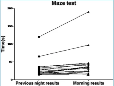
Procedural memory as evaluated by the maze test showed worse retrieval in OSA patients (Table 2) which re-mained significant after controlling for age, BMI and Beck depression scores (p=0.2). In patients with moderate/se-vere OSA, N3 sleep was correlated to delta results of the maze test (r=0.46, p=0.03). Results of the maze test before and after polysomnography are shown in Figures 1 and 2.
DISCUSSION
This study comparing memory retrieval after one night sleep shows that procedural memory, as evaluated by the maze test, is significantly impaired in moderate/se-vere OSA patients and these alterations are correlated with N3 sleep abnormalities. These findings corroborate the idea that healthy sleep fosters new memory consoli-dation. Previously, procedural memory has been shown to be impaired in subjects with insomnia(20) and in
pa-tients with depression and schizophrenia(21). However,
in those studies, a relationship between memory altera-tions and specific sleep abnormalities was not investi-gated. Our data indicate that N3 sleep reduction may produce crucial functional abnormalities in brain circuits relevant for memory. Previously, slow wave (N3) sleep abnormality has been related to memory consolidation in schizophrenia(22).
These results also highlight the importance of as-sessing memory consolidation using retrieval memory in overnight studies as opposed to cross-sectional evalua-tions. This approach will help to evaluate effects thera-peutic effects and interactions with comorbidities in OSA.
Previously, Genzel et al.(23) studying a small
procedur-al memory is a more sensitive and reliable measure to evprocedur-al- eval-uate memory consolidation still needs to be determined. Procedural memory is involved in storage of memories of
Figure 1. Maze test total time (s) in the following morning is un-changed or decreased when compared to values of the night before polysomnography in control subjects (N=10).
how to do something. This type of complex behavior and function invariably involves frontal cortex in association with structures of the diencephalon. On the other hand, declarative memory is considered a long term memory and involves the recall of either personal experience defined as episodic memory or semantic memory i.e. the meaning of words. It is generally believed that the hippocampus is the main structure involved in declarative memory. Stress and emotional content influence these recall of person-al experience and semantic memory: this fact points to the evidence that many structures such as thalamus, hy-pothalamus and amygdala also participate in declarative memory processes. In corroboration to our findings, in one study of epileptic patients, the evaluation of a de-clarative (paired-associate word list learning task) and a procedural (sequential finger tapping) task showed that an increase in the amount of slow-wave sleep only improved procedural memory. Those investigators showed that pro-cedural performance enhancement and slow wave sleep were correlated with very low-frequency hippocampal activity(24). These evidences are very initial considering the
complexity of the brain function and memory.
In our data, despite, the fact that declarative mem-ory was not different in patients with OSA as compared to control, it was found that the delta declarative memory, or the subtraction between overnight and morning results, were correlated to oxygen desaturation. One further step that must be tested is whether improving hypoxemia as-sociated with OSA can modify these findings.
Limitations of this study must be acknowledged. Controls were evaluated only with the Berlin question-naire and it must be considered that asymptomatic in-dividuals with mild OSA might have been undetected. However, abnormal findings were derived only from patients with OSA syndrome or moderate/severe OSA. Thus, in this study comparisons were obtained from pa-tients with OSA syndrome or moderate/severe OSA and individuals with low risk of apnea or asymptomatic mild OSA which by definition may not be classified as OSA syndrome.
In conclusion, we show that procedural memory consolidation is impaired in OSA patients and this is relat-ed to N3 sleep. It remains to be establishrelat-ed whether CPAP therapy and the recovery of N3 sleep improve patients
Figure 2. Maze test total time (s) in the following morning are higher when compared to values of the night before polysomnography in moderate/severe OSA subjects (N=20).
Table 2. Results of memory performance tests in the night before (encoding) and in the morning after polysomnography (retest) in patients with moderate/severe OSA and control subjects.
Controls (N=10) IAH≥15 (N=20)
p value
Encoding Retest Encoding Retest
Maze test (s) 34.2±14.8 24.1±11.8 31.8±24.1 40.7±39.7 <0.005*
Positive pictures 4.9±0.3 5.0±0.0 4.7±0.5 4.8±0.3 1.00
Negative pictures 5.0±0.0 4.9±0.4 4.9±0.3 4.8±0.4 0.76
Neutral pictures 4.6±0.5 4.5±0.7 4.3±0.9 4.4±0.9 0.40
Total pictures 14.5±0.5 14.5±0.7 14.0±1.2 14.1±1.2 0.76
False alarms pictures 0.5±0.9 0.6±1.0 2.9±2.8 1.95±2.3 0.10
PAW (IR) 11.8±2.8 15.9±4.0 15.1±2.7 20.0±3.1 0.44
PAW (DR) 5.9±1.4 6.8±1.3 5.8±1.6 6.9±1.4 0.64
processing capacities. Further studies, particularly using neuroimaging techniques may help to identify specific brain structures involved in memory impairment in OSA.
REFERENCES
1. Mendelson WB, Garnett D, Gillin JC, Weingartner H. The experi-ence of insomnia and daytime and nighttime functioning. Psychia-try Res. 1984;12(3):235-50.
2. Twigg GL, Papaioannou I, Jackson M, Ghiassi R, Shaikh Z, Jaye J, et al. Obstructive sleep apnea syndrome is associated with defi-cits in verbal but not visual memory. Am J Respir Crit Care Med. 2010;182(1):98-103.
3. Sforza E, Roche F, Thomas-Anterion C, Kerleroux J, Beauchet O, Celle S, et al. Cognitive function and sleep related breathing disor-ders in a healthy elderly population: the SYNAPSE study. Sleep. 2010;33(4):515-21.
4. Torelli F, Moscufo N, Garreffa G, Placidi F, Romigi A, Zannino S, et al. Cognitive profile and brain morphological changes in obstruc-tive sleep apnea. Neuroimage. 2011;54(2):787-93.
5. Kloepfer C, Riemann D, Nofzinger EA, Feige B, Unterrainer J, O’Hara R, et al. Memory before and after sleep in patients with mod-erate obstructive sleep apnea. J Clin Sleep Med. 2009;5(6):540-8. 6. Aviles-Reyes RX, Angelo MF, Villarreal A, Rios H, Lazarowski A,
Ramos AJ. Intermittent hypoxia during sleep induces reactive glio-sis and limited neuronal death in rats: implications for sleep apnea. J Neurochem. 2010;112(4):854-69.
7. Joo EY, Tae WS, Lee MJ, Kang JW, Park HS, Lee JY, et al. Reduced brain gray matter concentration in patients with obstructive sleep apnea syndrome. Sleep. 2010;33(2):235-41.
8. Joo EY, Tae WS, Han SJ, Cho JW, Hong SB. Reduced cerebral blood flow during wakefulness in obstructive sleep apnea-hypopnea syn-drome. Sleep. 2007;30(11):1515-20.
9. Schmidt C, Collette F, Leclercq Y, Sterpenich V, Vandewalle G, Berthomier P, et al. Homeostatic sleep pressure and respons-es to sustained attention in the suprachiasmatic area. Science. 2009;324(5926):516-9.
10. Joseph S, Zuriqat M, Husari A. Sustained improvement in cognitive and emotional status of apneic patients after prolonged treatment with positive airway pressure. South Med J. 2009. 102(6):589-94.
11. Netzer NC, Stoohs RA, Netzer CM, Clark K, Strohl KP. Using the Berlin Questionnaire to identify patients at risk for the sleep apnea syndrome. Ann Intern Med. 1999;131(7):485-91.
12. Johns MW. A new method for measuring daytime sleepiness: the Epworth sleepiness scale. Sleep. 1991;14(6):540-5.
13. Bergman H. Reliability of the Karolinska Rod-and-Frame Test. Percept Mot Skills. 1979;49(2):355-8.
14. Buysse DJ, Reynolds CF 3rd, Monk TH, Berman SR, Kupfer DJ. The Pittsburgh Sleep Quality Index: a new instrument for psychiat-ric practice and research. Psychiatry Res. 1989;28(2):193-213. 15. Beck AT, Steer RA. Internal consistencies of the original and revised
Beck Depression Inventory. J Clin Psychol. 1984;40(6):1365-7. 16. Wechsler D. WAlS-R: manual. San Antonio: The Psychological
Cor-poration; 1981.
17. Elwood RW. The Wechsler Memory Scale-Revised: psychomet-ric characteristics and clinical application. Neuropsychol Rev. 1991;2(2):179-201.
18. Mikels JA, Fredrickson BL, Larkin GR, Lindberg CM, Maglio SJ, Reuter-Lorenz PA. Emotional category data on images from the International Affective Picture System. Behav Res Methods. 2005;37(4):626-30.
19. Novelli L, Ferri R, Bruni O. Sleep classification according to AASM and Rechtschaffen and Kales: effects on sleep scoring parameters of children and adolescents. J Sleep Res. 2010;19(1 Pt 2):238-47. 20. Nissen C, Kloepfer C, Feige B, Piosczyk H, Spiegelhalder K,
Vo-derholzer U, et al. Sleep-related memory consolidation in primary insomnia. J Sleep Res. 2011;20(1 Pt 2):129-36.
21. Genzel L, Ali E, Dresler M, Steiger A, Tesfaye M. Sleep-dependent memory consolidation of a new task is inhibited in psychiatric pa-tients. J Psychiatr Res. 2010. 45(4):555-60.
22. Manoach DS, Thakkar KN, Stroynowski E, Ely A, McKinley SK, Wamsley E, et al. Reduced overnight consolidation of procedural learning in chronic medicated schizophrenia is related to specific sleep stages. J Psychiatr Res. 2010;44(2):112-20.
23. Genzel L, Dresler M, Wehrle R, Grözinger M, Steiger A. Slow wave sleep and REM sleep awakenings do not affect sleep dependent memory consolidation. Sleep. 2009;32(3):302-10.