Acoustic reflectance measurements in otosclerosis: case
study
Medidas de reflectância acústica na otosclerose: estudo de caso
Letícia Cortez Neto1, Bruna Carla Cibin1, Renata Mota Mamede Carvallo1, Seisse Gabriela Gandolfi Sanches1
ABSTRACT
This research aimed to analyze the reflectance measurements in a patient with otosclerosis. The patient, female, 51 years old; she complained of bilateral hearing loss and had a diagnosis hypothesis of otosclerosis. The following audiological tests were performed in both ears: tympanometry, pure-tone audiometry, and speech audiometry tests and acoustic reflec-tance measurements. Through acoustic wideband reflecreflec-tance analysis, it was possible to observe an increase in reflectance, such as is seen in otosclerotic ears, between the frequencies of 500 Hz and 1500 Hz and to differentiate the left and right ear. Acoustic reflectance measurements have the potential to yield results that allow the differentiation between the two ears; thus, the use of the wideband acoustic reflectance is sug-gested as a part of the otosclerosis diagnostic procedure.
Keywords: Otosclerosis; Ear, middle; Acoustic impedance tests; Hearing loss; Hearing tests
RESUMO
O objetivo deste trabalho foi analisar as medidas de reflectância em uma paciente com otosclerose. A paciente, do gênero feminino, 51 anos, apresentava queixa de hipoacusia bilateral e a hipótese diagnóstica de otosclerose. Foram realizados os seguintes testes de avaliação audioló-gica: imitanciometria, audiometria tonal, vocal e medidas de reflectância acústica, em ambas as orelhas. Por meio da análise dos resultados da reflectância acústica de banda larga, foi possível observar aumento na reflectância, característico de otosclerose, entre as frequências de 500 e 1500 Hz, e diferenciar a orelha esquerda e a orelha direita. As medidas de reflectância permitiram a obtenção de resultados detalhados por fre-quência que, em conjunto, possibilitaram a diferenciação entre as duas orelhas. O uso da reflectância de banda larga é sugerido para comple-mentar o diagnóstico da otosclerose.
Descritores: Otosclerose; Orelha média; Testes de impedância acústica; Perda auditiva; Testes auditivos
Human Hearing Investigation Laboratory, Department of Physical Therapy, Speech-Language and Audiology Sciences and Occupational Therapy, Faculty of Medicine, Universidade de São Paulo – USP – São Paulo (SP), Brazil.
(1) Department of Physical Therapy, Speech-Language and Audiology Sciences and Occupational Therapy, Faculty of Medicine, Universidade de São Paulo – USP – São Paulo (SP), Brazil.
Conflict of interests: No
Authors’ contribution: LCN data colllection, data analysis, and manuscript writing; BCC data analysis, writing and revision of the manuscript; RMMC data analysis, and final revision of the manuscript; SGGS conception and study design, data analysis, writing and final revision of the manuscript.
Correspondence address: Seisse Gabriela Gandolfi Sanches. R. Cipotânea, 51, Cidade Universitária, Butantã, São Paulo (SP), Brasil, CEP: 05360-160. E-mail: [email protected]
INTRODUCTION
During sound propagation, the emitted acoustic energy pas-ses through the external auditory canal and the middle ear, rea-ching the cochlea where the energy is absorbed by the system. The middle ear works as an amplifier, increasing the efficiency of sound transmission between the low-impedance air and the high-impedance fluid in the cochlea. Not all emitted energy reaches the cochlea. When the energy reaches the middle ear, some of it reaches the cochlea and is absorbed by the system, and some is reflected by the middle ear. This portion of energy that is reflected by the middle ear is called the reflectance(1).
Acoustic reflectance measurements have been explored in the last decade and allow the sound energy reflected by the middle ear to be quantified. Wideband reflectance (WBR) measures the transfer function of the middle ear over a wide frequency range and is being used as an alternative or comple-mentary diagnosis to tympanometry(2-5).
Reflectance is a real number between 0 and 1 (or 0 and 100%), where 0 means that all of the energy is absorbed and 1 means that all of the energy is reflected back to the external acoustic meatus.
The transmittance measurement is extracted from the reflec-tance values and decibel (dB) is the unit of measurement. This measurement quantifies the energy absorbed by the middle ear. The transmittance measurement facilitates the interpretation of the evaluation, as its values in dB decrease the intersubject variability at low and high frequencies, enabling a better cor-relation with audiometric values(6).
Otosclerosis, or otospongiosis, is a progressive autosomal dominant disorder that affects the middle ear. Otosclerosis is predominant in females and is manifested between adolescence and the fourth decade of life, most often in women between 20 and 30 years of age(7). In most cases, the disease manifests
bilaterally. In otosclerosis patients, reflectance measurements can identify the rigidity of the tympanic-ossicular system in more detail: during the disease progression, fixation of the stapes footplate in the oval window occurs, hindering the transmission of energy by the middle ear(3).
As WBR evaluates wide frequency bands, it provides more detailed and specific results on the tympanic-ossicular system. Thus, WBR is a tool that enables the differential diagnosis of otosclerosis. In many cases, immittance results in patients with otosclerosis reveal normal tympanometry (type A). To aid in differential diagnosis, reflectance conditions should be better studied in ears with otosclerosis. Therefore, the present study aimed to obtain and analyze the reflectance measurements of a patient with otosclerosis.
CLINICAL CASE PRESENTATION
The study was approved by the Research Projects Analysis Committee of the Clinical Hospital, Universidade de São Paulo
(Comitê de Análise de Projetos de Pesquisa do Hospital das Clínicas da Universidade de São Paulo - CAPPesq) (Protocol No 305/10). The Informed Consent Term was signed, enabling participation in this study.
The patient was female, 51 years old at the time of evalu-ation and was diagnosed with otosclerosis five years prior to the assessment presented in this study. The patient came to our department with reports of decreased hearing. According to the initial interview, the patient’s main complaints were not being able to understand conversations with other people and very low-volume hearing. She mentioned the presence of a constant acute tonal tinnitus and severe wheezing in both ears. She also complained about difficulty hearing, even in silence.
No other case of otosclerosis has been reported in her fa-mily. However, several people in the patient’s maternal family have developed hearing loss, without definitive diagnosis of the otologic disease. According to reports, the younger sister of the patient has tinnitus without hearing loss.
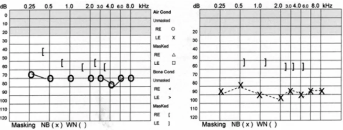
Air and bone conduction pure-tone audiometry was perfor-med at 250 to 8000 Hz frequencies, in octave intervals, with the addition of 3000 and 6000 Hz frequencies (GSI 61 audiometer - Grason-Stadler®, TDH 50P phones) and logoaudiometry. Pure-tone audiometry by bone conduction was performed as follows: first, a bone conduction free of masking was obtained with the vibrator positioned in the left ear. Then, the bone conduction of the left ear was obtained with approximately 20-25 dB SL contra-lateral effective narrowband noise. After this step, the bone vibra-tor was placed on the right mastoid, and the free bone conduction of the right ear was obtained. At the end, maximum equipment values with contralateral masking were applied, with no changes in thresholds obtained without the presentation of contralateral noise. The percentage rates of speech discrimination (SD) were obtained in both ears with contralateral masking (speech noise). Acoustic immittance measurements were performed using the AT 235H Interacoustics® Middle Ear Analyzer and consisted of tympanometry with a probe tone of 226 Hz and ipsilateral and contralateral acoustic reflexes research.
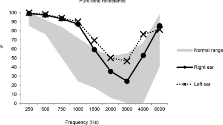
Reflectance measurements were performed using the MEPA 3 instrument (Middle Ear Power Analyzer - Mimosa Acoustics®, version 3.3). Before each reflectance test session, the probe was calibrated using a four-chamber set (CC4-V). Two measurements were made using the chirp stimulus and another measurement with pure tone stimulus. Data were collected with the chirp stimulus in 248 frequencies from 211 to 6000 Hz at intervals of 23 Hz, intensity of 60 dB sound pressure level (SPL), lasting from 0.1 to 10 seconds per point. For the pure tone stimulus, the reflectance was tested with the stimulus presented at 60 dB SPL punctually at the following frequencies: 250, 500, 750, 1000, 1500, 2000, 3000, 4000 and 6000 Hz. The WBR measurements considered for analysis included the reflectance energy (%) and the transmittance (dB).
According to the audiometry, the patient presented bilateral mixed hearing loss (Figure 1). The patient presented a type A tympanogram on the right ear and a type As tympanogram on the left ear, with a reduced admittance peak. She also showed no bilaterally ipsilateral and contralateral acoustic reflexes (Chart 1). With regards to logoaudiometry, the patient presen-ted Speech Reception Thresholds compatible with the mean speech frequencies (70 dB in the right ear and 90 dB in the left ear) and a PRSR 100 dB lower in the left ear (72%) compared with the right (100%).
According to these results, the transmittance was lower overall, indicating that less energy is being transmitted in the lower frequency (Chart 2). Moreover, the reflectance energy was higher than the normal range between frequencies of 500 and 1500 Hz in both ears, both for the chirp and the pure tone stimulus. The rates of overall mean reflectance were high in
both ears, 70.59% in the right ear and 78.50% in the left ear, and the difference was significant (Figure 2). The reflectance energy at medium frequencies was higher in the left ear than in the right ear, both for the pure tone and for the chirp stimulus (Figures 2 and 3). A paired Student’s t test was performed to compare the reflectance at frequencies of 1500 to 4000 Hz between the right and left ears, showing significant difference (p=0.011), whereas at lower frequencies (250 to 1000 Hz), there was no significant difference (p=0.083).
The diagnosis of otosclerosis was surgically confirmed in the patient months after the audiologic evaluation presented here.
DISCUSSION
The present study aimed to examine the reflectance measurements in a patient with otosclerosis. In most cases, otosclerosis is expressed bilaterally and presents well-cha-racterized symptoms. Otosclerosis, also called otospongiosis, presents characteristic evolution phases. In the case studied, Chart 1. Immittance results in both ears
Immitanciometry
Right ear Left ear
Meatoscopy --- ---
Equivalent volume of EAM 1.2 ml 0.8 ml
Peak pressure -6 daPa -27 daPa
Admittance peak (Ymt) 0.3 ml 0.2 ml
Acoustic Reflexes Survey Ipsi Contra
Af R/Ef L Hz Ipsi
Contra Af L/Ef R 500
1000 2000 4000 BB
Note: EAM = external acoustic meatus; Ipsi = ipsilateral acoustic reflex threshold; Contra Af R/Ef L = contralateral acoustic reflex threshold stimulus ear right/probe ear left; Contra Af L/Ef R = contralateral acoustic reflex threshold stimulus ear left/probe ear right
Note: RE = Right ear; LE = Left ear
Figure 1. Tone audiometry of the right and left ears, respectively
Chart 2. Transmittance results with pure tone and chirp stimuli in both ears
Frequency
Transmittance
Pure tone Chirp
LE RE LE RE
250 -35.21 -20.88 -28.73 -16.34
500 -17.38 -15.78 -17.73 -15.41
750 -12.30 -11.78 -12.74 -12.10
1000 -10.06 -9.02 -10.02 -8.76
1500 -5.16 -3.91 -5.00 -3.93
2000 -3.03 -1.89 -3.00 -1.91
3000 -2.73 -1.22 -2.74 -1.31
4000 -6.25 -3.28 -6.44 -3.60
6000 -7.31 -8.46 -8.20 -8.10
audiometry indicated a bilateral mixed hearing loss, which is not the typical presentation of early-stage disease. The presence of sensory impairment suggests a more advanced stage of the disease.
One of the advantages of using the WBR in relation to tympanometry is that it does not require the use of pressuriza-tion for the measurement, with little variability in the repeated measurements, unlike tympanometry. The positioning of the probe in the external acoustic meatus is not as crucial for the measurement as it is for the tympanometry (8); however, good
probe placement is important to obtain responses that are more reliable. The WBR is also advantageous because it is a fast measurement that covers a wide range of frequencies, allowing a more sensitive assessment of changes in the middle ear(1-4,9).
In this case studied, the WBR results were very similar, independent from the stimulus presented (chirp or pure tone),
showing high consistency among different evaluation results, in agreement with a previous study(10). In a study(10) conducted
with neonates, there were no differences in the reflectance rates measured with pure tone or chirp. The high consistency between the results makes WBR a reliable tool. The chirp stimulus seems more appropriate because it allows one to obtain measures not only of the frequencies at half-octave intervals but also a range of other frequencies at the same space (every 23 Hz). Therefore, more detailed results can be achieved.
According to the these results, the transmittance showed lower absorption of sounds between the 500-1500 Hz frequen-cies, indicating a rigidity pattern of the middle ear system. The transmittance result agrees with previous literature reports(2).
In the WBR results, a reflectance higher than the normal range(11) was observed at frequencies between 500 and 1500
Hz in both ears, consistent with results previously reported for otosclerosis(1-3). A study(3) conducted with 28 patients diagnosed
with otosclerosis showed higher reflectance values at frequen-cies below 1000 Hz (relative to the normal range), describing a typical pattern for ears with otosclerosis, suggesting rigidity of the system. In that study, 28 patients presented conductive hearing loss at low frequencies, suggesting initial involvement of the disease, unlike the case report discussed in the present study. The fact that the present study also detected differences at frequencies near 1500 Hz (relative to the normal range) may suggest that in more advanced stages of otosclerosis, there are not only changes in reflectance at regions below 1000 Hz but also at regions between 1000 and 1500 Hz.
In another study(2), aiming to present clinical applications
of reflectance and transmittance measurements, reflectance patterns detected for different diseases of the middle ear were described. The bilateral otosclerosis case presented by the re-searchers(2) follows the same reflectance pattern of the present
study, with higher reflectance than expected at frequencies between 500 and 1500 Hz, very different from the pattern described in cases of otitis media, eardrum perforation and disjunction of the ossicular chain presented in the study(2). What
these researchers(2) have postulated in relation to this typical
otosclerosis presentation is that the rigidity in the annular ligament of the stapes that occurs in ears with otosclerosis results in high system impedance at frequencies below 2000 Hz, thereby increasing the energy rate reflected to the frequency band between 500 and 1500 Hz, observed in the present study. Although the reflectance analysis is made by frequency--by-frequency visualization of the results compared to the normal range, we chose to generate a measure of the mean reflectance rate of each ear, considering all frequencies, with the aim of comparing one ear with the other, as both presented typical WBR of otosclerosis. The overall mean reflectance rates were 70.59% in the right ear and 78.50% in the left ear, with a difference between the ears. In the immitanciometry result, the difference between the left and right ears was very small. However, the WBR results show that the difference between the two ears is more significant than the difference observed in the immitanciometry. The difference between the ears could reveal a situation of greater stiffness in the left ear compared to the right. This result indicates which ear’s impedance might be more affected by the disease and may help to determine whi-ch ear to subject to a surgical procedure. Due to its precision and accuracy, the WBR allowed the comparison of results of both ears, highlighting differences detected between them. However, additional studies are necessary to understand how There was significant difference (p=0.021) when comparing the mean reflectance
rates (250 to 6000 Hz) between the right and left ears
Figure 2. Reflectance results with chirp stimulus in both ears
the asymmetry in WBR measurements between the ears can predict the disease.
The WBR is recommended for pre- and post-surgery follow--up in patients with otosclerosis. A previous study suggests that changes in WBR measurements observed in the pre- and post-surgical conditions could be a useful tool to monitor surgical success(12).
In this study, the WBR emerges as an alternative procedure, or even complementary, to immittanciometry for evaluation of the middle ear. The results from this study showed this tool as able to provide a detailed assessment of how the disease affects the function of the middle ear.
FINAL COMMENTS
The WBR measurements in the patient with otosclerosis allowed for a differentiation between the ears in relation to the absorption of sound by the middle ear. We suggest the use of WBR measurements to assist in a more detailed diagnosis of middle ear disorders.
ACKNOWLEDGEMENTS
We would like to thank Fundação de Amparo à Pesquisa do Estado de São Paulo (FAPESP) for the financial support (2009/06450-2).
REFERENCES
1. Feeney MP, Grant IL, Marryott LP. Wideband energy reflectance measurements in adults with middle-ear disorders. J Speech Lang Hear Res. 2003;46(4):901-11. http://dx.doi.org/10.1044/1092-4388(2003/070) 2. Allen JB, Jeng PS, Levitt H. Evaluation of human middle ear function via an acoustic power assessment. J Rehabil Res Dev. 2005;42(4 Suppl 2),63-78.
3. Shahnaz N, Bork K, Polka L, Longridge N, Bell N, Westerberg BD. Energy reflectance and tympanometry in normal and otosclerotic ears. Ear Hear. 2009;30(2):219-33. http://dx.doi.org/10.1097/ AUD.0b013e3181976a14
4. Beers AN, Shahnaz N, Westerberg BD, Kozak FK. Wideband reflectance in normal Caucasian and Chinese school-aged children and in children with otitis media with effusion. Ear Hear. 2010;31(2):221-33. http://dx.doi.org/10.1097/AUD.0b013e3181c00eae
5. Silva KAL, Urosas JG, Sanches SGG, Carvallo RMM. Wideband reflectance in newborns with present transient-evoked otoacoustic emissions. CoDAS. 2013;25(1):29-33. http://dx.doi.org/10.1590/S2317-17822013000100006
6. Merchant GR, Horton NJ, Voss SE. Normative reflectance and transmittance measurements on healthy newborn and 1-month-old infants. Ear Hear. 2010;31(6):746-54. http://dx.doi.org/10.1097/ AUD.0b013e3181e68e68
7. Salomone R, Riskalla PE, Vicente AO, Boccalini MCC, Chaves AG, Lopes R et al. Pediatric otosclerosis: case report and literature review. Rev Bras Otorrinolaringol. 2008;74(2):303-6. http://dx.doi.org/10.1590/ S0034-72992008000200024
8. Voss SE, Allen JB. Measurement of acoustic impedance and reflectance in the human ear canal. J Acoust Soc Am. 1994;95(1):372-84. http://dx.doi.org/10.1121/1.408329
9. Hunter LL, Bagger-Sjoback D, Lundberg M. Wideband reflectance associated with otitis media in infants and children with cleft palate. Int J Audiol. 2008;47 Suppl 1:S57-61. http://dx.doi. org/10.1080/14992020802294057
10. Silva KAL. Reflectância de banda larga em recém-nascidos: uso combinado de procedimentos eletroacústicos [doctor’s thesis]. São Paulo: Faculdade de Medicina da Universidade de São Paulo; 2011.
11. Shahnaz N, Bork K. Wideband reflectance norms for Caucasian and Chinese young adults. Ear Hear. 2006;27(6):774-88.