Obliquity of the Stapes in Otosclerosis: A New
Radiological Sign
Veluswamy Anand
1H. N. Udayabhanu
1B. Siva Subramaniam
21Department of Otorhinolaryngology and Head and Neck Surgery,
MCV Memorial ENT Trust Hospital, Pollachi, Tamil Nadu, India
2Department of Radiology, SRL, Diagnostic-Hitech Scan Center,
Coimbatore, Tamil Nadu, India
Int Arch Otorhinolaryngol 2016;20:94–98.
Address for correspondenceDr. Veluswamy Anand, MS, Department of Otorhinolaryngology and Head and Neck Surgery, MCV Memorial ENT Trust Hospital, Palaghat Road 115, Pollachi, TN 642001, India (e-mail: [email protected]).
Introduction
Otosclerosis is a two stage, metabolic bone remodelling process of the otic capsule characterized by bone resorption (otospongiosis) and recalcification (otosclerosis). The term otosclerosis was coined by Adam Politzer in 1894.1Anson and Bast etiologically related the histologically unstable cartilage in thefissular region to the development of otosclerosis. A mass of newly formed bone remodels itself and becomes an
active focus of otosclerosis.2–4Malleoincudal dislocation may occur when there is regrowth of otosclerotic bone above the oval window. Secondary torsional stresses on the stapes pulling the incus down is believed to be responsible for Malleoincudal dislocation, which can be diagnosed by High Resolution Computed Tomography (HRCT) temporal bone imaging.5After stapes surgery, the biologically remodeled incus would revert back to its original position due to the absence of torsional forces of stapes and subsequent pull by
Keywords
►
obliquity
►
otosclerosis
►
stapes
►
temporal bone
Abstract
Introduction
Observing the obliquity of stapes by closely scrutinizing the HRCT
temporal bone in otosclerosis revealed a reliable and consistent
fi
nding. This
fi
nding
can add to the existing radiological criteria in diagnosis of otosclerosis.
Objective
The objective of this study is to establish the obliquity of stapes in
otosclerosis by radiological measurements using HRCT temporal bone by comparing:
(a) the distance between the horizontal (tympanic) segment of facial nerve and stapes
head in otosclerotic ears (study group) with non-otosclerotic ears (control group); and
(b) the angle subtended by stapes with promontory in the study and control groups.
Methods
This is a prospective study performed after the institutional Ethics
Commit-tee clearance (IEC 3/2013).
Results
An increased mean distance between the horizontal segment of facial nerve
and stapes head in otosclerotic patients (i.e., 2.49mm
þ
/
0.24mm SD), when
compared with the non-otosclerotic patients (i.e., 1.46mm
þ
/
0.16mm SD) is noted.
There is a change in angle (i.e., 64.550
þ
/
7.190 SD) subtended by the stapes toward
the promontory in otosclerotic ears when compared with that of controls (i.e.,
99.700
þ
/
40 SD). We applied the Mann-Whitney U non-parametric test and
consid-ered
p
value of
<
0.0001 highly signi
fi
cant.
Conclusions
Obliquity of stapes in otosclerosis referred to as a
“
Pisa
”
sign by the senior
author has diagnostic value as a new radiological sign in imaging of otosclerosis. This
obliquity explains the torsional effect of otosclerosis on the ossicular chain. The
fi
ndings
correlate with late complications and failures in stapes surgery.
received
December 10, 2015 accepted
December 16, 2015 published online March 9, 2016
DOI http://dx.doi.org/ 10.1055/s-0036-1579743. ISSN 1809-9777.
Copyright © 2016 by Thieme Publicações Ltda, Rio de Janeiro, Brazil
the superior incudal ligament. This can lead to late compli-cations in stapes surgery. The purpose of this study is to objectively evaluate the obliquity of stapes toward promon-tory and the torsional effect of otosclerosis on the stapes and rest of the ossicular chain by radiological imaging. Though observed by surgeons, no radiological study has been done to evaluate and utilize this critical information.
Objectives
The objectives of this study are to document the obliquity and inferior displacement of stapes observed during stapedotomy surgery for otosclerosis. We performed a radiological evaluation to a) calculate the distance between the tympanic segment of facial nerve and stapes head and b) to establish the torsional effect of otosclerosis on stapes and ossicular chain by measuring the change in angle subtended by the stapes with the promon-tory in both the study and control groups.
Method
This is a prospective study done in our center from January 2013 to December 2014. This study was comprised of 20 patients divided into two groups. The study group consisted of 10 otosclerotic patients and the control group of 10 non-otosclerotic patients who presented to us with sudden sensorineural hearing loss. All patients with sudden sensorineural hearing loss (SSNHL) underwent tympanometry with reflex study. Only those with either ipsilateral or contralateral reflex positive were selected to exclude any otosclerosis in this group. After getting informed consent from patients and clearance from insti-tutional ethics committee (IEC 3/2013), we examined patients radiologically by HRCT temporal bone imaging. The scans were obtained with identical protocols. With the patient lying in the neutral position, we performed HRCT temporal bone imaging in both axial and coronal planes with 1 mm slice thickness with table feed of 1mm, perpen-dicular to the orbito meatal line using Siemens Somotome Duo (Siemens, Erlangen, Germany). We took high resolu-tion temporal bone CT scans of 1mm contiguous coronal sections with no gap between two slices at the level of lateral semicircular canal. The same radiologist read the images. Measurement of the length and also the angle were done only in the coronal plane using syngo software.
Length
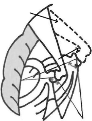
A straight line was drawn to measure distance between the mid horizontal (tympanic) segment of facial nerve and stapes head in coronal sections using syngo software in both the study and control groups (►Fig. 1).
Angle
The angle subtended by the stapes with promontory in the otosclerotic and non-otosclerotic patients was measured using syngo software between two lines, one from the oval window to head of stapes and second line from the promon-tory to stapes head (►Fig. 2).
Result
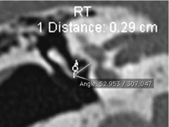
We compared the radiological measurements obtained in the otosclerotic and non-otosclerotic patients (i.e., SSNHL) and ran a statistical analysis with SPSS 17.0 software using the Mann-Whitney U non-parametric test. The mean distance between the facial nerve and the stapes head in High resolution CT scans, 1 mm contiguous coronal sections, taken at the level of the lateral semicircular canal was 2.49 mm þ/ 0.24 mm SD in otosclerotic ears and 1.46 mmþ/ 0.16 mm SD in non-otosclerotic ears. In the otosclerosis subjects, the mean angle measured was 64.55°þ/ 7.19° SD, whereas in the non-otosclerotic ears it was 99.70°þ/ 4° SD. The Man-Whitney U non-parametric test showed that the test statistic value was greater than the table value for length and angle measurements in both the groups and thepvalue was highly significant at level<0.0001. This indicates a torsional effect and downshift of the stapes toward the promontory (►Tables 1,2,3and►Figs. 3–8).
Fig. 1 Illustration showing the increase in length from facial nerve to stapes head due to obliquity of stapes in otosclerosis (dotted line).
Discussion
Histologically, otosclerosis is characterized by a wave of abnormal bone remodelling with resorption of the otic capsule bone and replacement with a hyper cellular woven bone that, over a long time, undergoes further remodelling, resulting in sclerotic mosaic architecture.6The most common area of stapesfixation is the anterior crus in the region of the embryonicfissula antefenestram.7–9Fissula antefenestram is unique to humans.10Because of the metamorphosis of the lining cartilage, fissulae in general and large fissulae in particular are areas of histological instability.2An underlying genetic defect of COL1A1 gene in collagen metabolism results in the generation of an unstable extracellular matrix with a high propensity for remodelling.11It is quite clear from the above that biological remodelling is the norm in oval window area in otosclerosis.
Other researchers have observed that, in otosclerosis, differences in the tilt of stapes superstructure and malposi-tion of the incus are common.12The obliquity of the stapes toward the promontory and the torsional effect of otosclerosis
on the ossicular chain is understood by comparing the distance between the stapes and facial nerve canal as well as the angulations of stapes with promontory in both study and control groups in this study. The torsional effect of otosclerosis, leading to the obliquity of stapes, also affects the incus and rest of the ossicular chain. These observations have not yet been objectively evaluated and documented.
Radiology of Otosclerosis
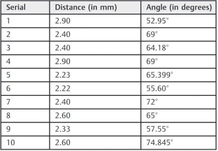
The usual radiological findings in case of otosclerosis is low attenuation in fissula antefenestram, thickening of the oval window membrane, plaque or new bone formation, and reduced surface area of the stapes.5 The mean Hounsfield unit (HU) values in the area anterior to the oval window (A-OW) are significantly lower in otosclerosis than in others. Based on receiver operating characteristic (ROC) analysis, the cut-off HU value in A-OW was 2,187.3 HU.13Malleoincudal dislocation can occur, secondary to torsional stresses, particularly when there is growth of otosclerotic bone above the oval window, which is diagnosed by CT criteria.5In addition to these, there is increased distance between the stapes head and the horizontal segment of Table 1 HRCT temporal bone measurements in otosclerotic
patients
Serial Distance (in mm) Angle (in degrees)
1 2.90 52.95°
2 2.40 69°
3 2.40 64.18°
4 2.90 69°
5 2.23 65.399°
6 2.22 55.60°
7 2.40 72°
8 2.60 65°
9 2.33 57.55°
10 2.60 74.845°
Table 2 HRCT temporal bone measurements in non-otosclerotic patients
Serial Distance (in mm) Angle (in degrees)
1 1.30 103°
2 1.20 99°
3 1.72 100.029°
4 1.52 93.729°
5 1.60 101°
6 1.40 101°
7 1.59 93.217°
8 1.54 97.369°
9 1.48 105.325°
10 1.30 103.45°
Table 3 Mann-Whitney U Test
i) Ranks
Group
N Mean
Rank
Sum of Ranks
CT.OTO.L 1 10 15.50 155.00
CT.NON-OTO.L 2 10 5.50 55.00
Total 20 – –
OTO.ANG 1 10 5.50 55.00
NON-OTO.ANG 2 10 15.50 155.00
Total 20 – –
ii) Test Statisticsa
Test Distance
statistic value for both groups
Angle statistic value for both groups
Mann-Whitney U 0.000 0.000
Wilcoxon W 55.000 55.000
Z -3.790 -3.782
Asymp. Sig. (2-tailed) 0.000 0.000
Exact Sig. [2 (1-tailed Sig.)]
0.000b 0.000b
Exact Sig. (2-tailed) 0.000 0.000
Exact Sig. (1-tailed) 0.000 0.000
Point Probability 0.000 0.000
Abbreviations: CT. OTO.L, CT otosclerosis length; CT.NON-OTO, CT otosclerosis length; OTO.ANG, otosclerosis angle; NON.OTO ANG, non-otosclerosis angle; Asymp., asymptomatic; Sig., signal.
Group 1, Otosclerosis; Group 2, Non-otosclerosis. aGrouping Variable: group.
facial nerve in otosclerosis when compared with the non-otosclerotic patients. There is also a difference in the angle subtended by the stapes with promontory between otosclerotic and non-otosclerotic patients. Thesefindings documented in our study after HRCT temporal bone imaging can be used as new
radiological sign in the diagnosis of otosclerosis. The normal non-protruding middle portion of the facial canal runs superior to the oval window and inferior to the lateral SCC. Only 1mm of bone separates the tympanic segment of the facial nerve canal from the vestibule.5
Fig. 3 HRCT Temporal bone coronal image showing length and angle measured in Otosclerotic patient.
Fig. 4 HRCT Temporal bone coronal image showing angle measured in otosclerotic patient.
Fig. 5 HRCT Temporal bone length measured in non-otosclerotic patient.
Fig. 6 HRCT Temporal bone showing angle measured in non-oto-sclerotic patient.
Fig. 7 Graph showing (Scatter dot) HRCT length between stapes head and facial nerve in otosclerosis and non-otosclerosis patients.
In a study by Zhu F. et al, the distance between head of stapes and facial nerve is 1.18 (0.42 mm).14In a study by Nicoleta Maru et al on intra temporal course and morphometric features of the facial nerve, the distance between the tympanic portion of the facial nerve and the stapes head is 1.58 mm.15
In our study, the mean distance between stapes head and facial nerve in non-otosclerotic control group is 1.46mm (0.16 mm SD), which correlates with the literature. How-ever, in otosclerosis we found that there is an increase in mean distance between the facial nerve and the stapes head (i.e., 2.49 mm0.24 mm SD). The mean angle subtended by the stapes with the promontory in otosclerotic patients is around 66.55° when compared with the non-otosclerotic, for which it is around 99.70°.
New Radiological Sign in Otosclerosis
Imaging plays an important role in conductive deafness cases for anatomical detailing, differential diagnosis, surgical planning, and assessment of post-operative complications.8 High-resolu-tion CT (HRCT) of the temporal bone using 1-mm (or less) thick sections is the modality of choice for assessment of the labyrin-thine windows and cochlear capsules.16High-resolution CT can detect an otosclerotic focus in up to 85% of patients with clinical otosclerosis.17On CT, the detection of otosclerosis can be difficult to the inexperienced eye because the spread of the disease is often symmetrical. A small lucency at thefissula antefenestram is typical for otosclerosis, although diagnosis of otosclerosis is based on clinical and audiological criteria. CT scan is useful to differentiate Pagets’disease, superior semicircular canal dehis-cence, middle ear malformations, congenital stapesfixation, and tympanosclerosis.18–20The mean HU value alone in the area anterior to the oval window (A-OW) is not very reliable in early detection of otosclerosis. Distances between horizontal segment of facial nerve and stapes head, along with stapes-promontory angle in HRCT as described will further improve radiologic diagnosis of otosclerosis especially to differentiate other similar conditions.
The senior author suggested that, for this description of obliquity of stapes in otosclerosis, the term“Pisa”eluding to the notion of the leaning tower of Pisa is ubiquitous. The “Pisa”sign, as described here, can be used in preoperative CT assessment of otosclerosis without using specialized instru-ments like telemanipulator systems used in other studies.18
Conclusion
The obliquity of stapes increases the distance between the stapes and horizontal portion of facial nerve and also causes a change in the angle subtended by stapes with promontory in otosclerosis. This obliquity, referred to as the“Pisa” sign, has a diagnostic value as a new radiological sign in otosclerosis imaging. It helps to understand the torsional effect of otosclerosis on the ossicular chain and to analyze late complications and failures in stapes surgery and ways to avoid them. Our objective assessment and documentation of“Pisa”sign is a simple and reliable method, using existing CT scan technology and software.
Financial Disclosure
Allfinancial and material support by MCV Memorial ENT Trust Hospital. The authors declare no conflict of interest.
References
1 Politzer A. Uber primare Erkrankung der Knocheren Labyrinth-Kapsel. Johrenheilk 1894;25:309–327
2 Anson BJ, Cauldwell EW, Bast TH. Thefissula ante fenestram of the human otic capsule; developmental and normal adult structure. Ann Otol Rhinol Laryngol 1947;56(4):957–985
3 Bast TH. Development of otic capsule. Residual cartilages and defective ossification and their relation to otosclerotic foci. Arch Otolaryngol 1940;32:771–782
4 Anson BJ, Cauldwell EW, Bast TH. Thefissula ante fenestram of the human otic capsule; aberrant form and contents. Ann Otol Rhinol Laryngol 1948;57(1):103–128
5 Swartz DJ, Loevener AL. Imaging of the Temporal bone. 4th ed. New York, USA: Thieme; 2009
6 Shea JJ Jr, Shea PF. Stapedectomy for otosclerosis. In: Glasscock ME, Gulya AJ, eds. Surgery of the ear. Ontario, Canada: Elsevier; 2003: 517–531
7 Ruedi L. Pathogenesis of otosclerosis. Arch Otolaryngol 1963; 78:469–477
8 Valvassori GE. Otodystrophies. In: Berrett A, Brunner S, Valvassori GE, eds. Modern Thin Section Tomography. Illinois, USA:
Spring-field; 1973:109–117
9 Valvassori GE. Radiologic diagnosis of cochlear otosclerosis. Laryngoscope 1965;75(10):1563–1571
10 Gulya AJ. Developmental anatomy of the temporal bone and skull base. In: Glasscock ME, Gulya AJ, eds. Surgery of the ear. Ontario, Canada: Elsevier; 2003:3–33
11 Iruela-Arispe ML, Vernon RB, Wu H, Jaenisch R, Sage EH. Type I collagen-deficient Mov-13 mice do not retain SPARC in the extra-cellular matrix: implications for fibroblast function. Dev Dyn 1996;207(2):171–183
12 Myers EN, Carrau RL, Eds. Operative otolaryngology: Head and Neck surgery. 2nd ed. Philadelphia: Elsevier; 2008
13 Kawase S, Naganawa S, Sone M, Ikeda M, Ishigaki T. Relationship between CT densitometry with a slice thickness of 0.5 mm and audiometry in otosclerosis. Eur Radiol 2006;16(6):1367–1373 14 Zhu F, Sun M, Zhang J, Sun D, Jiang Y. [Location of tympanic
segment and mastoid segment of facial nerve and prevention of prosopoplegia in operations]. Lin Chung Er Bi Yan Hou Tou Jing Wai Ke Za Zhi 2011;25(7):314–316
15 Măru N, Cheiţă AC, MogoantăCA, Prejoianu B. Intratemporal course of the facial nerve: morphological, topographic and mor-phometric features. Rom J Morphol Embryol 2010;51(2):243–248 16 Purohit B, Hermans R, Op de Beeck K. Imaging in otosclerosis:
A pictorial review. Insights Imaging 2014;5(2):245–252 17 Naumann IC, Porcellini B, Fisch U. Otosclerosis; incidence of
positivefindings on high resolution computed tomography and their correlation with audiological test data. Ann Otol Rhinol Laryngol 2005;114(9):709–716
18 Mafee MF, Henrikson GC, Valvassori GE et al. Use of CT in stapedial otosclerosis. Radiology 1985;156:709–714
19 Belden CJ, Weg N, Minor LB, Zinreich SJ. CT evaluation of bone dehiscence of the superior semicircular canal as a cause of sound and/or pressure induced vertigo. Radiology 2003;226:337–343 20 JuusoKujala. Modern Surgical treatment of otosclerosis [dissertation].
Helsinki, Finalnd: Helsinki University Printing House; 2009:24 21 Maier T, Strauss G, Bauer F, Grasser A, Hata N, Lueth TC. Distance