RBCCV 44205-1458 DOI: 10.5935/1678-9741.20130029
Mediastinitis: mortality rate comparing single-stage
surgical approach and preconditioning of wound
Mediastinite: mortalidade comparando abordagem cirúrgica em tempo único e o pré-condicionamento
da ferida operatória
Marcelo Curcio Gib
1, Juglans Souto Alvarez
2, Orlando Carlos Belmonte Wender
31. MSc; Cardiovascular surgeon at Cardiovascular Surgery Department of the Hospital de Clínicas de Porto Alegre, Universidade Federal do Rio Grande do Sul (UFRGS), Porto Alegre, RS, Brazil.
2. Cardiovascular surgeon at Cardiovascular Surgery Department of the Hospital de Clínicas de Porto Alegre, UFRGS, Porto Alegre, RS, Brazil. 3. PhD, Professor at Faculty of Medicine, UFRGS, Porto Alegre, RS, Brazil.
Research performed at Cardiovascular Surgery Department of the Hospital de Clinicas de Porto Alegre UFRGS - Graduate Program in Medicine: Surgery, Porto Alegre, RS, Brazil.
Correspondence address: Marcelo Curcio Gib
Travessa Pedra Redonda, 450 – casa 9 – Porto Alegre, RS, Brazil Zip code: 91760-630.
E-mail: [email protected]
Financial support: FAPERGS e CAPES
Article received on November 17th, 2012 Article accepted on January 28th, 2013 Abstract
Objective: This study aims to compare hospital mortality rate of surgical debridement followed by primary wound closure versus surgical debridement with closure after preconditioning of the wound.
Methods: A historical cohort of 43 patients with postoperative mediastinitis type III and IV between 2000 and 2008. The diagnosis of mediastinitis was based on physical examination and laboratory tests. Patients were divided into two groups: patients who received the protocol of preconditioning of the wound (Group 2) and those who did not (Group 1).
Results: Of the 43 patients, 15 received the protocol and were assigned to Group 2, and 28 patients to Group 1. Myocardial revascularisation was the surgical intervention most affected by infection, accounting for 69.8% of patients in Group 1 and 64.3% in Group 2.Staphylococcus aureus was the predominant pathogen, accounting for 58.1% of all cases, 50% in Group 1 and 73.3% in Group 2. Hospital mortality rate was 42.9% in Group 1 and 20% in Group 2 (P=1.86), with relative risk of 2.14 and CI [0.714-6.043]. Among the 28 (65.1%) patients who underwent single-stage surgical approach, 12 (27.9%) underwent primary wound closure with irrigation, seven (16.3%) only primary
closure, six (14%) omental lap, and three (7%) pectoralis muscle lap.
Conclusion: Due to the lack of established guidelines, the choice of the surgical approach is based largely on low-level evidence references. Preconditioning of the wound appears to
lead to a reduction in mortality in these patients, being a good surgical option.
Descriptors: Mediastinitis. Mortality. Infection. Surgical wound infection.
Resumo
Objetivo: Este estudo tem por objetivo comparar a taxa de mortalidade intra-hospitalar do debridamento cirúrgico seguido de fechamento da ferida operatória, com a do debridamento cirúrgico com fechamento após pré-condicionamento da ferida. Métodos: Coorte histórica composta por 43 pacientes portadores de mediastinite pós-operatória tipo III e IV entre os anos de 2000 e 2008. O diagnóstico de mediastinite foi feito com base em exames físico e laboratoriais. Os pacientes foram divididos em dois grupos, os que seguiram o protocolo de pré-condicionamento da ferida operatória (Grupo 2) ou não (Grupo 1).
(P=1,86), com risco relativo de 2,14 e IC [0,714-6,043]. Entre os 28 (65,1%) pacientes do estudo que seguiram a abordagem cirúrgica em um único tempo, 12 (27,9%) foram submetidos a
fechamento primário com irrigação, sete (16,3%), a fechamento primário isolado, seis (14%), rotação de retalho de epíplon, e três (7%), interposição de retalho de músculo peitoral.
Conclusão: Na ausência de uma diretriz bem estabelecida, a escolha do tipo de intervenção cirúrgica é feita utilizando-se referências com baixo nível de evidência. O pré-condicionamento da ferida operatória parece levar a redução da mortalidade nesses pacientes, sendo uma boa alternativa cirúrgica.
Descritores: Mediastinite. Mortalidade. Infecção. Infecção da ferida operatória.
Abbreviations, Acronyms & Symbols
INTRODUCTION
Median sternotomy is the technical approach most used in the surgical treatment of cardiopathies. Mediastinitis is a severe complication leading to an increase in hospital costs, morbidity and mortality [1,2]. The treatment, however, has progressed with new antibiotics, along with technical and surgical care.
Postoperative incidence of mediastinitis ranges from 0.5% to 5% [3-5]. However, mortality associated with such surgical complications, even after appropriate treatment, is extremely high, ranging between 14% and 47% [1,6,7]. Several studies
have identiied risk factors such as obesity, diabetes, reopera
-tion, smoking, prolonged operative time, bilateral use of the
internal thoracic artery, and postoperative bleeding [4,5,8,9]. Surgical management of postoperative mediastini-tis counts on several techniques described in the literature [5,10-17]. Treatment may include single- or multiple-stage
procedures, with or without the use of muscle or omental lap
[11,12,18]. Therapeutic modalities encompass two options: preconditioning of the wound, leaving the wound open for a better cleaning and mediastinal drainage with dressings, or using one of the several closure techniques available. Single-stage closure shows a recurrence rate between 5%-50% in comparison to two-stage closure whose rate ranges from 2%-30% [19,20], however the long term exposure of the medias-tinum enhances the morbidity.
The present study aims to compare inpatient mortality rate from surgical debridement followed by primary wound closure with that from surgical debridement with closure after preconditioning of the wound.
METHODS
From January 2000 to December 2008, at Hospital de Clínicas de Porto Alegre (HCPA), southern Brazil, 3,166 car-diac surgeries were performed in adults using median ster-notomy and extracorporeal circulation (ECC). A historical cohort of patients who had postoperative mediastinitis was
followed up during hospital stay after the irst surgery and
reintervention(s). From 2007, we implemented the protocol of preconditioning of the wound for all patients. Data were abstracted from the patient’s medical records. The entire patient sample that showed mediastinitis during this period
was identiied through the Cardiovascular Surgery Division
records in combination with those from the Commission on Hospital Infection Control (CCIH) of HCPA. Mediastinitis
was deined as deep surgical wound infection with clinical
and microbiologic evidence of compromised retrosternal space. During this period, 49 patients developed mediastini-tis, an incidence of 1.55%. Of these, 43 patients met the
clas-siication criteria (El Oakley) [3] as type III or IV and were
included in the study.
The diagnosis of mediastinitis was based on physical examination and laboratory tests. Three diagnostic criteria were elected during initial evaluation: sternal instability;
leukocytosis of more than 15,000; and wound secretion. All
patients were evaluated by members of the Cardiovascular Surgery Division at the moment of diagnosis. After the es-tablished diagnosis, the following data were collected from all patients: blood cultures, bedside culture of wound secre-tion, and culture of this same secretion during surgical pro-cedure. After initial data collection, a double intravenous antibiotic regimen, 500 mg of vancomycin every 12 hours and 1000 mg of cefepime every 8 hours, was started until
the identiication of the pathogen, with subsequent adjust -ment of treat-ment according to antibiogram results.
Patients were divided into two groups:
• Group 1 – Composed of 28 patients who underwent single-stage surgical intervention without preconditioning of the wound, regardless of closure technique;
• Group 2 – Composed of 15 patients who received the protocol of preconditioning of the wound.
Protocol of preconditioning of the wound
Radical subcutaneous and sternal edges debridement with complete removal of foreign materials (steel wires,
skin and subcutaneous sutures, hemostatic foam). Pericar -dial cavity wash with 5 L of 0.9% saline solution and 0.5% polyvinylpyrrolidone-iodine (PVPI) solution. Compression
CCIH ECC HCPA ICU PRBC PVPI
Commission on Hospital Infection Control Extracorporeal circulation
Hospital de Clínicas de Porto Alegre Intensive care unit
Packed red blood cells
dressings and clinical follow-up until improvement of the
following parameters: fall in leukocyte blood count, negative blood cultures, end of fever peaks, and beginning of wound
granulation. Dressing changes three times per day, or more, in the case of abundant secretion, which were performed by the HCPA nursing team specialized in complex dressings and scar care. Clinical evaluation guided the surgical team in relation to the solution to be used: papain, if chemical de-bridement was needed, 0.5% PVPI, when with purulent as-pect, or, if none of these situations was present, only saline solution was used. After clinical stabilization and
improve-ment of wound conditions, the patient was then taken to the surgical block for deinitive closure of sternotomy wound.
For closure, we evaluated criteria for viability of the sternal bone, mediastinum aspect, and subcutaneous granulation. Whenever necessary, a plastic reconstruction procedure was
associated: pectoralis muscle or omental lap.
Statistical analysis and ethical aspects
The present study was approved by HCPA Graduate and Research Group and the Research Ethics Committee of this
institution (Project 08-588 approved in 15/12/2008), as well
as the use of medical records. Patients with mediastinitis type
I, II and V were excluded from the study, as well as those pa-tients with incomplete inpatient follow-up information in the
medical records. The inal sample comprised all cases that fulilled the eligibility criteria for inclusion in the study.
Fisher’s exact test and the chi-square test were used for
statistical analysis. The level of signiicance was set at 5% and 95% conidence interval. Data were entered into a data -base converted into a 2007 Excel spreadsheet for Windows and subsequently exported and analyzed using the Statistical
Package for the Social Sciences software, version 17.0. The
results obtained were expressed as mean ± standard deviation for quantitative variables and frequency and percentage for categorical variables.
RESULTS
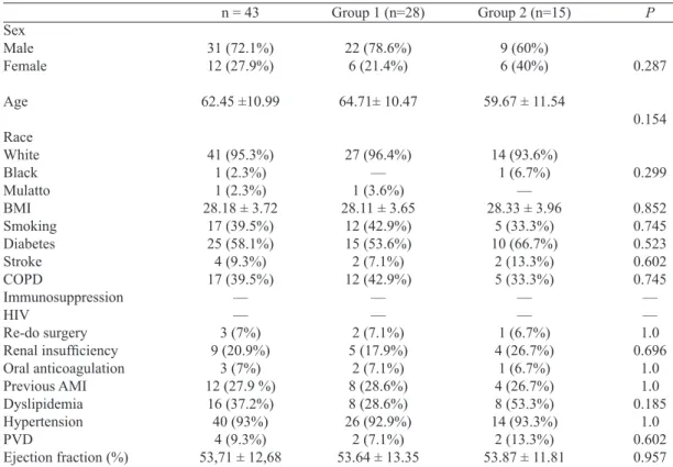
Of the 43 patients with mediastinitis included in the study, 15 received the protocol of preconditioning of the wound (Group 2) and the remaining 28 patients composed Group 1. Both groups were matched for the preoperative variables
analyzed (Table 1). Classic risk factors for mediastinitis were
homogeneously distributed between groups, the most rele-vant being diabetes, obesity and systemic hypertension.
Table 1. Demographic variables.
Sex Male Female
Age
Race White
Black
Mulatto BMI
Smoking
Diabetes
Stroke
COPD
Immunosuppression HIV
Re-do surgery
Renal insuficiency
Oral anticoagulation Previous AMI Dyslipidemia Hypertension PVD
Ejection fraction (%)
n = 43
31 (72.1%) 12 (27.9%)
62.45 ±10.99
41 (95.3%) 1 (2.3%) 1 (2.3%) 28.18 ± 3.72
17 (39.5%) 25 (58.1%) 4 (9.3%) 17 (39.5%)
–– –– 3 (7%) 9 (20.9%)
3 (7%) 12 (27.9 %)
16 (37.2%) 40 (93%)
4 (9.3%) 53,71 ± 12,68
Group 1 (n=28)
22 (78.6%) 6 (21.4%)
64.71± 10.47
27 (96.4%) –– 1 (3.6%) 28.11 ± 3.65
12 (42.9%) 15 (53.6%) 2 (7.1%) 12 (42.9%)
–– –– 2 (7.1%) 5 (17.9%)
2 (7.1%) 8 (28.6%) 8 (28.6%) 26 (92.9%)
2 (7.1%) 53.64 ± 13.35
Group 2 (n=15)
9 (60%) 6 (40%)
59.67 ± 11.54
14 (93.6%) 1 (6.7%)
–– 28.33 ± 3.96
5 (33.3%) 10 (66.7%)
2 (13.3%) 5 (33.3%)
–– –– 1 (6.7%) 4 (26.7%)
1 (6.7%) 4 (26.7%) 8 (53.3%) 14 (93.3%)
2 (13.3%) 53.87 ± 11.81
P
0.287
0.154
0.299
0.852 0.745 0.523 0.602 0.745 –– –– 1.0 0.696
1.0 1.0 0.185
1.0 0.602 0.957
BMI = body mass index; COPD = chronic obstructive pulmonary disease; HIV = human immunodeiciency virus;
Myocardial revascularization was the surgical intervention most affected by infection, accounting for 69.8% of patients in Group 1 and 64.3% in Group 2. The left internal thoracic artery was used in 94.7% of patients in Group 1 and 100% in Group 2. None of the patients underwent bilateral mammary grafting. In Group 1, 21.4% of patients were admitted to the
intensive care unit (ICU) for some time before the irst proce -dure, whereas in Group 2 the rate was 33.3% (Table 2). One patient in Group 1 needed intra-aortic balloon pump. In none of the groups reintervention was needed due to bleeding.
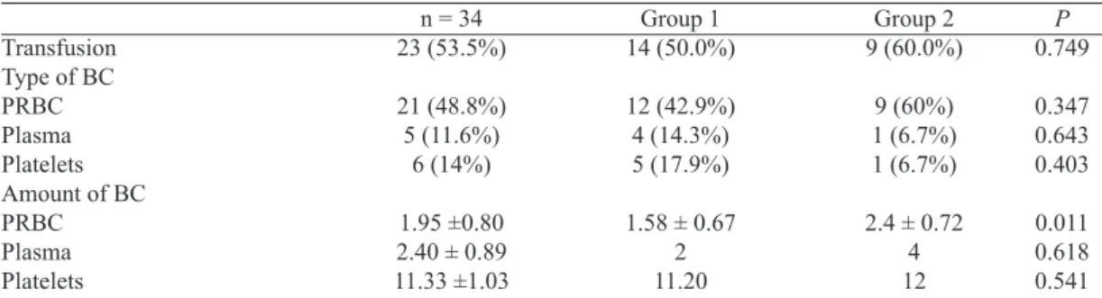
In 23 (53.5%) patients, blood-derived concentrates were necessary during the surgery of underlying pathology. Of these,
14 patients belonged to Group 1 and 9 to Group 2. Packed
red blood cells were the blood component most used in both
groups. However, Group 2 used a signiicantly greater num -ber of transfused units than Group 1 (1.58±0.67 vs. 2.4±0.72). There were no statistical differences in the number of other blood component units transfused, as described in Table 3.
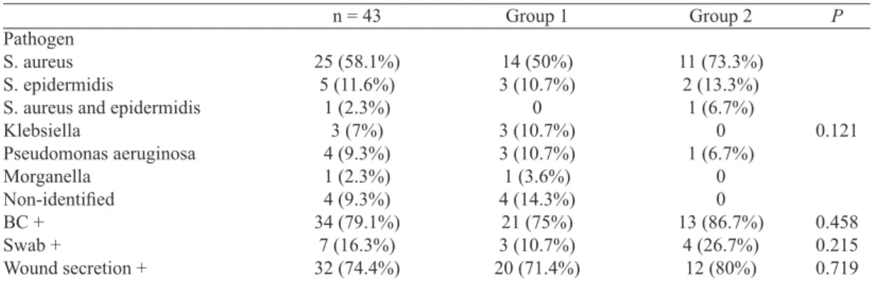
Several pathogens were identiied as causing mediasti -nitis (Table 4). Staphylococcus aureus was the predominant
Table 2. First procedure.
LPHS (days) Admission via ER ER stay (days) ICU admission ICU stay (days) URG procedure Baseline surgery CABG AVR MVR MV repair Aortic dissection Others Combined approach
Use of internal mammary artery n=31 No. of anastomoses
CPB time Cross clamp time Procedure time
Re-exploration for bleeding Intubation time
IABP
n = 43 8.07 ± 11 11 (25.6%) 2.64 ± 1.69
–– 9.17 ± 5.38
3 (%) 30 (69.8%) 7 (16.3%) 3 (7%) 1 (2.3%) 1 (2.3%) 1 (2.3%) 4 (9.3%) 30 (96.8%) 2.97 ± 0.85 85.44 ± 45.52 60.98 ± 37.06 247.91± 69.62
–– 23.19 ±43.47
1 (%)
Group 1 (n=28) 5.75 ± 6.61
6 (21.4%) 3 ± 2.1
–– 8 ± 6.93 2 (7.1%) 18 (64.3%) 6 (21.4%) 2 (7.1%) 1 (3.6%) 1 (3.6%) –– 3 (11.9%) 18 (94.7%) 2.83 ± 0.78 90.54 ± 51.33 64.64 ± 41.93 251.46 ± 81.32
–– 18.96 ± 16.14
1 (3.6%)
Group 2 (n=15) 12.4 ± 15.75
5 (33.3%) 2.20 ± 1.1
–– 10.33 ± 4.51
1 (6.7%) 12 (80%) 1 (6.7%) 1 (6.7%) –– –– 1 (6.7%) 1 (6.7%) 12 (100%) 3.17 ± 0.94 75.93 ± 31.42 54.13 ± 25.53 241.27 ± 41.40
–– 31.07 ± 71.17
–– P 0.137 0.473 0.464 –– 0.651 1.0 0.467 0.768 1.0 0.301 0.322 0.382 0.653 –– 0.526 1.0
AVR = aortic valve replacement; CABG = coronary artery bypass grafting; CPB = cardiopulmonary bypass; ER = emergency room; IABP = intra-aortic balloon pump; ICU = intensive care unit; LPHS = length of preoperative hospital stay; MV repair = mitral valve repair; MRV = mitral valve replacement; URG = urgent; Preop = preoperative; Reop = reoperation
Table 3. Use of blood components.
Transfusion Type of BC PRBC Plasma Platelets Amount of BC PRBC Plasma Platelets
n = 34 23 (53.5%)
21 (48.8%) 5 (11.6%)
6 (14%)
1.95 ±0.80 2.40 ± 0.89 11.33 ±1.03 Group 1 14 (50.0%) 12 (42.9%) 4 (14.3%) 5 (17.9%)
1.58 ± 0.67 2 11.20 Group 2 9 (60.0%) 9 (60%) 1 (6.7%) 1 (6.7%)
pathogen, accounting for 58.1% of all cases, 50% in Group 1 and 73.3% in Group 2. Blood culture was positive in 79.1% of cases. Wound secretion collected during reintervention en-abled to identify the causal agent in 74.4% of cases. In four patients (14.3%) in Group 1, none of the diagnostic means used in the study enabled to identify the pathogen.
Among 28 (65.1%) study patients who underwent single-stage surgical approach, 12 (27.9%) received primary wound closure with irrigation, seven (16.3%) only primary closure,
six (14%) omental lap, and three (7%) pectoralis muscle lap. Inpatient mortality rate was 42.9% in Group 1 and 20%
in Group 2 (P=1.86), with relative risk of 2.14 and CI [0.714-6.043], as described in Table 5. After deinitive treatment, the
need for new interventions was similar in both groups, 13 (46.4%) patients in Group 1 and 7 (46.7%) in Group 2.
DISCUSSION
The incidence of postoperative mediastinitis after car-diac surgery is low [3-5]. However, mortality and morbidity rates during treatment are high [1,6,7]. Much has been done
to identify and treat its risk factors, which are multiple and,
when two or more factors occur concomitantly, its action seems to be enhanced [4,5,8]. When analyzed alone, there
seems not to be a most important risk factor in its genesis.
Classic factors of surgical site infection such as trichotomy, infection in other sites, antibiotic prophylaxis, antisepsis, personal experience, and experience of medical and nursing team remain important.
In our cohort, demographic variables as well as risk fac -tors were homogenously distributed between both groups. Myocardial revascularization using the internal thoracic ar-tery remains the surgery most affected by mediastinitis. In none of the cases the bilateral use of the internal thoracic artery showed infection.
The use of blood-derived concentrates is high, being used in more than half the patients who developed mediastinitis.
Packed red blood cells (PRBC) were the most predominant
blood component. The preconditioning group received a
sig-niicantly higher amount of PRBC during baseline surgery.
Erythrocyte transfusion causes immunomodulation, although
its extension and deiciency type are variable [21]. Random -ized clinical trials, analyzing ICU inpatients, demonstrated
that the use of non-leukocyte-reduced erythrocyte concen -trates leads to multiple organ failure and death in up to 10%
of patients, against 5% in those who used leukocyte-reduced
erythrocyte concentrates [22]. This is the complication most
often associated with blood transfusion and lung injury. Currently, risk of infection associated with PRBC trans -fusion is 1:2,000 for bacterial infection; 1:58,000-149,000
Table 4. Pathogens.
Pathogen S. aureus S. epidermidis
S. aureus and epidermidis Klebsiella
Pseudomonas aeruginosa Morganella
Non-identiied
BC + Swab +
Wound secretion +
n = 43
25 (58.1%) 5 (11.6%)
1 (2.3%) 3 (7%) 4 (9.3%) 1 (2.3%) 4 (9.3%) 34 (79.1%)
7 (16.3%) 32 (74.4%)
Group 1
14 (50%) 3 (10.7%)
0 3 (10.7%) 3 (10.7%) 1 (3.6%) 4 (14.3%)
21 (75%) 3 (10.7%) 20 (71.4%)
Group 2
11 (73.3%) 2 (13.3%)
1 (6.7%) 0 1 (6.7%)
0 0 13 (86.7%)
4 (26.7%) 12 (80%)
P
0.121
0.458 0.215 0.719 BC = blood culture; Swab = wound secretion collected before reintervention
Table 5. Management.
Surg-reint time ATB time (days) Postop ICU time (days) Mortality
Postop time (days) New interventions
n = 43 18.65 ±15.45 27.19 ±24.64 18.40 ±30.75 15 (34.9%) 32.40±41.50
20 (46.5%)
Group 1 16.75 ± 15.29 24.75 ± 25.44 14.25 ± 29.26 12 (42.9%) 26.67 ± 43.27
13 (46.4%)
Group 2 22.20 ± 15.64 31.73 ± 23.31 26.13 ± 32.99
3 (20%) 55.33 ± 27.79
7 (46.7%)
P 0.276 0.372 0.232 0.186 0.301 1.0
ATB = antibiotics; ICU = intensive care unit; Postop = postoperative; Surg-reint time = time between irst surgery
for hepatitis B; and 1:872,000-1,700,000 for hepatitis C [23]. Analyzing these data, we may consider Group 2 as possibly more severe than Group 1.
The attempt to identify a causal agent and a rapid surgi-cal intervention accompanied by a comprehensive antibiotic coverage remains as the best treatment approach to medias-tinitis. Staphylococcus aureus was the most frequent causal agent, leading to infections with rapid clinical course and more aggressive characteristics. Its elimination during the preoperative period should have top priority, in addition to strengthened care by the surgical team during the intraop-erative period. Brazilian studies indicate Staphylococcus au-reus as the most predominant causal agent in mediastinitis, whereas international reports demonstrate a predominance of
Staphylococcus epidermis [9,10,24,25].
Using the preconditioning wound can reduce mortality by approximately 22% compared to the procedures of time only. This improvement in survival seems mainly related best clin-ical conditions of the patient and the wound at the time of
inal closure of the chest. Despite the clinical relevance moti
-vating obtained statistical signiicance. However, most mod -ern techniques for the treatment of mediastinitis and still not available in our country, such as vacuum therapy, is worth the preconditioning of the wound, which leads us to believe that this really is the best way to combat mediastinitis.
A single approach that may be successfully applied to
all mediastinitis cases remains unknown [3]. Several reports
have failed to differentiate or classify the type of associat-ed infection always using the same management approach.
Acute infection that develops during the two irst postopera
-tive weeks have a disease mechanism different from that of chronic or recurrent infection, which may take months or
even years to develop after baseline pathology repair surgery [3]. Management approach in these cases, therefore, is
dif-ferent. The lack of knowledge on these facts is likely to be the reason why there are so many conlicting reports in the
literature regarding the best approach to poststernotomy me-diastinitis.
Mediastinitis type I, which was not included in this study, seems to respond well to debridement with primary resuture and use of irrigation. Wound debridement with two-stage
closure using or not laps is considered, for some authors,
as the ideal treatment for chronic mediastinitis type V. Pai-rolero & Arnold [16] reported excellent results with the use of preconditioning of the wound and subsequent
reconstruc-tion with laps. In that case series, 38 consecutive cases of
mediastinitis type V were treated with no deaths during the
irst 30 postoperative days and ive deaths during the mean
study follow-up period of 24.8 months, none of them related to reconstruction or sepsis. Infection recurrence rate in those patients was 13.2%.
Prolonged antibiotic therapy alone is associated with mor-tality and morbidity rates currently unacceptable, and its use
has been abandoned in early years [6]. An important advance in the treatment of mediastinitis occurred in 1963 when, for
the irst time, the use of continuous mediastinal irrigation
with saline solution using routine chest drainage tubes af-ter closure of the saf-ternum was proposed [14]. Afaf-terward, the addition of antibiotics in the irrigation was suggested, which was associated with an increase in bacterial resistance and fungal contamination, especially of the genus Candida [13]. As an alternative, then, the use of PVPI was started for con-tinuous mediastinal cavity wash since it is a fungicidal and bactericidal solution associated with low toxicity. The use of primary resuture with or without mediastinal irrigation offers the advantage of a procedure that produces a closed wound with stable sternum, but studies have reported high therapy failure associated with a high mortality rate [20].
An approach often used in the presence of compromised
sternal edges is the use of muscle and omental laps. Lee et al. [11], in 1976, were the irst to describe the use of omental laps for illing the substernal dead space. The use of pecto
-ralis muscle lap was described for the irst time in 1980, but recent studies have reported conlicting results, and some au
-thors still defend the use of laps of rectus abdominus muscle
as the technique of choice [12]. Reconstruction performed
with the use of laps, which aims at illing anterior medias -tinal dead space and an increase in blood supply, has shown low mortality rates. Nevertheless, disadvantages associated with this method include an increase in surgical trauma,
per-sistent pain in the lap, muscle weakness, and hernias [7].
Moreover, there are several series exhibiting poor long-term results with these techniques. Moreschi et al. [18] reported 81 consecutive mediastinitis cases treated with different
mo-dalities, early intervention with the use of laps being a good
treatment option with low mortality rates.
Therapy failure in mediastinitis leading to infection
re-currence aggravates signiicantly an already dificult situa -tion, resulting in an even higher mortality rate [3,10].
In an attempt to reduce high recurrence and treatment failure rates associated with primary resuture, several
alter-natives and technical reinements have been suggested over
the past decades. Among them, we point out plastic
recon-struction procedures using pectoralis muscle or omental lap
[11,12]. More recently, the use of vacuum has been
intro-duced as an intermediate stage between irst-approach surgi
-cal debridement and deinitive wound closure, but this ap
-proach is yet to be commercially spread in our ield [17,20].
Based on our experience, preconditioning of the wound seemed to be a good therapeutic option in severe patients
who develop postoperative mediastinitis. Despite the difi -culty in maintaining a patient with the chest open, it seems an
important technical improvement, which enables a deinitive
REFERENCES
1. Loop FD, Lytle BW, Cosgrove DM, Mahfood S, McHenry MC, Goormastic M, et al. J. Maxwell Chamberlain memorial paper. Sternal wound complications after isolated coronary artery bypass grafting: early and late mortality, morbidity, and cost of care. Ann Thorac Surg. 1990;49(2):179-86.
2. Nelson RM, Dries DJ. The economic implications of infection in cardiac surgery. Ann Thorac Surg. 1986;42(3):240-6.
3. El Oakley RM, Wright JE. Postoperative mediastinitis: classiication
and management. Ann Thorac Surg. 1996;61(3):1030-6.
addition to technical care. Despite showing morbidity, rou-tine dressing changes seem to reduce inpatient mortality.
Although without statistical signiicance, the results herein
obtained seem encouraging and in agreement with clinical
indings.
After the wound preconditioning period, patients were
re-evaluated and the deinitive procedure was performed ac -cording to sternum conditions. In cases of viable bone, de-bridement of sternal edges was performed with resuture and use of irrigation. In cases of compromised bone, sternectomy
was performed with illing of dead space. In cases in which
the upper two thirds of the sternum were compromised, we
used the rotation of the pectoralis major muscles; however, when the defect was in the lower third, omental lap was per -formed and, in some cases, both techniques were per-formed. However, our study has some limitations. First, this was a retrospective study and this is not the best design to evalu-ate treatment. Due to small number of patients, the different techniques of late esternal closure could not be analyzed in-dividually. Finally, the low annual incidence of this compli-cation may cause small changes in the surgery process along the time.
CONCLUSION
Due to the lack of established guidelines, the choice of
the mediastinitis procedure is based largely on low-level evi-dence references. Our study encourages the search for an ef-fective intervention, there seems to be a reduction in hospital mortality with preconditioning of the wound although this
difference was not statistically signiicant. The precondition -ing wound has been in our midst, a good alternative in the treatment of severe mediastinitis. Further studies involving greater standardization of case reports and patient random-ization are warranted to better understand the interventions applied to this severe affection.
4. Abboud CS, Wey SB, Baltar VT. Risk factors for mediastinitis after
cardiac surgery. Ann Thorac Surg. 2004;77(2):676-83.
5. Eklund AM, Lyytikäinen O, Klemets P, Huotari K, Anttila VJ, Werkkala KA, et al. Mediastinitis after more than 10,000 cardiac
surgical procedures. Ann Thorac Surg. 2006;82(5):1784-9.
6. Serry C, Bleck PC, Javid H, Hunter JA, Goldin MD, DeLaria GA,
et al. Sternal wound complications. Management and results. J Thorac Cardiovasc Surg. 1980;80(6):861-7.
7. Milano CA, Kesler K, Archibald N, Sexton DJ, Jones RH.
Mediastinitis after coronary artery bypass graft surgery. Risk
factors and long-term survival. Circulation. 1995;92(8):2245-51.
8. Gummert JF, Barten MJ, Hans C, Kluge M, Doll N, Walther T,
et al. Mediastinitis and cardiac surgery--an updated risk factor
analysis in 10,373 consecutive adult patients. Thorac Cardiovasc Surg. 2002;50(2):87-91.
9. Ridderstolpe L, Gill H, Granfeldt H, Ahlfeldt H, Rutberg H. Superficial and deep sternal wound complications:
incidence, risk factors and mortality. Eur J Cardiothorac Surg.
2001;20(6):1168-75.
10. Sjögren J, Malmsjö M, Gustafsson R, Ingemansson R.
Poststernotomy mediastinitis: a review of conventional surgical treatments, vacuum-assisted closure therapy and presentation of the Lund University Hospital mediastinitis algorithm. Eur J Cardiothorac Surg. 2006;30(6):898-905.
11. Lee AB Jr, Schimert G, Shaktin S, Seigel JH. Total excision
of the sternum and thoracic pedicle transposition of the greater omentum; useful strategems in managing severe mediastinal infection following open heart surgery. Surgery. 1976;80(4):433-6.
12. Jurkiewicz MJ, Bostwick J 3rd, Hester TR, Bishop JB, Craver
J. Infected median sternotomy wound. Successful treatment by
muscle laps. Ann Surg. 1980;191(6):738-44.
13. Sarr MG, Gott VL, Townsend TR. Mediastinal infection after cardiac surgery. Ann Thorac Surg. 1984;38(4):415-23.
14. Shumacker HB Jr, Mandelbaum I. Continuous antibiotic irrigation
in the treatment of infection. Arch Surg. 1963;86:384-7.
15. Jones G, Jurkiewicz MJ, Bostwick J, Wood R, Bried JT, Culbertson
J, et al. Management of the infected median sternotomy wound
with muscle laps. The Emory 20-year experience. Ann Surg.
1997;225(6):766-76.
16. Pairolero PC, Arnold PG. Management of recalcitrant median sternotomy wounds. J Thorac Cardiovasc Surg. 1984;88(3):357-64.
17. Argenta LC, Morykwas MJ. Vacuum-assisted closure: a new
18. Moreschi AH, Macedo Neto AV, Barbosa GV, Saueressig MG.
Aggressive treatment using muscle laps or omentopexy in
infections of the sternum and anterior mediastinum following sternotomy. J Bras Pneumol. 2008;34(9):654-60.
19. Berg HF, Brands WG, van Geldorp TR, Kluytmans-VandenBergh FQ, Kluytmans JA. Comparison between closed drainage techniques for the treatment of postoperative mediastinitis. Ann Thorac Surg. 2000;70(3):924-9.
20. Catarino PA, Chamberlain MH, Wright NC, Black E, Campbell
K, Robson D, et al. High-pressure suction drainage via a polyurethane foam in the management of poststernotomy mediastinitis. Ann Thorac Surg. 2000;70(6):1891-5.
21. Society of Thoracic Surgeons Blood Conservation Guideline
Task Force, Ferraris VA, Ferraris SP, Saha SP, Hessel EA
2nd, Haan CK, Royston BD, et al; Society of Cardiovascular
Anesthesiologists Special Task Force on Blood Transfusion.
Perioperative blood transfusion and blood conservation in cardiac surgery: the Society of Thoracic Surgeons and The Society of
Cardiovascular Anesthesiologists clinical practice guideline. Ann Thorac Surg. 2007;83(5 Suppl):S27-86.
22. Bilgin YM, van de Watering LM, Eijsman L, Versteegh MI, Brand
R, van Oers MH, et al. Double-blind, randomized controlled trial
on the effect of leukocyte-depleted erythrocyte transfusions in
cardiac valve surgery. Circulation. 2004;109(22):2755-60.
23. Yomtovian R, Lazarus HM, Goodnough LT, Hirschler NV, Morrissey AM, Jacobs MR. A prospective microbiologic surveillance program to detect and prevent the transfusion of bacterially contaminated platelets. Transfusion. 1993;33(11):902-9.
24. Arruda MV, Braile DM, Joaquim MR, Suzuki FA, Alves
RH. The use of the vancomycin paste for sternal hemostasis and mediastinitis prophylaxis. Rev Bras Cir Cardiovasc. 2008;23(1):35-9.