RBCCV 44205-1454 DOI: 10.5935/1678-9741.20130025
Doppler echocardiographic criteria in patency
assessment of composite grafts from left internal
thoracic artery
Parâmetros Dopplerluxométricos de perviedade do enxerto composto de artéria torácica interna
esquerda
Maria Claudia A. Leitão
1, José Glauco Lobo Filho
2, Tiago Magalhães Freire
3, Marília Leitão
Montenegro
4, Francisco Vagnaldo Fechine Jamacuru
5, Eduardo Rebouças de Carvalho
6, Amanda
Ximenes Couto Bem
7, Heraldo Guedis Lobo Filho
8, Manuel Odorico de Moraes Filho
91. Cardiologist echocardiographer, master’s degree from the Department of Physiology and Pharmacology, School of Medicine, Federal University of Ceará, Professor of Medicine at Christus University, Fortaleza, CE, Brazil. Author of the master's thesis that gave rise to this paper. Echocardiographer of this study.
2. Professor of Cardiovascular Surgery, School of Medicine, Federal University of Ceará, Fortaleza, CE, Brazil. Head of the surgical team of this study.
3. Medical student, Federal University of Ceará, Fortaleza, CE, Brazil. Assistance in data collection, literature review and drafting work. 4. Medical student, Christus University, Fortaleza, CE, Brazil. Responsible
for data collection, assistance in literature review and drafting work. 5. Research doctor and statistician, Clinical Pharmacology Unit, Federal
University of Ceará, Fortaleza, CE, Brazil. Responsible for coordinating the statistical analysis.
6. Medical doctor graduated from Federal University of Ceará, Fortaleza, CE, Brazil. Responsible for the statistical analysis.
Assistance in data collection, literature review.
8. Cardiovascular Surgeon - Specialist Member of Brazilian Society of Cariovascular Surgery. MS at the Federal University of Ceará, Fortaleza, CE, Brazil. Research.
9. PhD in Oncology from the University of Oxford, UK, Professor of the Federal University of Ceará, Fortaleza, CE, Brazil. Master’s thesis supervisor.
Work carried out at Federal University of Ceará, Fortaleza, CE, Brazil.
Correspondence address: Maria Claudia de Azevedo Leitão
Rua Canuto de Aguiar, 500 – ap. 1600 – Fortaleza, Ceará, Brazil Zip code 60160-120
E-mail: [email protected] / [email protected]
Article received on November 9th, 2012 Abstract
Objectives The purpose of our study was to establish, with an entirely noninvasive method, transthoracic Doppler echocardiography, criteria for patency of composite left internal thoracic artery grafts when placed on the left anterior descending artery and other branches of the left coronary system.
Methods: The control group comprised 20 patients with single graft and 20 patients with composite graft; all forty having their patency conirmed by coronary angiogram (CA). In this control group, two Doppler echocardiographic variables, diastolic mean velocity-time and integral diastolic peak velocity to systolic peak velocity ratio were recorded. For each variable, established cut-off points were established, using the ROC (Receiver Operator Characteristic) curves, to identify criteria which could differentiate the composite grafts. Only patients
with composite grafts were included in the 159-patients study group. The criteria established by the cut-off points in the control group were then applied to detect patency using a diastolic fraction of ≥ 0.5 as the gold standard. The sensitivity, speciicity, and positive and negative predictive values of these two criteria were determined.
168
Conclusion: Values reaching the criteria established for each variable indicate high probability of composite graft patency. Lower values have a large proportion of false negatives and are not conclusive as patency criteria.
Keywords: Coronary artery bypass. Doppler echocardiography. Mammary arteries. Myocardial ischemia. Myocardial revascularization.
Resumo
Objetivo: O objetivo deste estudo é estabelecer parâmetros preditores de perviedade, avaliados por Dopplerluxometria, do enxerto composto de artéria torácica interna esquerda, quando
revasculariza a artéria interventricular anterior e outro ramo do sistema esquerdo.
Métodos: O grupo controle foi formado por 20 pacientes com enxerto simples e 20 pacientes com enxerto, composto cuja perviedade foi conirmada por cineangiocoronariograia. No grupo controle, as variáveis de luxo relação velocidade pico diastólico/velocidade pico sistólico e integral da velocidade média/tempo na diástole foram registradas. Para cada variável, estabeleceram-se pontos de corte para identiicar enxertos compostos, usando-se curvas ROC (receiver operator characteristic). No grupo estudo, foram avaliados 159 pacientes com enxerto composto, determinando-se os dois parâmetros de luxo. Pontos de cortes estabelecidos no grupo controle foram usados para determinar sensibilidade, especiicidade, valores preditivos positivo e negativo de cada variável relacionada à perviedade dos enxertos, tomando-se como referência a fração diastólica ≥ 0,5.
Resultados: No grupo controle, os pontos de corte estabelecidos para as variáveis velocidade pico diastólico/ velocidade pico sistólico e integral velocidade média/tempo na diástole foram, respectivamente, 0,71 e 0,09m. No grupo estudo, a sensibilidade para a velocidade pico diastólico/velocidade pico sistólico e integral da velocidade média/tempo na diástole, considerando seus pontos de corte, foi de 36,4% e 40%, respectivamente. Os respectivos valores preditivos negativos foram 11% e 10.48%, enquanto especiicidade e valor preditivo positivo foram de 100% para os dois parâmetros.
Conclusão: Valores maiores ou iguais aos estabelecidos para cada variável indicam alta probabilidade de perviedade do enxerto composto. Valores inferiores apresentam grande proporção de falsos negativos, não sendo conclusivos quanto à perviedade.
Descritores: Ponte de artéria coronária. Ecocardiograia Doppler. Isquemia miocárdica. Artéria torácica interna. Revascularização miocárdica.
Abbreviations, Acronyms & Symbols
INTRODUCTION
Anatomic confirmation of patency and functional evaluation of the coronary grafts is crucial in the coronary artery bypass graft surgery (CABG). The most commonly used method for assessing bypass patency in CABG is coronary angiography (CA). The routine tests for myocardial ischemia
are often dificult to interpret in patients who underwent
CABG. Therefore, a method that directly analyzes the graft and is less invasive than CA is needed [1]. In recent years, coronary angiography by multidetector computed tomography
has been used with good sensitivity and speciicity [2], but
these two methods are expensive, and the patients are exposed to radiation [3-5]. The transthoracic Doppler echocardiography (TDE) is a particularly interesting tool.
The irst descriptions of using Doppler applications in
CABG were reported by Fusejima [6], in 1987, and Takagi et
al. [7], in 1993. It is a noninvasive and low cost method, and of quick completion even at bedside setting. It can identify left
internal thoracic artery (LITA) graft, measure its blood low
[8], and it can be carried out in every routine echocardiogram requested in the follow up appointments. Furthermore, it can be used to provide information on a patient’s physiological response to increased myocardial oxygen demand, the
coronary reserve low [9,10].
The low of the in situ LITA has systolic predominance. When anastomosed to a coronary artery, the low begins to show an increment of the diastolic component. This is justiied because the coronary arteries inlow occurs primarily during
diastole [11]. Thus, the LITA starts to assume the pattern of
the coronary low, including varying its diastolic component
according to demand [12].
Several measurements can be recorded by Doppler spec-tral imaging such as: peak velocities during systole and
dias-CA CABG CPB DPV DVTI FN FP LAD LCS LITA NPV PPV ROC SMV SPV SVTI TDE TN TP VTI
Coronary angiography
Coronary artery bypass graft surgery Cardiopulmonary bypass
Diastole peak velocity Diastole velocity-time integral False-negative
False-positive
Left anterior descending coronary artery Left coronary system
Left internal thoracic artery Negative predictive value Positive predictive value Receiver Operator Characteristic Saphenous magna vein Systole peak velocity Systole velocity-time integral
Transthoracic Doppler echocardiography True-negative
tole (SPV and DPV), mean velocities, mean velocity-time in-tegrals during systole (SVTI) and diastole (DVTI), diastolic
fraction, cross-sectional vessel area, and blood low at rest
and under stress. Using these recorded measurements, the
low reserve can also be measured by comparing low under stress to low at rest [13].
When LITA revascularizes the left anterior descending coronary artery (LAD), the presence of a diastolic compo-nent is a promising sign of graft patency. In a composite graft, when two coronary arteries are revascularized by LITA, de-termining their patency is a challenge. TDE has a high
sensi-tivity and speciicity when performed on left internal thoracic arteries (LITA) grafted to the LAD. It is known that the low
is greater in composite grafts than in single grafts since there is a naturally increased demand at rest. This observation throught 10 years of experience was the motivation for our
research. This difference has not yet been quantiied [14,15]. How much low increment would it be necessary to suggest that both grafts in coniguration of the composite graft were
patent?
The purpose of this study is to contribute to the ield of
physiology and cardiology. The clinical relevance of this
pa-per is to ind out if transthoracic Doppler echocardiographic
criteria could estimate the patency of the composite LITA graft when it revascularizes LAD and another branch of the left coronary system (LCS).
The overall objective of this study was to establish at rest, Doppler echocardiographic criteria for assessing the patency of a LITA composite graft, when it is revascularizing LAD
and another LCS branch. The speciic objective was to iden
-tify the sensitivity, speciicity, and positive and negative pre -dictive values (PPV and NPV, respectively) of the DPV/SPV and DVTI variables for determining composite graft patency,
using a diastolic fraction (DF) ≥ 0.5 as the reference value.
METHODS
This study was carried out in two groups: control and study group. None of the patients included or excluded from the study group were part of the control group.
Control Group
In the control group, 40 bypass patients were studied; 20 had single grafts, and 20 had composite grafts. A single graft is when LITA is anastomosed to LAD only and a composite graft is when a segment of the valveless saphenous magna vein (SMV) is anastomosed in “Y” shape in a single graft with a distal anastomosis to another branch of the LCS. Pa-tients with simple and composite grafts, who had their graft
patency conirmed by angiography, underwent TDE in order to record two low variables: the DVTI, and the ratio of di -astolic peak velocity to systolic peak velocity (DPV/SPV).
For each low variable, cutt-off points were established to
identify composite grafts by analyzing the Receiver Operator Characteristic (ROC) curves. Patients whose LITA was not adequately seen due to technical limitations were excluded.
Study Group
In the study group, 159 examinations were evaluated, corresponding to 159 subjects, 27% of which were female patients. The mean age was 63.4 years, ranging from 36 to 82 years. All patients had undergone CABG with a composite graft in which the LITA was grafted to the LAD and to another LCS artery by a saphenous vein segment in “Y” anastomosis without cardiopulmonary bypass (CPB).
All Doppler studies were performed by one echocardiographer (MCAL). The average time interval between surgery and examination was approximately 12.9 months, ranging from 2 to 96 months. The examinations were done and collected for the last 10 years and the preparation of the database occurred between December 2010 and January 2011.
Patients were excluded from the study group because of the following reasons: a LITA that could not be visualized,
when LITA provided blood low to more than two vessels of
the LCS; and patients who were submitted to the Vineberg procedure, LITA single grafts, LITA sequential grafts, and
on-pump operations. Tests performed during the irst two months
after CABG were also excluded because as outpatients it’s easier to avoid unstable conditions that could be presented during hospitalization period. The concern was to get an uniform group. In this phase, the Doppler criteria obtained from the cutoff points in the control group were applied to these 159 patients. They were tested in the real world. The rate of true-positives, false-true-positives, false-negatives and true-negatives were calculated. Sensitivity, speciicity, PPV and NPV, were
determined with their respective conidence intervals of 95%.
The reference pattern for patency was the diastolic fraction
of ≥ 0.5 which is considered by meta-analysis the Doppler
gold standard for assessment of single graft patency with LITA. There is no one criterion yet for patency of composite grafts with LITA. The fact that all patients in the control group had
FD ≥ 0.5 supports its use.
The Doppler examinations were conducted at the Unimed Regional Hospital (Hospital Regional da Unimed). The operations were performed at these four hospitals: Unimed Regional Hospital (Hospital Regional da Unimed), Monte Klinikum Hospital (Hospital Monte Klinikum), São Raimundo Hospital (Hospital São Raimundo), and the Walter Cantídio Hospital at the Federal University of Ceará (Hospital Walter Cantídio da Universidade Federal do Ceará). This study was approved by the Research Ethics Committee of Federal University of Ceará (CONEP - Comitê de Ética em Pesquisa) on October 12, 2010, protocol no. 315/10 and letter no. 349/10.
Description of technique
transducer was a nonlinear 5 MHz frequency. The patient was placed in the dorsal decubitus position and the trans-ducer placed on the left supraclavicular region. Using color Doppler, the LITA leaving the subclavian artery was visual-ized. Subsequently, a pulsed Doppler was applied to obtain the spectral curve and measurement of the variables. Angle adjustment was not necessary for aligning the Doppler cursor
with LITA low [8].
Statistical analysis
The data were analyzed using the Statistical Package for the Social Sciences® for Windows (v.16, SPSS Inc. Chicago, IL) statistical package. None of the quantitative variables had normal distributions according to the Kolmogorov-Smirnov test. As a result, parametric tests could not be used and the variables were described by medians and interquartile ranges. The nonparametric Mann-Whitney test was used to compare the medians in relation to the type
of graft coniguration (single or composite) in the control
group phase.
The cutoff points for the DPV/SPV and DVTI variables
during the standardization phase were deined using the ROC
curve. In the study group phase, TDE criteria for composite graft patency determined by the cutoff points were put in a 2x2 contingency table, designed using the DF as the reference
for graft patency. Subsequently, sensitivity, speciicity, PPV, and NPV were calculated, with 95% conidence intervals.
The results were expressed in tables. A probability of type I error of 5% was established 5% for every analysis, being
considered statistically signiicant the value of P<0.05.
RESULTS
Control Group
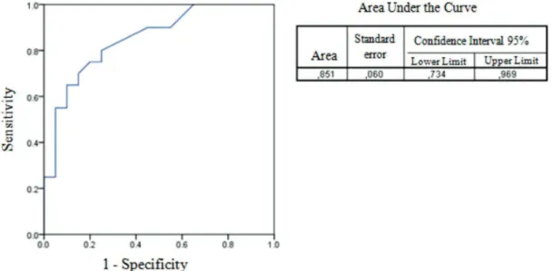
The cutoff points obtained from the ROC curves for iden-tifying composite grafts were 0.71 for DPV/SPV (Figure 1) and 0.09m for DVTI (Figure 2). The median values and the respective interquartile ranges for DPV/SPV and DVTI were also calculated. In the control group, the patients with
com-posite grafts had signiicantly higher values (P< 0.0001) than those with single grafts (Table 1). All patients of the control group with composite graft showed FD>0.5.
Study Group
Table 1 shows the median values and interquartile range of the variables in the control group and study group consid-ering the type of graft performed.
Table 2 shows the performance of our criterion DSV/SPV
≥0.71 in the assessment of patency of LITA composite grafts.
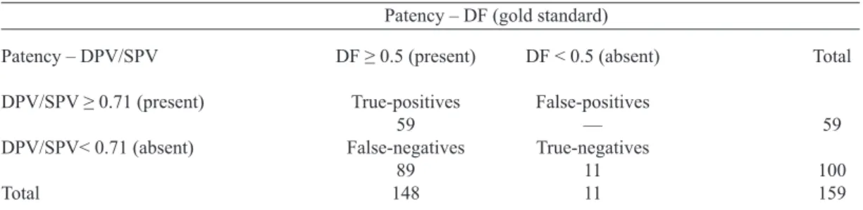
Because DF is the most accurate criterion according to the literature of LITA graft, it was our reference standard for val-idation of our two patency criteria for composite grafts. This table displays the rates of true-positive (TP), false-positive
(FP), false-negative (FN), and true-negative (TN) indings for the criterion DPV/SPV ≥ 0.71. Among the 159 patients of the study group, 59 were TP: they had DSV/SPV ≥ 0.71 and patency was conirmed by DF ≥ 0.05. Eleven were TN,
they neither reached the patency criterion for LITA compos-ite graft we were testing nor achieved the reference pattern. 89 were FN: our criterion had detected as non-patent or
non-composite graft but they had DF ≥ 0.05.
Fig. 2 – ROC curve in the control group to determine the best cutoff point for the variable VTId in evaluating the patency of composite graft
Table 1. The medians and interquartile ranges (lower and upper quartiles) of the variables analyzed during the standardization and validation phases by type of graft used.
Variables
DPV/SPV DVTI (m)
Variables
DPV/SPV DVTI (m)
Median
0.64 0.06
Interquartile range
0.45 – 0.69 0.04 – 0.08
Median
0.87 0.12
Interquartile range
0.69 – 1.05 0.09 – 0.16
Statistical signiicance
P<0.0001***
P<0.0001*** Control Group
Simple graft (n=20) Composite graft (n=20)
Study Group
Median
0.64 0.08
Interquartile range
0.45 – 0.69 0.06 – 0.10 Composite graft (n=159)
DPV/SPV = Peak diastolic velocity/peak systolic velocity; DVTI = Diastolic velocity integral
Table 2. Detecting composite graft patency (present or absent) using DPV/SPV, with DF as the gold standard. Data obtained from 38 patients.
Patency – DPV/SPV
DPV/SPV ≥ 0.71 (present)
DPV/SPV< 0.71 (absent)
Total
DF ≥ 0.5 (present)
True-positives 59 False-negatives
89 148
DF < 0.5 (absent)
False-positives –– True-negatives
11 11
Total
59
100 159 Patency – DF (gold standard)
Table 3 shows the same for the criterion DVTI ≥ 0.09m. The number of TP was 54: they had DVTI ≥ 0.09m and pa
-tency was conirmed. There were also eleven TN. 94 patients,
out of a total of 159, were FN, meaning our criterion had detected as non-patent or non-composite graft but they had
DF ≥ 0.05. There was no FP with our two criteria: no graft
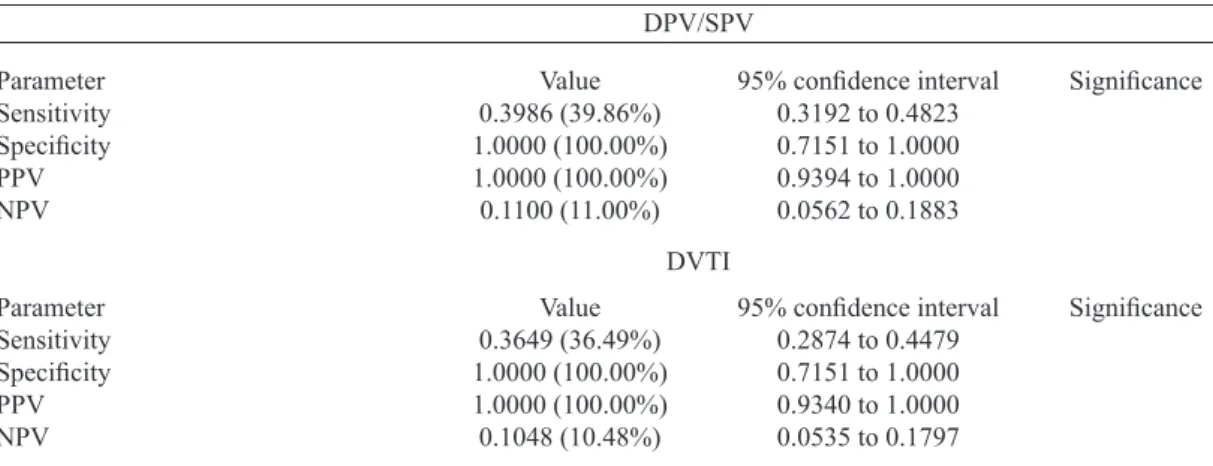
detected as patent composite graft had DF lower than 0.05. In Table 4, we can see the percentages of sensitivity,
spec-iicity, positive and negative predictive values calculated for
our patency criteria of the composite grafts, all of them with
conidence interval of 95%. For both criteria, DPV/SPV ≥ 0.71 and DVTI ≥ 0.09m, the speciicity and positive predic -tive value were 100%.
DISCUSSION
The prognosis of the CABG patients depends on
demon-stration of an eficient by pass function. This study is one of
Table 3. Detecting composite graft patency (present or absent) using DVTI, with DF as the gold standard. Data obtained from 38 patients.
Patency – DVTI
DVTI ≥ 0.09 m (present)
DVTI< 0.09 m (absent)
Total
DF ≥ 0.5 (present)
True-positives 54 False-negatives
94 148
DF < 0.5 (absent)
False-positives –– True-negatives
11 11
Total
54
105 159 Patency – DF (gold standard)
DVTI = Diastolic average velocity-time integral; DF = Diastolic velocity integral
Table 4. Validation criteria for using DPV/SPV to detect composite graft patency, with DF as the gold standard.
Sensitivity, speciicity, positive predictive value (PPV), negative predictive value (NPV), and the associated 95% conidence intervals were calculated.
Parameter Sensitivity
Speciicity
PPV NPV
Parameter Sensitivity
Speciicity
PPV NPV
Value 0.3986 (39.86%) 1.0000 (100.00%) 1.0000 (100.00%) 0.1100 (11.00%)
Value 0.3649 (36.49%) 1.0000 (100.00%) 1.0000 (100.00%) 0.1048 (10.48%)
95% conidence interval
0.3192 to 0.4823 0.7151 to 1.0000 0.9394 to 1.0000 0.0562 to 0.1883
95% conidence interval
0.2874 to 0.4479 0.7151 to 1.0000 0.9340 to 1.0000 0.0535 to 0.1797
Signiicance
Signiicance
DPV/SPV
DPV/SPV = Peak diastolic velocity/peak systolic velocity; DVTI = Diastolic velocity integral; PPV = Positive Predictive Value; NPV = Negative Predictive Value
DVTI
the irst contributions to inding criteria to establish patency
of composite graft of LITA and SMV revascularizing LAD and another branch of the LCS, using TDE at rest; a totally noninvasive diagnostic method which is part of the routine clinical practice [17].
It is important to mention that all patients in the control group
had their graft patency conirmed by CA. This is meaningful because it corroborates the eficiency of the criteria we applied
in the study group. In the study group phase, an actual practical application of these criteria occurred in a considerable sample of 159 patients with composite graft. This sample is consistent with the reality of the routine follow-up of CABG patients. This evaluation is not easy because these patients can present
atypi-cal symptoms and nonspeciic, or even inconclusive, changes in
some exams, such as exercise stress test for example.
The VTI is calculated by using the mean velocity, map-ping the Doppler spectral curve point to point rather than at
punctual time. That’s why it quantiies the low with more re -liability than the peak velocity. It’s simple and reproducible.
Peak velocity is affected by the intravascular resistance and pressure and is a measure of the maximum velocity at one moment during systole or diastole. Even so, Shimizu et al. [18] and Dubey et al. [19] have shown that the graft
diam-eter, its degree of obstruction, and its low, are proportional to
DPV [20]. A DPV/SPV ratio greater than 1 [21] is associated
with good angiographic indings and can provide sensitivity up to 100% and a speciicity of 58% for detecting graft pa -tency [12].
The DF expresses how much the diastolic low represents in relation to the total (systolic plus diastolic) low and it can be calculated using VTI (m) or the blood low (ml/min).
When VTI is used, the diastolic VTI fraction is obtained by dividing the DVTI by the sum of the VTI in systole plus VTI
in diastole. When blood low is used, the diastolic fraction is the diastolic blood low divided by the sum of diastolic and systolic blood low. Blood low is obtained from the prod -uct of three variables: VTI, cross-sectional vessel area, and
heart rate. For this reason, blood low is more susceptible to
interoperator variations and to the status of the patient at the time of the exam. The diastolic fraction calculated using the velocity integral has shown a good correlation with cardiac catheterization results. A DF value lower than 0.5 is predic-tive of graft stenosis [16,22].
The speciicity of the two criteria evaluated in this
study was 100%, meaning that all of the composite grafts
that were not patent were correctly classiied by the cutoff
points. Thus, DPV/SPV and DVTI values greater than or equal to the cutoffs were a strong indicators of composite
graft patency. Despite the 100% speciicity, the possibility of
false-positives cannot be ruled out. During the surgical report collection phase, it was observed that some single grafts had
a surprisingly high diastolic low to LAD at rest. In these
cases, patients even with obstruction of the SMV graft to the other LCS branch would meet the patency criteria for patent composite grafts established in this study.
A large number of patients did not meet our criteria for patency of the composite graft, but they had DF ≥ 0.5; the
FN. LITA low was suggestive of obstruction at rest. Several factors can affect the LITA graft low and may explain these data. One of the grafts in the coniguration of the composite
graft could be obstructed; however, what we could observe
more frequently was the latent low. The latency of the low
when there is no obstruction in the graft but rather a reduced
demand is described in the literature. Understanding low
competition is extremely important to grasp the complex
anatomy and pathophysiology of coronary low. LAD low
competition is associated with a reduction in LITA diastolic
velocity. Invasive studies have shown that residual low in
the recipient artery, when the obstruction is not severe, can
compete with the low of the patent LITA, thereby reducing blood low in the graft. In addition, collateral circulation re -sulting from the total obstruction of a vessel, also called a
physiological bridge, can lead to low competition within the
LITA bypass [18].As the coronary disease progress es in the
native or collaterals, the low of the graft increases consider -ably. It is described in the literature and we can notice it in everyday practice. LITA has the ability to restore its patency after apparent occlusion in the progression of the coronary
disease. Other conditions can inluence LITA low, includ -ing severe dysfunction of the left ventricle [6], coronary
mi-crocirculation disease, hypertrophy, ibrosis and myocardial
viability [24,25].TDE at stress can be very helpful in these situations and it can avoid a more invasive test [23]. The
in-creased demand will enhance the velocity of the graft low and conirm that it was a false negative and the graft is patent.
The realization that there is graft malfunction with TDE can’t
be done unless the graft low has been previously registered
and its patency established.
There was a sensitivity of 39.86% for DPV/SPV and 36.49% for DVTI. One may think that the detection rate is low, yet as sensitivity represents the ability to identify patent composite grafts among those that are truly patent, it is not a low rate. It is even audacious if in 40% of CABG patients with composite grafts coming for an echocardiogram with
three more minutes we are able to afirm that their grafts are
patent. Moreover, TDE is not expensive and totally nonin-vasive. It can be repeated frequently in routine or forward to a change in clinical status, providing the possibility of com-parison of data during follow ups. Actually, it is much more important to compare previous Doppler measurements in a given patient than isolated ones.
Final considerations
Studying resting coronary graft low with Doppler
echocardiography in a completely noninvasive manner is
challenging. The coronary vessel pattern is similar to a in -gerprint and then, there are unlimited normal anatomical variations. Coronary heart disease involves a large number of
other pathophysiological conditions that can affect the low
through the coronary arteries and their grafts. Similar to oth-er exams, Transthoracic Dopploth-er echocardiography requires accurate interpretation, and the multifactorial context must be considered.
low reserve and notice its potential increase. Values equal or
greater than the cutoffs are probably indicators of composite graft patency. These cases do not require additional testing be-yond the resting TDE, although the medical decision making always depends on the clinical setting. One of the mains
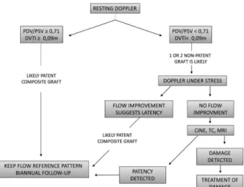
con-tributions of this study is briely illustrated in Figure 3.
REFERENCES
1. Cerqueira Neto FM, Guedes MA, Soares LE, Almeida GS, Guimarães AR, Barreto MA, et al. Flowmetry of left internal thoracic artery graft to left anterior descending artery: comparison between on-pump and off-pump surgery. Rev Bras Cir Cardiovasc. 2012;27(2):283-9.
2. Nieman K, Pattynama PM, Rensing BJ, Van Geuns RJ, De Feyter PJ. Evaluation of patients after coronary artery bypass surgery: CT angiographic assessment of grafts and coronary arteries. Radiology. 2003;229(3):749-56.
3. Tello R, Hartnell GG, Costello P, Ecker CP. Coronary artery bypass graft low: qualitative evaluation with cine single-detector row CT and comparison with indings at angiography. Radiology. 2002;224(3):913-8.
4. Rozen WM, Alonso-Burgos A, Murray AC, Whitaker IS. Is there a need for preoperative imaging of the internal mammary recipient site for autologous breast reconstruction? Ann Plast Surg. 2013;70(1):111-5.
5. Brenner DJ, Hall EJ. Computed tomography: an increasing source of radiation exposure. N Engl J Med. 2007;357(22):2277-84.
6. Fusejima K. Noninvasive measurement of coronary artery blood flow using combined two-dimensional and Doppler echocardiography. J Am Coll Cardiol. 1987;10(5):1024-31.
7. Takagi T, Yohikawa J, Yoshida K, Akasaka T. Noninvasive assessment of left internal mammary artery graft patency using duplex Doppler echocardiography from supraclavicular fossa. J Am Coll Cardiol. 1993;22(6):1647-52.
8. Crowley JJ, Shapiro LM. Noninvasive assessment of left internal mammary artery graft patency using transthoracic echocardiography. Circulation. 1995;92(9 Suppl):II25-30.
9. Gaudino M, Serricchio M, Tondi P, Glieca F, Giordano A, Trani C, et al. Non-invasive evaluation of mammary artery low reserve and adequacy to increased myocardial oxygen demand. Eur J Cardiothorac Surg. 1998;13(4):404-9.
10. Chirillo F, Bruni A, Balestra G, Cavallini C, Olivari Z, Thomas JD, et al. Assessment internal mammary artery and saphenous vein graft patency and low reserve using transthoracic Doppler echocardiography. Heart. 2001;86(4):424-31.
11. Arruda A, Campos Filho O, Ribeiro E, Petrizzo A, Andrade JL, Carvalho AC, et al. Assessment of left internal thoracic artery anastomosis with left anterior descending coronary artery by Doppler echocardiography. Arq Bras Cardiol. 1997;69(6):413-9.
12. De Simone L, Caso P, Severino S, Scherillo M, D’Andrea A, Varricchio A, et al. Noninvasive assessment of left and right internal mammary artery graft patency with high-frequency transthoracic echocardiography. J Am Soc Echocardiogr. 1999;12(10):841-9.
Fig. 3 – Follow-up algorithm for bypass patients with composite LITA grafts that are grafted to the LAD and another LCS branch
(1100% speciicity for both, 39.86% sensitivity for DPV/SPV, 36.49% sensitivity for DVTI, 100% PPV for both, 11% NPV for DPV/SPV, and 10.48% NPV for DVTI)
Study and technique limitations
The Doppler imaging may not have visualized the LITA for anatomical or operator-dependent reasons. The
anatom-ical reasons include unfavorable conigurations of the rib
cage, the position and diameters of the LITA graft, and the mobility of the heart [6-22]. The overlap of vessels in the su-praclavicular block may also have impaired the visualization of LITA.
In the quantitative evaluation of the graft low, the LITA di -ameter was considered to be equal during systole and diastole
because of the technical dificulty in measuring this difference.
This study did not obtain data on comorbidities, the medications used by the patients, previous coronary anatomy, or the segmental and overall myocardial contractility, all of
which are factors that can affect LITA low.
CONCLUSION
Values equal or greater than those established by the criteria
DPV/SPV ≥ 0.71 and DVTI ≥ 0.09m suggest high probability
13. Pezzano A, Fusco R, Child M, Riccobono S, Milazzo A, Recalcati F, et al. Assessment of left internal mammary artery grafts using dipyridamole Doppler echocardiography. Am J Cardiol. 1997;80(12):1603-6.
14. Lobo Filho JG, Leitão MCA, Lobo Filho HG, Silva AA, Machado JJA, Forte AJV, et al. Revascularização miocárdica com enxerto composto de artéria torácica interna esquerda em Y: análise de luxo sanguíneo. Rev Bras Cir Cardiovasc. 2004;19(1):1-8.
15. Lobo Filho JG, Leitão MCA, Forte AJV, Lobo Filho HG, Silva AA, Bastos ES, et al. Flow analysis of left internal thoracic artery in myocardial revascularization surgery using Y graft. Tex Heart Inst J. 2006;33(4):430-6.
16. Jones CM, Athanasiou T, Tekkis PP, Malinovski V, Purkayastha S, Haq A, et al. Does Doppler echography have a diagnostic role in patency assessment of internal thoracic artery grafts? Eur J Cardiothorac Surg. 2005;28(5):692-700.
17. Lobo Filho JG, Leitão MCA, Lobo Filho HG, Soares JPH, Magalhaes GA, Leão Filho CSC, et al. Cirurgia de revascularização coronariana esquerda sem CEC e sem manuseio da aorta em pacientes acima de 75 anos: análise das mortalidades imediata e a médio prazo e das complicações neurológicas no pós-operatório imediato. Rev Bras Cir Cardiovasc. 2002;17(3):208-14.
18. Shimizu T, Hirayama T, Suesada H, Ikeda K, Ito S, Ishimaru S. Effect of low competition on internal thoracic artery graft: postoperative velocimetric and angiographic study. J Thorac Cardiovasc Surg. 2000;120(3):459-65.
19. Dubey B, Bhan A, Choudhary SK, Sharma S, Sharma R, Airan B, et al. Assessment of internal mammary artery graft patency:
angiography or Doppler? Asian Cardiovasc Thorac Ann. 2000;8(4):325-9.
20. Cagli K, Emir M, Kunt A, Ergun K, Muharrem T, Murat T, et al. Evaluation of low characteristics of the left internal thoracic artery graft: perioperative color Doppler ultrasonography versus intraoperative free-bleeding technique. Tex Heart Inst J. 2004;31(4):376-81.
21. Hata M, Raman JS, Shiono M, Sezai A, Negishi N, Sezai Y, et al. What can Doppler wave forms of the left internal thoracic artery teach us? The eficacy of apical transthoracic approach of Doppler echocardiography. Ann Thorac Cardiovasc Surg. 2002;8(2):92-6.
22. El-Masry MM, Salama MM, Darwish AZ, El-Aziz OA. Assessment of left internal mammary artery graft patency by transthoracic Doppler echocardiography. Clin Cardiol. 2002;25(11):511-6.
23. Kitamura S, Kawachi K, Seki T, Sawabata N, Morita R, Kawata T. Angiographic demonstration of no-low anatomical patency of internal thoracic-coronary artery bypass grafts. Ann Thorac Surg. 1992;53(1):156-9.
24. Krams R, Sipkema P, Zegers J, Westerhof N. Contractility is the main determinant of coronary systolic low impediment. Am J Physiol. 1989;257(6 Pt 2):H1936-44.