RBCCV 44205-1467 DOI: 10.5935/1678-9741.20130038
Clinical and ultramicroscopic myocardial
randomized study of beating versus arrested heart
for mitral surgery
Estudo randomizado clínico e ultramicroscópico na cirurgia valvar mitral com o coração com atividade
elétrica versus sem atividade elétrica
Clotario Neptali Carrasco Cueva
1, Maiara dos Santos Rocha
2, Carlos Maurício Cardeal Mendes
3,
Luiz Antonio Rodrigues de Freitas
4, José Augusto Baucia
5, Roberto Badaró
61. Adjunct IV Professor of the Medical School of the Universidade Federal da Bahia (UFBA), teacher and cardiovascular surgeon, Salvador, BA, Brazil. Participated in all stages from project design, execution, analysis of results and writing of the article for publication.
2. Student of the Medicine Course of the UFBA, Salvador, BA, Brazil. Participated in the entire project with emphasis on the collection and organization of data.
3. Professor of Postgraduate Interactive Processes of Organs and Systems/ ICS/UFBA, Salvador, BA, Brazil. Performed the statistical analysis of the database.
4. Adjunct Professor of the Medical School of the UFBA, Salvador, BA, Brazil. Senior reseacher of FIOCRUZ. Performed histological analysis with electron ultramicroscopy.
5. Adjunct Professor of the Medical School of the UFBA, Salvador, BA, Brazil, Salvador, BA, Brazil. Participated in the project design, the execution of operations and in the writing.
6. Professor of the Medical School of the UFBA, Salvador, BA, Brazil. Participated in all stages from project design, execution, analysis of results and writing of the article for publication.
This study was carried out at Hospital Ana Nery and Centro de Pesquisas Gonçalo Moniz – Fiocruz, Bahia, Salvador, BA, Brazil.
Correspondence address: Clotario Neptali Carrasco Cueva
Departamento de Anestesiologia e Cirurgia, Disciplina de Cirurgia Torácica e Cardiovascular da Faculdade de Medicina da Universidade Federal da Bahia Av. Augusto Viana, S/N – Canela – Salvador, BA, Brasil – Zip code: 40110-160. E-mail: [email protected]
Article received on February 24th, 2013 Article accepted on May 6th, 2013 Abstract
Introduction: Beating heart surgery on normothermic by-pass simulates physiologic cardiac status.
Objectives: This study compared clinical and transmission electron microscopic aspects of myocardial protection during mitral valve replacement using warm retrograde perfusion in empty beating versus arrested heart with cold blood antero-grade cardioplegia.
Methods: Randomized study to evaluate myocardial cellular ischemia-reperfusion of both techniques to replace the mitral valve. Thirty-four patients were randomly assigned into group A (beating heart) and group B (arrested heart). The following parameters were assessed: echocardiography, blood chemistry, hemoglobin, lactate. During the surgical procedure a total of 102 myocardial biopsies were performed for ultrastructural analysis from anterior left ventricular wall: before cardiopulmonary by-pass, before aortic desclamping and 10 minutes after reperfusion.
Results: Elevation of lactate at 3 hours during the proce-dure was higher in group A, but similar at the end of surgery (P=0.06). Cardioversion was necessary in 5/17 (A) vs. 13/17 (B) P=0.07. Median intraoperative systemic temperature was
sig-niicantly lower in the group B compared to A (32ºC vs. 36ºC),
P<0.001. There was no signiicant difference of the ultramicro -scopic aspects of the heart biopsies before, during and after surgery in both groups. Cellular and mitochondrial transient abnormalities such as mitochondrial swelling, glycogen loss and cytosol swelling were detected independently of the moment of the biopsies.
Conclusion: Myocardial protection and ultrastructural ab-normalities were similar for both types of mitral valve replace-ment beating or arrested heart techniques.
Abbreviations, acronyms & symbols
advocating one or the other technique should be taken into consideration to decide between either a beating or arrested heart surgery procedure.
In this report we present the results of clinical and transmission electron microscopy (TEM) myocardial assessment in the immediate and postoperative period in a randomized prospective study to compare mitral valve replacement using arrested versus on-pump empty beating heart surgery.
METHODS
Design of study
This was a randomized prospective study to compare mitral valve replacement using arrested versus on-pump beating heart surgery. The objective of this study was to
evaluate the immediate clinical indings and cellular ischemia
and reperfusion injury of both techniques to replace the mitral valve. Herein we will report the clinical and the preliminary
cellular and mitochondrial TEM indings of biopsies of the
myocardium performed during the surgery heart procedure in both groups.
Local of study
This study was performed at the Hospital Ana Neri, the reference Center for Cardiac surgery of the Federal University
ATC ATP BMI CPB ECG FDR Fiocruz ICU KCl LAD MVR NYHA TEM
Activated time coagulation Adenosine triphosphate Body mass index Cardiopulmonary bypass Electrocardiogram False discovery rates Osvaldo Cruz Foundation Intensive care unit Potassium chloride
Left anterior descendent artery Mitral valve replacement New York Heart Association Transmission electron microscopy
INTRODUCTION
During open-heart surgery prevention of ischemia and reperfusion following cardioplegic arrest are essential for myocardial protection [1]. It has been demonstrated that empty beating heart surgery simulates a physiologic cardiac status and is a good method for myocardial protection [2]. On the other hand, most surgeons prefer the conventional
hypothermic arrested heart surgery justiied by the low
risk of air embolism and less blood in the surgical site that improves visualization [3,4]. Several complications are well documented in arrested heart surgery such as increasing the need for inotropic drug support, intra-aortic balloon pumping, prolonging of the length of intensive care, prolonging of hospital stays, increasing cost and mortality rates [5]. Also, low cardiac output syndrome and severe arrhythmias are associated with postoperative morbidity and mortality [6]. Although beating heart valve replacement with continuous coronary sinus perfusion is a good method for myocardial protection because eliminate the use of cardioplegia corollary risk of ischemic reperfusion injury [7,8]. In addition, beating heart surgery produces less cardiac arrhythmia events and no major differences in cerebrovascular events [9,10]. Beating heart technique is being demonstrated to produces less alterations on the biochemistry markers so, have better myocardium protection [11]. The pros and cons for
Resumo
Introdução: A cirurgia valvar mitral pode ser realizada com o coração com atividade elétrica, vazio e normotérmico com pinça-mento aórtico, perfusão sanguínea no seio coronário, simulando
um estado isiológico.
Objetivos: Comparar as manifestações clínicas e ultrami-croscópicas do miocárdio, na cirurgia valvar mitral, com o coração com atividade elétrica versus sem atividade elétrica.
Métodos: Estudo randomizado constituído de 34 pacientes:
grupo A (batendo) e grupo B (parado). Os parâmetros foram:
hematológico, bioquímico, ecocardiográico, lactato. Foram re -alizadas 102 biopsias da parede anterior do ventrículo esquerdo preparadas para análise ultraestrutural: antes da circulação ex-tracorpórea, antes do despinçamento aórtico e 10 minutos após a interrupção da circulação extracorpórea.
Resultados: Veriicou-se elevação do lactato 3 horas após o início do procedimento, que foi maior no grupo A (P=0,06),
todavia semelhantes no inal da cirurgia. A cardioversão foi
necessária em (A) 5/17 vs. (B) 13/17, P=0,07. A temperatura
in-traoperatória média foi signiicativamente menor no grupo B em
relação ao grupo A (32oC vs. 36oC), P<0,001. A análise ultrami-croscópica das amostras das biopsias do coração antes da circu-lação extracorpórea, ao término do pinçamento aórtico e após a saída da circulação extracorpórea, revelou anormalidades tran-sitórias semelhantes no citoplasma, núcleos e mitocôndrias em ambos os grupos, independentemente do momento das biopsias. Conclusão: A proteção miocárdica na cirurgia valvar mitral apresentou aspectos semelhantes na preservação da integridade ultraestrutural dos cardiomiócitos quando realizada com o coração com ou sem atividade elétrica.
of Bahia, Brazil and the transmission electron microscopy studies were performed at Osvaldo Cruz Foundation (Fiocruz), Bahia, Brazil.
Data collection period
The patients were enrolled from April 2010 to March 2011. Demographic, history of dyslipidemia, hypertension personal habits, history of rheumatic fever and other valvulopathies. History of stroke, hematological and laboratory chemistry
proiles. Body mass index (BMI) was calculated using the
height in relation to weight formula.
Population of study
A total of 34 patients were selected to have mitral valve substitution.
Inclusion criteria: a) all patient included into the study were adults 18-60 years old, b) with an echocardiography diagnosis of mitral and/or tricuspid valve disease due to
inlammatory acquired diseases, c) no previous history
of cardiac surgery and d) elective indication for valve replacement.
Exclusion criteria: a) with metabolic diseases such
as diabetes mellitus and uremia, b) with coronary artery diseases, c) dilated myocardiopathy, d) with severe chronic pulmonary obstructive diseases, e) with present or past history of malignances diseases, f) acute endocarditis, g) with severe pre-operatory laboratory parameters such as creatinine levels
>3mg/dL, hemoglobin ≤ 7.0 g/dL, prothrombin time/activity ≤70% and clotting time ≥ 10 minutes.
Randomization procedure
We used a simple method that consisted in use of a randomization list for the enrollment of the patients: This list was provided by the biostatistician to consecutively enroll into group A or B according to the following random sequence: AAAAAAABBABABBAAABABAABBBBA BBBBABB. A Runs Test of validation for randomization was applied Standard Normal = -0.6966, P-value = 0.486; alternative hypothesis: two sided, this test accepted the null hypothesis that this sequence was random. As recommended
by CONSORT 2010 [12] a low diagram was constructed to
depict the enrollment of the patients (Figure 1). Surgical risk was measured using EuroScore formula available on web: euroscore.org/calc.htlm
Surgery techniques
Anesthetic protocol was identical for all patients. All procedures were performed with median sternotomy. Prior to aortic and venous cannulation for the institution of CPB, 400 U/kg heparin sulphates was administrated to the patients in order to maintain the activated time coagulation (ATC) values above 480 seconds. During cardiopulmonary bypass
(CPB), non-pulsatile low was kept at 2.0 L/m2/min to 2.5
L/m2/min with a roller pump and hollow-iber membrane
oxygenator were used. Mean arterial pressure during CPB was maintained between 60 mmHg and 70 mmHg. Oxygen
low was maintained during CBP a saturation of 90%
concentration.
Group A: All patients received normothermic blood cardioplegia delivered retrograde via coronary sinus [13]. After cross-clamping aorta to avoid air embolism the left heart was kept vented in the ascendant aorta and right pulmonary vein. Surgery was performed under a temperature of 36oC using an actively warmer system for blood perfusion.
Systemic temperature was monitored with a catheter placed
into nasopharyngeal. The low rate was kept between 250-300
mL/min and a peak perfusion pressure of 40-60 mmHg [14]. Myocardial function was monitored continuously during the intraoperative procedure with 5-lead electrocardiogram (ECG). Occurrences of bradycardia or ST-segment changes were considered as sign of myocardial ischemia and were immediately corrected.
Group B: surgery was performed under intentionally actively cooled moderate hypothermia of 32oC and
monitored as same as in group A also a beating alarm sound when threshold limit of 31-34oC were reached. Myocardial
protection was performed using blood cardioplegia 4:1, anterograde, intermittent. With the following components of the solution; for induction: Potassium chloride (KCl)
15 ml, sodium bicarbonate 60 ml, glucose 5% 350 ml,
sodium aspartate 13.5 ml, sodium glutamate 13,5 ml. For maintenance: KCl 5 ml, Sodium bicarbonate 60 ml, glucose
5% 350 ml, sodium aspartate 13.5 ml, sodium glutamate
13.5 ml. In all patients, the heart was arrested with 300 ml/min of the induction solution delivered at 4oC during 2
minute with an interval of 15 minutes into the aortic root at a pressure of 60 mmHg measured directly through a separate port of the cardioplegic cannula. After arrest, low potassium cardioplegia solution (maintenance) was infused via anterograde cannula and the aortic root was perfused through the cannula with oxygenated blood at the same rate as for the induction. Measurement of intramyocadial temperature was not performed. Inotropic support was done using a dopamine solution at the dose of 5 mcg/kg/min when blood pressure was inferior to 50 mmHg.
In both groups, the right atrium was opened longitudinally and another incision was made into the fossa ovale: superiorly toward the superior vena cava and inferiorly behind coronary
sinus. Mitral valve replacement (MVR) was performed using a metallic or bioprostheses substitution by interrupted suture. The tricuspid valve repair was done following De Vegas’ technique in both groups and the CPB was disconnected [15].
Hemodynamic studies
Echocardiogram was performed before and after the surgery procedure in both groups using a transthoracic technique to measure ejection fraction and the systolic pressure of pulmonary artery. New York Heart Association (NYHA) functional class protocol was adopted to classify the degree of cardiac dysfunction of each patient before and after surgery.
Briely, Class I: cardiac disease, but no symptoms and no
limitation in ordinary physical activity, e.g. shortness of breath when walking, climbing stairs. Class II: mild symptoms (mild shortness of breath and/or angina) and slight limitation during ordinary activity. Class III: marked limitation in activity due to symptoms, even during less-than-ordinary activity, e.g. walking short distances (20–100 m). Comfortable only at rest Class IV: severe limitations. Experiences symptoms even while at rest. Mostly bedbound patients.
Myocardial biopsy
During the surgery biopsies were performed as full thickness transmural specimen of three small fragments measuring 0.5 to 15 mm2 from the left ventricle anterior free wall, near to the
apex and between distal left anterior descendent artery (LAD) and the diagonal branch. Biopsies were taken with Tru-Cut biopsy needle (Travenol Laboratories Inc.™) at three different times: (a) before ischemia (b) at the end of ischemia and (c) 10
min after reperfusion. The samples were immediately ixed in cold 2.5% glutaraldehyde diluted in a 0.1-M cacodilate buffer
at pH of 7.4. After rinsing in a buffer, fragments were
post-ixed in a 1% osmium tetroxide solution mpost-ixed with the same
buffer during one hour at 4o
C. Fragments were dehydrated with increasing concentration solutions of acetone and then
iniltrated with polibed resin. Ultra-ine sections measuring
between 50-70 nm were obtained using an automatic ultra-microtome, Leica EM UC7™ and collected with 200-mesh copper grids. Histological semi-thin sections and ultra-structural photomicrographs were performed after samples analysis, registering repetitive patterns. Photomicrographs were obtained using a Zeiss EM-109 TEM operated at 80 KV.
Classiication of the microscopic alterations
The assessment of the ultramicroscopic photography’s was based of the system grade 0-4. It was observed the mitochondria, cytoplasmic and nuclear changes according to the following criteria: Grade 0: no structural changes, Grade
1: mild intermyoibrillar and intramitochondrial edema,
lost of glycogen. Grade 3: severe edema, sarcolemma bled, myocardial contraction band, mitochondrial amorphous densities, and disruption of sarcolemma membrane. Grade 4: lost of cytoplasmic architecture, myocardial contraction bands, mitochondrial amorphous densities, and disruption of sarcolemma membrane. All analysis of these subcellular ultramicroscopic alteration of the myocytes was done blinded by the pathologist without known which study group were the sample.
Anatomopathology of the replaced valve
All valve that was replaced, immediately after removal was sent to the laboratory for study.
Statistical analysis
A data bank was constructed and analyzed using the R statistical analysis program. Each individual received a code number and the bank was accessed through an individual password. Because this was a randomized trial of superiority hypothesis tests we performed a comparison for proportions using Fisher’s exact test. For the comparison of distribution
of non-normal quantitative variables between groups we performed the Mann-Whitney test. For quantitative variables with repeated measurement throughout the study we use the analysis of variances. Due to simultaneous analysis of numerous hypotheses we calculated the recommended false discovery rates (FDR) by direct transformation of the P values according to the method proposed by Rosner [16].
Ethical issues
Written informed consent was obtained from each individual patient prior to enrollment into the study. The study was approved by the Ethics Committee of the University Hospital Professor Edgard Santos under the number 098/2009.
RESULTS
Table 1 summarized the preoperative patients proile data
obtained in both groups. Of note, the degree of functional
classiication of the extent of heart failure according NYHA was signiicantly different between the two groups.
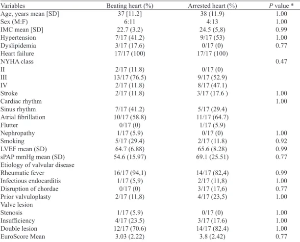
Table 1. Preoperative clinical proiles of studied patients in both groups.
Variables
Age, years mean [SD] Sex (M:F)
IMC mean [SD] Hypertension Dyslipidemia Heart failure NYHA class II III IV Stroke Cardiac rhythm Sinus rhythm Atrial ibrillation Flutter Nephropathy Smoking LVEF mean (SD) sPAP mmHg mean (SD) Etiology of valvular disease Rheumatic fever
Infectious endocarditis Disruption of chordae Prior valvuloplasty Valve lesion Stenosis Insuficiency Double lesion EuroScore Mean
Beating heart (%)
37 [11.2] 6:11 22.7 (3.2) 7/17 (41.2) 3/17 (17.6) 17/17 (100) 2/17 (11.8) 13/17 (76.5) 2/17 (11.8) 2/17 (11.8) 7/17 (41.2) 10/17 (58.8) 0/17 (0) 1/17 (5.9) 5/17 (29.4) 64.7 (6.88) 54.6 (15.97) 16/17 (94,1) 1/17 (5,9) 0/17 (0) 2/17 (11,8) 1/17 (5.9) 4/17 (23.5) 12/17 (70.6) 3.03 (2.22)
Arrested heart (%)
38 (11.9) 4:13 24.5 (5,8) 9/17 (53) 0/17 (0) 17/17 (100) 0/17 (0) 9/17 (52.9) 8/17 (47.1) 3/17 (17.6 )
5/17 (29.4) 11/17 (64.7) 1/17 (5.9) 0/17 (0) 2/17 (11.8) 65.6 (8.28) 69.1 (25.51) 14/17 (82,4) 2/17 (11,8) 3/17 (17,6) 4/17 (23,5) 0/17 (0) 3/17 (17.6) 14/17 (82.4) 3.8 (2.42)
Patients included in the arrested heart group had more severe heart failure. However, heart failure did not correlate with more alteration in the cardiac rhythms. There was no
signiicant difference in cardiac rhythms between the two
groups. Also the rate of rheumatic mitral valve disease was found to be similar in both groups.
Intraoperatory aspects
Table 2 summarized all variables that could be compared. Basically, surgery time, CPB and aorta cross clamp time
were almost identical. But, the need for deibrillation was marginally signiicant higher in group B than group A. Also temperature was signiicant lower in group B as expected
because the technique requirement P=0.005). Group A required less use of inotropic drugs during and after surgical procedure than group B since the heart was constantly beating (P=0.005). The most common association procedure was tricuspid annuloplasty (8/17 group A and 14/17 group B), P=0, 07. The median size of the biological or mechanical prosthetic valve was 31 mm in both groups. Only one patient was submitted to comissurotomy mitral valve in group A. Blood loss analysis was confounded by a single patient in
group A that had a blood clotting dysfunction and accounted for total blood lost in this group.
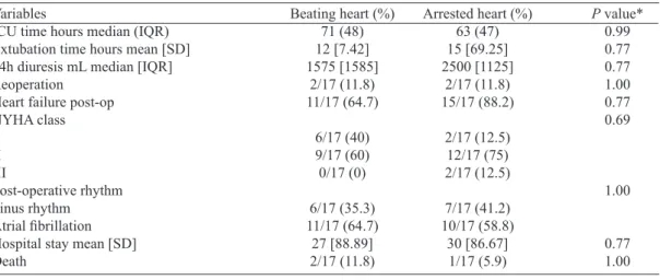
The follow up variable analysis are shown in Table 3. There was no difference between both groups for intensive care unit (ICU) time for recovery also occurrence of heart failure independently of the NYHA functional class before and after surgery. Also, the frequencies of arrhythmias were similar in both groups.
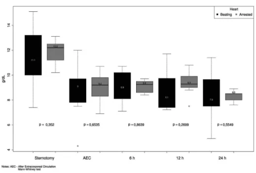
Overall, the outcomes of the patients after surgery were similar for both groups. Only one patient died in the arrested heart group ten days after surgery due to septic shock and two in the beating heart group ten and 42 days after surgery due to septic shock and endocarditis respectively. Figure 2 shows the monitoring of hemoglobin values during and immediately after the surgery procedure. As expected the hemoglobin levels in the blood dropped off in both
group without signiicant difference related with the type
of surgical procedure. Figure 3 demonstrates the lactate monitoring levels in both groups during the surgery. There
was a signiicant elevation of lactate at 3 hours during the
procedure in group A compared with group B, but at the end of the surgery in both groups the levels were similar.
Table 2. Comparison of the intraoperatory parameters between the two surgery procedures of mitral valve replacement replacement.
Variables
Surgery time min mean [SD] Cardiopulmonary bypass (IQR) (min) Aorta cross clamp time (min) Median (IQR)
Deibrillation. n
Temperature oC median (IQR)
Blood lost mL median (IQR) Mean arterial pressure Inotropic requirement Tricuspid annuloplasty
Beating heart (%)
N=17 214 [SD=36] 85 [SD=30] 60 [SD=14] 5/17 (29.5) 36 [SD=0.5] 1150 [SD=600] 60 (SD=10) 4/17 (23.5) 8/17 (47.1)
Arrested heart (%)
N=17 234 [SD=72] 95 [SD=27] 60 [SD=27] 13/17 (76.5) 32 [SD=0] 1100 [SD=1400] 64 [SD=18] 13/17 (76.5) 14/17 (82.4)
P value*
0.94 1.00 0.94 0.63 0.00 1.00 1.00 0.12 0.63
Min = minutes; SD = standard deviation; IQR = median; n = number of deibrillation episodes; mL - milliliters; * P value: FDR *FDR
Table 3. Clinical and after surgery characteristics of mitral valve replacement in both group.
Variables
ICU time hours median (IQR) Extubation time hours mean [SD] 24h diuresis mL median [IQR] Reoperation
Heart failure post-op NYHA class I II III Post-operative rhythm Sinus rhythm Atrial ibrillation
Hospital stay mean [SD] Death
Beating heart (%)
71 (48) 12 [7.42] 1575 [1585] 2/17 (11.8) 11/17 (64.7) 6/17 (40) 9/17 (60) 0/17 (0) 6/17 (35.3) 11/17 (64.7) 27 [88.89] 2/17 (11.8)
Arrested heart (%)
63 (47) 15 [69.25] 2500 [1125] 2/17 (11.8) 15/17 (88.2) 2/17 (12.5) 12/17 (75) 2/17 (12.5) 7/17 (41.2) 10/17 (58.8) 30 [86.67] 1/17 (5.9)
Fig. 2 – Hemoglobin levels of the patients of both groups during procedure
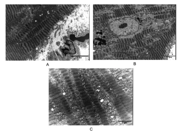
TEM myocardial biopsy indings
Figures 4 (Group A) and 5 (Group B). The igures
A, B and C represent the ultramicroscopic aspects of the
mitochondria and cytoplasm: igure A (biopsy 1) - before the bypass; igure B (biopsy 2) - at the end of ischemic arrest; and igure C (biopsy 3) - 10 minutes after reperfusion pre, during
and post cardiac surgery of the three performed biopsies for both groups respectively. Figures 4A and 5A are biopsies performed before ischemic arrest that demonstrates cytosolic edema with mitochondria swelling grade 1 and glycogen loss seen in the biopsy before ischemic arrest. Figure 4B and 5B are biopsies performed at the end of ischemic arrest that show a sarcoplasmic reticule and mitochondrial swollen with
Fig. 4 – Myocardial ultramicroscopic representative pictures of the biopsies during mitral valve replacement of group A (beating heart). Figures 4A is a biopsy before ischemia, 4B is a biopsy at the end of ischemia, and igures 4C is a biopsy 10 minutes during reperfusion
focal cristae disruption grade 1 and 2. Figure 4C and 5C are biopsies performed 10 minutes post CPB, after reperfusion shows the mitochondrial swelling with cristae disruption and condensation, sarcomeres were intact. Counting the frequencies of that alteration was performed a quantitative analysis. In Table 4 we summarized the frequencies of those cellular and mitochondria alterations studied. At the end of ischemic arrest there was an increasing in cellular and mitochondria alterations in both groups. The statistical analysis of the comparison of both groups revealed no
signiicant difference; however, a more detailed description is under preparation and will be subject of another speciic
Table 4. Cellular and mitochondrial quantitative frequencies of alterations in both groups.
Ultraestrutural abnormalities (type of alteration)
1st biopsy (cellular)
1st biopsy (mitochondrial)
2nd biopsy (cellular)
2nd biopsy (mitochondrial)
3rd biopsy (cellular)
3rd biopsy (mitochondrial)
Beating heart (%)
Group A N=17 Present Absent
Arrested heart (%)
Group B N=17 Present Absent
*P value
0.94 0.77 0.94 0.77 0.77 0.77 7/17 (41.2)
3/17 (17.6) 8/17 (47.1) 9/17 (52.9) 4/17 (23.5) 3/17 (17.6)
10/17 (58.8) 14/17 (82.4) 9/17 (52.9) 8/17 (47.1) 13/17 (76.5) 14/17 (82.4)
10/17 (58.8) 7/17 (41.2) 11/17 (64.7) 13/17 (76.5) 8/17 (47.1) 8/17 (47.1)
7/17 (41.2) 10/17 (58.8)
6/17 (35.3) 4/17 (23.5) 9/17 (52.9) 9/17 (52.9) N: number of patients; (%) percentage of patients; *FDR: false Discovery rates
Fig. 5 – Myocardial ultramicroscopic representative pictures of the biopsies during mitral valve replacement of group B (arrested heart). Figures 5A is a biopsy before ischemia, 5B is a biopsy at the end of ischemia, and igures 5C is a biopsy 10 minutes during reperfusion
DISCUSSION
In this comparative randomized prospective study of mitral valve replacement using arrested heart versus on-pump empty beating heart surgery we demonstrated that
there is no signiicant difference in terms of clinic aspects
between the two surgical procedures independently of
the NYHA heart failure classiication before and after the
surgical intervention. Also, preliminary studies of TEM myocardial biopsies performed pre and post procedure do not favor one method over the other particularly when looking at
mitochondria, nuclei or cytoplasmic alterations related to the type of surgical method.
surgical procedure. In a systematic review of beating-heart valve surgery, published by Salhiyyah & Taggart [10], 39 reports were reviewed, however only two were randomized control trials. Indeed, the report of Matsumoto et al. [9] that studied 50 patients (25 each group ) concluded that the perfusion technique of either retrograde warm blood or blood perfusion on beating had similar outcome including mortality and complications even though ultra microscopy study were not performed, the biochemical markers of
myocardial injury were signiicantly lower in the retrograde
technique. Despite of our study limitation present incomplete biochemistry analysis, were similar for the mortality rates and complications. Otherwise, in the report of Karadeniz et al. [20] that focused on the comparison of neurological parameters for the three groups with a total of 50 patients, even though the beating heart technique did not cross-clamp the aorta to avoid embolism, the results of neurological
indings were similar. In our study all 34 patients had none
neurological alteration at the immediately post surgery follow up. Also, in the 31 remain patients during 12 moth follow up. Since 1971 [21] when a warm blood cardioplegia surgery was introduced and routinely used most studies that compared with hypothermic cardioplegia, despite of the majority were observational studies, all of them agree that the major advantage of this technique is that heart beating protects against ischemia reperfusion injury in addition to reduced workload and cardiopulmonary bypass time, special for mitral valve replacement that the repair are done close to physiological conditions in the state of left ventricular beating tonus [10,21,22]. This knowledge has been extrapolated, but not proven by most these studies that beating heart protects against mitochondrial and cellular damage [6,10,23]. Even though our study had the limitation of small number of patients included into the study, the analysis of 2400 microphotographies from 102 biopsies performed among those 34 patients studied patients, do not support the idea of more injury occur in one technique than the other. The preliminary results presented here do not support the idea of beating heart surgery better protects the myocytes of ultrastructure alterations induced by the type of reperfusion, at least in terms of myocardial necrosis, cellular and mitochondrial alterations.
Indeed, suggesting that the myocardial protection for subcellular damages is independent of keep the heart beating or arrested. In the experimental work reported by Kamlot et al. [24] also they reported that dog heart submitted to 180 minutes of cardiopulmonary bypass either with continuous warm or intermittent cold cardioprotection, no difference was noted in the electron microscopy damage with both cardioplegic protections. Interesting, adenosine triphosphate (ATP), creatine phosphate and lactates levels were slightly degraded in both cardioplegia groups, but the adenosine
triphosphate levels were signiicantly reduced in the right
ventricular of the warm compared to the cold group. In our study, the time of cardiopulmonary bypass was around 85-95 minutes. Even though we have not analyzed the ATP levels in our study further we will present details of the qualitative and quantitative ultra structural aspects of the myocardium biopsies compared in both groups. As we have
presented here, there are no signiicant differences cellular
and mitochondrial alterations between both groups. This is similar to the work of Schaper et al. [1] who reported an ultra-structural morphometric study of myocardial biopsies performed in 31 patients submitted to aortic valve replacement with cardiopulmonary bypass after induction of cardiac arrested surgery which revealed similar results to those we have encountered in our study. Biopsies performed before and after clamping show cellular and mitochondria alteration compatible with ischemia, which apparently are reversible after desclamping.
It has been suggested that the coronary sinus pressure should be maintain 60-80 mmHg when beating heart procedures are
selected. Also, the optimal low rate are not yet determined, it
has been suggested that between 150-250ml/min is the minimal
necessary to warrant 4-5% of the total cardiac output [6,9]. A
limitation of our study was a small number of patients in both groups to careful evaluate this variable. Another important point is the degree of severity of the damage of the valve. Simultaneous antegrade/retrograde warm blood perfusion with a beating heart has been used independent of the severity of valve disease [13]. Even, we used the NYHA class IV cardiac
failure score no signiicant difference was noted using one
or another technique. On the other hand studies monitoring
troponin I levels monitoring shown no signiicant difference
on the cardiac troponin I concentration of warm reperfusion to cold blood cardioplegia [8,25]. The need of more utilization of dopamine in the arrested group in our study was due to the routine procedure adopted by our anesthesiologist of keep the blood pressure above 50mmHg all time during the surgery. Another limitation of our study was not having done the biopsy of the right ventricle, which could provide better evaluation of this ventricle.
CONCLUSION
We conclude that no signiicant cellular and mitochondrial
damage occurs with heart surgery for valve replacement using arrested or on-pump empty beating heart surgery. Also, both techniques are safe and have minimal risk of death direct related to the procedure.
ACKNOWLEDGEMENT
in this work. Also, we thank Maria Lucia Vieira for her strong technical support on the Ultramicroscopic tissue sections preparation. We thank Dr. Sanjay Mehta for English revision of the manuscript.
DISCLOSURE
There is no conlict of interest and no participation or
approvals from manufactures companies.
REFERENCES
1. Schaper J, Schwarz F, Kittstein H, Stämmler G, Winkler B, Scheld H, et al. The effects of global ischemia and reperfusion on human myocardium: quantitative evaluation by electron microscopic morphometry. Ann Thorac Surg. 1982;33(2):116-22.
2. Ricci M, Macedo FI, Suarez MR, Brown M, Alba J, Salerno TA. Multiple valve surgery with beating heart technique. Ann Thorac Surg. 2009;87(2):527-31.
3. Kaplon RJ, Pham SM, Salerno TA. Beating-heart valvular surgery: a possible alternative for patients with severely compromised ventricular function. J Card Surg. 2002;17(2):170-2.
4. Cressoni ES, Avanci LE, Braile DM, Cicogna AC, Lima-Oliveira AP, Gerez MA, et al. Myocardial protection to the hypertrophied heart: the eternal challenge. Rev Bras Cir Cardiovasc. 2008;23(1):97-107.
5. Salerno TA, Houck JP, Barrozo CA, Panos A, Christakis GT, Abel JG, et al. Retrograde continuous warm blood cardioplegia: a new concept in myocardial protection. Ann Thorac Surg. 1991;51(2):245-7.
6. Babaroglu S, Yay K, Parlar AI, Ates C, Mungan U, Cicekcioglu F, et al. Beating heart versus conventional mitral valve surgery. Interact Cardiovasc Thorac Surg. 2011;12(3):441-7.
7. Chambers DJ, Fallouh HB. Cardioplegia and cardiac surgery: pharmacological arrest and cardioprotection during global ischemia and reperfusion. Pharmacol Ther. 2010;127(1):41-52.
8. Evora PR, Pearson PJ, Seccombe JF, Schaff HV. Ischemia-reperfusion lesion. Physiopathologic aspects and the importance of the endothelial function. Arq Bras Cardiol. 1996;66(4):239-45.
9. Matsumoto Y, Watanabe G, Endo M, Sasaki H, Kasashima F, Kosugi I. Eficacy and safety of on-pump beating heart surgery for valvular disease. Ann Thorac Surg. 2002;74(3):678-83.
10. Salhiyyah K, Taggart D. Beating-heart valve surgery: A systematic review. Asian Cardiovasc Thorac Ann. 2009;17(6):650-8.
11. Mizuno T, Arai H. On-pump beating-heart mitral valve plasty
without aortic cross-clamping. Jpn J Thorac Cardiovasc Surg. 2006;54(10):454-7.
12. Schulz KF, Altman DG, Moher D; CONSORT Group. CONSORT 2010 statement: updated guidelines for reporting parallel group randomized trials. Ann Intern Med. 2010;152(11):726-32.
13. Salerno TA, Panos AL, Tian G, Deslauriers R, Calcaterra D, Ricci M. Surgery for cardiac valves and aortic root without cardioplegic arrest (“beating heart”): experience with a new method of myocardial perfusion. J Card Surg. 2007;22(6):459-64.
14. Ikonomidis JS, Yau TM, Weisel RD, Hayashida N, Fu X, Komeda M, et al. Optimal low rates for retrograde warm cardioplegia. J Thorac Cardiovasc Surg. 1994;107(2):510-9.
15. Bara C, Zhang R, Haverich A. De Vega annuloplasty for tricuspid valve repair in posttraumatic tricuspid insuficiency: 16 years experience. Int J Cardiol. 2008;126(3):e61-2.
16. Rosner B. Fundamentals of biostatistics. 6a ed. New York: Duxbury Press; 2006.
17. Hannan EL, Wu C, Smith CR, Higgins RS, Carlson RE, Culliford AT, et al. Off-pump versus on-pump coronary artery bypass graft surgery: differences in short-term outcomes and in long-term mortality and need for subsequent revascularization. Circulation. 2007;116(10):1145-52.
18. Karolak W, Hirsch G, Buth K, Légaré JF. Medium-term outcomes of coronary artery bypass graft surgery on pump versus off pump: results from a randomized controlled trial. Am Heart J. 2007;153(4):689-95.
19. Kloner RA, Jennings RB. Consequences of brief ischemia: stunning, preconditioning, and their clinical implications: part 1. Circulation. 2001;104(24):2981-9.
20. Karadeniz U, Erdemli O, Yamak B, Genel N, Tutun U, Aksoyek A, et al. On-pump beating heart versus hypothermic arrested heart valve replacement surgery. J Card Surg. 2008;23(2):107-13.
21. Follette DM, Steed DL, Foglia RP, Fey KH, Buckberg GD. Reduction of postischemic myocardial damage by maintaining arrest during initial reperfusion. Surg Forum. 1977;28:281-3.
22. Gersak B. Mitral valve repair or replacement on the beating heart. Heart Surg Forum. 2000;3(3):232-7.
23. Botta L, Cannata A, Bruschi G, Fratto P, Martinelli L. Beating heart mitral valve surgery: innovation or back to the past? J Card Surg. 2010;25(3):318.
24. Kamlot A, Bellows SD, Simkhovich BZ, Hale SL, Aoki A, Kloner RA, et al. Is warm retrograde blood cardioplegia better than cold for myocardial protection? Ann Thorac Surg. 1997;63(1):98-104.