110 111
110 111
INTRODUCTION
Periodontitis is a multi-factorial disease with microbial dental plaque as the initiator of
peri-Clinical and microbial evaluation of dental scaling associated with
subgingival minocycline in chronic periodontitis subjects
Avaliação clínica e microbiana da raspagem dental associada à
minociclina subgengival em indivíduos com periodontite crônica
Silvia Maria Rodrigues Querido* Sheila Cavalca Cortelli**
Marcelo Werneck Barata de Araújo*** José Roberto Cortelli****
* Research Assistant, Dental Research and Graduate Studies Division; **Assistant Professor, Microbiology and Periodontics Research and Graduate Studies Division, Department of Dentistry; ****Professor and Chairman, Preventive Dentistry and Periodontics Research and Graduate Studies Division, Department of Dentistry – University of Taubaté.
*** Assistant Professor, Dental Research and Graduate Studies Division, Department of Periodontology, Guarulhos University.
ABSTRACT: The aims of this double-blind randomized clinical trial were to evaluate the presence of periodontal pathogens and the clinical response of periodontal pockets treatment to scaling and root planing (SRP) associated with subgingival minocycline (SM). A total of 36 subjects, 26 to 60 years old (40.7 ± 9.1), who had been previously diagnosed with chronic periodontitis, were included in the present study. Eighteen subjects were selected for the test group (TG), who were treated with SRP plus SM (new treatment), and 18 subjects for the control group (CG) who received SRP plus vehicle (current treatment). Two homologous sites in each subject with a probing depth (PD) ≥ 6 mm were chosen. To evaluate the clinical response after treatment, PD was measured at baseline and at 90 days. Microbiological evaluation was performed to detect 7 periodontal pathogens using polymerase chain reac-tion at baseline, 30, and 120 days. A mean reducreac-tion in PD of 2.8 and 2.1 mm was observed in the TG and CG, respectively. At baseline, P. gingivalis was the most prevalent organism in both test (65.8%) and control (48.6%) groups. After 120 days it fell to 30.8% in TG and to 23.1% in CG. There were no statistically significant differ-ences between the test and control groups concerning PD (p > 0.05 by Wilcoxon test) or presence of periodontal pathogens (p > 0.05 by Wilcoxon and chi-square; p > 0.01 by Signal test). The results observed showed that the new treatmentwas as effective as the current treatmentin reducing periodontal pathogens and PD among chronic periodontitis subjects.
DESCRIPTORS: Bacteria; Dental scaling; Minocycline.
RESUMO: O objetivo deste estudo randomizado duplo-cego foi avaliar a presença de periodontopatógenos e o comportamento clínico de bolsas periodontais tratadas com raspagem e aplainamento radicular (RAR) associa-do à minociclina subgengival (MS). Foram incluíassocia-dos no presente estuassocia-do 36 indivíduos de 26 a 60 anos de idade (40,7 ± 9,1), previamente diagnosticados com periodontite crônica. Dezoito indivíduos foram selecionados para o grupo teste (GT), tratado com RAR e MS (novo tratamento), e 18 indivíduos, para o grupo controle (GC), que recebeu RAR e veículo (tratamento convencional). Foram selecionados dois sítios homólogos em cada indivíduo com profundidade de sondagem (PS) ≥ 6 mm para testar a hipótese proposta. Para avaliar o comportamento clí-nico após o tratamento, a mensuração da PS foi realizada inicialmente e após 90 dias. A avaliação microbiana foi realizada para detecção de 7 periodontopatógenos através de reação em cadeia da polimerase, inicialmente e após 30 e 120 dias. Observou-se redução média de 2,8 e 2,1 mm na PS nos grupos teste e controle, respectivamente. Inicialmente, P. gingivalis foi o microrganismo mais prevalente tanto no GT (65,8%) quanto no GC (48,6%). Após 120 dias, houve redução para 30,8% no GT e 23,1% no GC. Não houve diferenças estatisticamente significativas entre os grupos em relação à PS (teste Wilcoxon - p > 0,05) e presença dos periodontopatógenos (teste Wilcoxon e qui-quadrado - p > 0,05 e teste do sinal - p > 0,01). Os resultados observados demonstraram que o novo trata-mento foi tão efetivo quanto o tratatrata-mento convencional na redução de periodontopatógenos e de PS em indivíduos com periodontite crônica.
DESCRITORES: Bactérias; Raspagem dentária; Minociclina.
110 111
110 111
variety of determinants and factors including sub-ject characteristics, social and behavioral factors, systemic factors, genetic factors, tooth-level fac-tors, microbial composition of dental plaque and other emerging risk factors12.
There is plenty of evidence that over 300 dif-ferent species of bacteria are recognized in the oral cavity, but only a few are directly associated with periodontal disease11. These include P. gingivalis, P. intermedia, A. actinomycetemcomitans, T. forsy-thensis (formerly B. forsythus), F. nucleatum, Sele-nomona species, C. rectus and E. corrodens18.
Mechanical therapy (scaling and root planing) aimed at the removal of pathogens and their prod-ucts, has been shown to be effective in reducing bacterial colonization to sub-threshold values, in order to control the progression of periodontal dis-ease (current treatment). However, scaling and root planing (SRP) may not eradicate species that have invasive properties, i.e., bacterial species that can reach epithelial cells and subepithelial connective tissues of the periodontium1, leading to the use of
antimicrobial agents, which may help to eliminate the pathogens3.
Several locally delivered antimicrobial agents currently used are designed to enhance the heal-ing process and improve periodontal health. In diseased sites that are more difficult to control, the application of local drug delivery devices may be a valid approach for treating infected sites9.
However, research has focused on the devel-opment of a new treatment to appropriately de-liver the drug into the periodontal pockets. For instance, a method of microencapsulating mino-cycline hydrochloride in a bioabsorbable polymer (polyglycolide-co-dl-lactide) has been developed, resulting in microspheres that are administered into sites with periodontal disease22.
The aim of this study was to evaluate the presence of periodontal pathogens and the clini-cal response to SRP periodontal pocket treatment associated with subgingival minocycline (SM) in chronic periodontitis subjects.
MATERIALS AND METHODS
Thirty-six subjects with generalized severe chronic periodontitis were recruited to participate in this double-blind randomized clinical trial, af-ter informed consent (Institutional Committee on Research Involving Human, protocol n. 013/02) was obtained from each subject. To be included in the study, subjects had to meet requirements for
inclusion (non-treated chronic periodontitis, and patient had to be in good general health) and exclu-sion criteria (according to Cortelli et al.8, 2003).
At baseline, periodontal and radiographic ex-aminations determined the periodontal status of all subjects, based on the criteria presented by the American Academy of Periodontology2 (1999).
During the periodontal examination, one trained, calibrated examiner measured periodontal probing depth (PD) at 6 sites per tooth, using a manual periodontal probe (PQWBR – Hu Friedy Mfg. Inc. Chicago, IL, USA). The reproducibility of these measurements were analyzed using kappa statis-tics, and obtained kappa = 0.83. Two homologous sites with PD ≥ 6 mm without furcation involve-ment were selected from each subject to test the hypothesis proposed. After randomization, two groups were formed; one considered a test group (SRP plus SM) and one a control group (SRP plus vehicle). All subjects were examined at baseline and at 90 days for measurement of PD. In addition, a trained examiner evaluated supragingival plaque control (Oral Hygiene Index - OHI13) and oral
hy-giene compliance at baseline and at 90 days. Subgingival samples were taken from each of the two sites from each subject using sterile paper points (Tanari, Tanariman Industrial Ltda., Manacapuru, Brazil) inserted to the depth of the pockets after the removal of supragingival plaque. Paper points were removed after 15 seconds, and immediately placed in phosphate-buffered saline (Promega, Madison, WI, USA) (pH 7.4) on ice. These microbiological samples were collected at baseline, 30, and 120 days.
112 113
112 113
Avila-Campos et al.4 (1999) (F. nucleatum), Saiki et al.14 (1988) (T. forsythensis, C. rectus, E. corrodens, P. gingivalis and P. intermedia) and Cortelli et al.8
(2003) (A. actinomycetemcomitans).
PCR products were analyzed using 1.5% agar-ose gel (Sigma, Dorset, UK) electrophoresis per-formed at 4 V/cm in tris-acetate EDTA buffer (Pro-mega, Madison, WI, USA). The gel was stained with 0.5 µg/ml ethidium bromide (Amershan, Airlington Heights, USA) and photographed under 300 nm ultraviolet light.
Each subject received local anesthesia followed by full-mouth SRP both supra- and subgingivally according to individual needs. Following SRP, the test group was treated with minocycline micro-spheres (Arestin, OraPharma, Inc., Warmister, PA, USA), while the control group was treated with vehicle (polymer without minocycline - Byofórmula, manipulation pharmacy, São Paulo, Brazil).
The microspheres were dispensed subgingival-ly to the base of the periodontal pocket by means of a disposable plastic cartridge (OraPharma Inc., Warmister, PA, USA) affixed to a stainless-steel handle. Each cartridge contains 1 mg of mino-cycline microencapsulated in Poly (glycolie-co dl-lactide) dry powder and it is enough for one peri-odontal pocket. At 90 days, test and control groups received an additional application of the assigned treatment.
Statistical analysis
Sample size calculation was performed us-ing the PS – Power Sample Size program (Biostat, Englewood, NJ, USA), version 2.1.30. The sample size of 36 subjects was reached when α = 0.05, power = 0.80, and an initial difference between mean pocket depth of 0.1 mm among groups, and a 1:1 proportion of subjects in each group were established.
The changes between the baseline and the 90-day measurement of PD for test and control groups were examined by Wilcoxon test (p < 0.05). Differences in the OHI scores between groups and exams were analyzed by analysis of variance, New-man-Keuls and HSD – Tukey (Honest Significant Differences) tests (p < 0.05). The prevalence of each pathogen at baseline, 30, and 120 days was ana-lyzed by Wilcoxon test (p < 0.05). The proportional distribution of the bacteria between test and con-trol groups was analyzed by Signal test (p < 0.01). In addition, a chi-square test was used to deter-mine a positive association between two bacterial species (p < 0.05).
RESULTS
Thirty-six subjects (17 female and 19 male), aged 26 to 60 years old (mean 40.7 ± 9.1), par-ticipated in the present study. Data related to the mean of PD indicates that, at baseline, the val-ues for test and control groups were not different, 7.8 ± 0.9 mm and 7.9 ± 0.8 mm, respectively. After 90 days, the mean of PD decreased to 5.0 ± 1.4 mm among test group, and 5.8 ± 2.1 mm for the control group. The results did not show a statistically sig-nificant difference between the test and the control groups (p > 0.05 by Wilcoxon test).
Median values of the OHI scores were 93.5% for the test group and 95.5% for the control group at baseline, and after 90 days it decreased to 67.5% and 72.0% respectively for the test and control groups. A statistically significant difference was observed between baseline and 90 days for the OHI scores within each of the groups. However, when the difference between the test and control groups was investigated, no statistically significant differ-ences were observed between each time point.
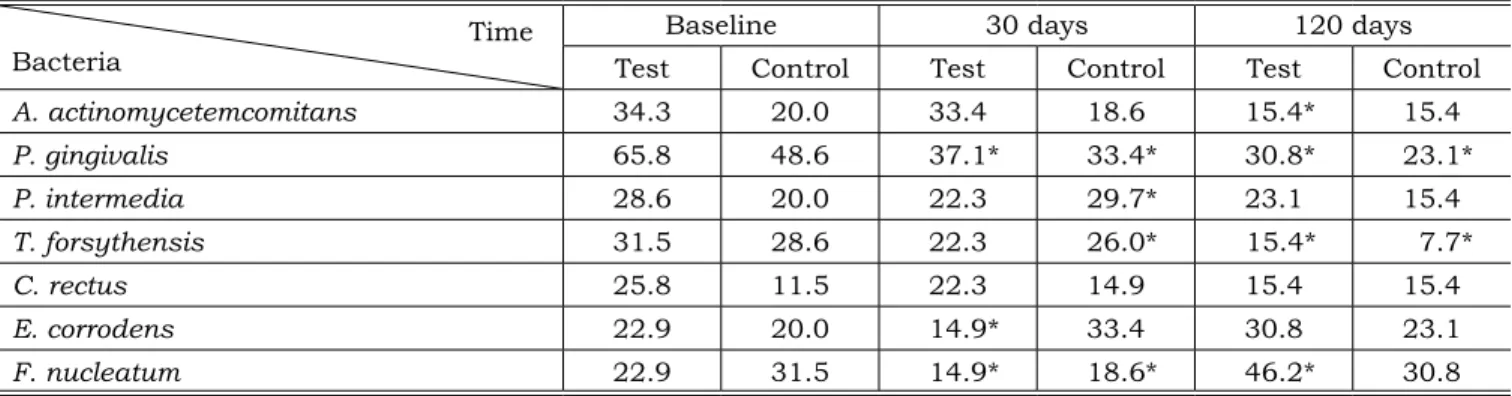
The prevalence of analyzed bacteria present in the 36 subjects, for the test and control groups at baseline, 30, and 120 days is indicated in Table 1. In the test group, the prevalence of A. actinomy-cetemcomitans (p = 0.01), P. gingivalis (p = 0.00) and T. forsythensis (p = 0.02) significantly de-creased after 120 days (p < 0.05). There was a sig-nificant increase in F. nucleatum (p = 0.01). The prevalence of bacterial species was higher among test than among controls at all three time-points. However, data indicated that prevalence decreased as time increased. At baseline, prevalence of bacte-ria ranged from 22.9 - 65.8% among the test group, and from 11.5 - 48.6% among controls. At 30 days, the prevalence among the test group ranged from 14.9 - 37.1% and from 14.9% - 33.4% among con-trols. At 120 days, bacterial prevalence among the test group had increased, compared to the 30-day time point (range: 15.4 - 46.2%). However, the val-ues were still below the levels observed at baseline. Among controls, prevalence at 120 days was lower than at the two previous time-points, ranging from 7.7% to 30.8%.
The Signal test did not show significant dif-ferences in the proportional distributions of peri-odontal pathogens between the test and control groups (p > 0.01).
acti-112 113
112 113
nomycetemcomitans and P. intermedia (p = 0.0049, χ2 = 7.93), A. actinomycetemcomitans and C. rectus
(p = 0.0176, χ2 = 5.64), A. actinomycetemcomitans
and P. gingivalis (p = 0.0485, χ2 = 3.89), and A. ac-tinomycetemcomitans and E. corrodens (p = 0.0211, χ2 = 5.32), all in the test group.
Among controls, statistically significant positive associations were found between P. gin-givalis and C. rectus (p = 0.0288, χ2 = 4.78 and
p = 0.0050, χ2 = 7.88 at baseline and at 120
days, respectively); P. gingivalis and T.
forsythen-sis (p = 0.0186, χ2 = 5.54); C. rectus and T. forsy-thensis (p = 0.0290, χ2 = 4.77); P. gingivalis and P. intermedia (p = 0.0370, χ2 = 4.35); A. actino-mycetemcomitans and P. gingivalis (p = 0.0050, χ2 = 7.88); and A. actinomycetemcomitans and C. rectus (p = 0.0003, χ2 = 13.00).
DISCUSSION
Mechanical therapy is usually the first mode of treatment recommended for most periodontal TABLE 1 - Prevalence (%) and distribution of periodontal pathogens among test and control groups (n = 36).
Time
Bacteria TestBaseline Control Test30 days Control Test120 days Control
A. actinomycetemcomitans 34.3 20.0 33.4 18.6 15.4* 15.4
P. gingivalis 65.8 48.6 37.1* 33.4* 30.8* 23.1*
P. intermedia 28.6 20.0 22.3 29.7* 23.1 15.4
T. forsythensis 31.5 28.6 22.3 26.0* 15.4* 7.7*
C. rectus 25.8 11.5 22.3 14.9 15.4 15.4
E. corrodens 22.9 20.0 14.9* 33.4 30.8 23.1
F. nucleatum 22.9 31.5 14.9* 18.6* 46.2* 30.8
*Statistically significant difference (p < 0.05) – Wilcoxon test.
FIGURE 1 - Chi-square test of associations among examined species in test and control groups at baseline, 30, and 120 days.
Microorganism Aa Pg Pi Cr Tf Ec
Pg NI - TG
NI - CG
Pi NI - TG NI - CG
Cr NI - TG NI - CG
NI - CG NI - CG
Tf NI - CG NI - CG
Ec
NI - TG/CG
Fn
NI = not-independent; TG = Test group; CG = Control group. Aa = A. actinomycetemcomitans; Pg = P. gingivalis; Pi = P. interme-dia; Cr = C. rectus; Fn = F. nucleatum; Ec = E. corrodens; Tf = T. forsythensis.
114 115
114 115
infections, as the mean probing depth reduc-tion ranges from 0.03 in pocket depths between 1-3 mm, to 2.16 mm in pocket depths greater than 6 mm7. In scientific investigations, probing depth
is a method frequently used to evaluate both treat-ment and disease progression21. Badersten et al.5
(1984) showed a reduction in probing depth of approximately 2.3 mm in a study of nonsurgical therapy of subjects with advanced chronic peri-odontitis. The results of this study are in agree-ment with the previous literature, as subjects in both groups responded favorably to initial treat-ment, with a reduction in PD of 2.9 mm among test and 2.4 mm among control groups.
In the present study, there were no significant differences between test and control groups with regard to probing depth. Likewise, Timmerman et al.19 (1996) showed no additional benefit in probing
depth when SRP was associated with SM, com-pared to controls.
It has been shown that the root surface itself harbors viable bacteria that may provide niches for subgingival bacterial recolonization1,10. The root
may become a bacterial reservoir from which peri-odontal pathogenic bacteria can recolonize previ-ously treated pockets and contribute to the failure of therapy or the recurrence of the disease6. The
present study analyzed the presence of periodon-tal pathogens at 30 and 120 days, time points established on the attempt to evaluate possible microbial recolonization, always 30 days after the subgingival administration of minocycline. Sbor-done et al.15 (1990) suggested that the microbiota
might reestablish itself within 40-60 days following subgingival debridement with poor supragingival plaque control. Deep pockets are particularly dif-ficult to control, since recolonization of the subgin-gival microbiota occurs at around 120-240 days, despite meticulous supragingival plaque control and multiple sessions of subgingival debridement9.
The subjects enrolled in our study received oral hygiene instructions and were closely followed by a trained examiner who evaluated plaque control and oral hygiene compliance. Despite the close plaque control, some of the severely periodontally-involved participants of the present study showed a similar pattern of recolonization previously ob-served by other investigators.
P. gingivalis in our study was the most preva-lent bacteria both in the test (65.8%) and con-trol (48.6%) groups at baseline. This bacteria was found in a high percentage in the test and control groups after 30 (37.1% and 33.4%, respectively)
and 120 days (30.8% and 23.1%, respectively). P. gingivalis has been considered as one of the most important pathogens in advanced periodontitis based on its high prevalence in progressive peri-odontitis lesions16,17. After 30 days, the prevalence
of only P. intermedia, C. rectus and E. corrodens increased in the control group. A reduction in bac-terial prevalence was observed in all species except for C. rectus, E. corrodens and F. nucleatum, which did not decrease after 120 days. The PCR method was used to characterize not only the specific bac-terial types, but also the possible intercorrelations that may occur. The results of the present study confirm that the studied bacterial correlations do not follow a unique pattern. These results may be of use in the explanation of bacterial interassocia-tion, one of the objectives of molecular biology. The present study also demonstrated no significant statistical differences between the test and control groups with regard to microbiological parameters. The high sensitivity of PCR may partially explain these findings, and the differences in the micro-bial profiles could be observed if a quantitative microbiological instead of a PCR approach had been chosen. Similarly, Timmerman et al.19 (1996)
showed a significant reduction in the prevalence of periodontal pathogens over a 15-month period, but no significant reduction was observed when local delivery of minocycline was used.
In contrast, van Steenberghe et al.20 (1999)
demonstrated that SM as an adjunct to SRP re-sulted in the reduction of P. gingivalis, P. inter-media, C. rectus, E. corrodens, F. nucleatum and T. denticola, a reduction in PD greater than that seen in the control. Although this study protocol20
did not allow optimal oral hygiene instruction and reinforcement, this was emphasized in the pres-ent study.
Williams et al.22 (2001) reported that SRP
plus SM was effective in reducing PD in chronic periodontitis individuals as shown in the present study. However, their results demonstrated that adjunctive therapy with minocycline microspheres provided significantly greater PD reduction than SRP alone.
114 115
114 115
minocycline. However, subgingival debridement in combination with oral hygiene instruction by itself has been shown to be an effective treatment.
CONCLUSION
In conclusion, the results of this double-blind randomized clinical trial indicated that SRP associ-ated with SM (new treatment) was as effective as mechanical therapy alone (current treatment) in reducing both the prevalence of periodontal
patho-REFERENCES
1. Adriaens PA, de Boever JA, Loesche WJ. Bacterial invasion in root cementum and radicular dentin of periodontally diseased teeth in humans. J Periodontol 1988;59(4):222-30.
2. American Academy of Periodontology. Ann Periodontol 1999;4(1):1-6.
3. American Academy of Periodontology. The role of con-trolled drug delivery for periodontitis. J Periodontol 2000;71(1):125-40.
4. Avila-Campos MJ, Sacchi CT, Whitney AM, Steigerwart AG, Mayer LW. Arbitrarily primed-PCR for identification and epidemiologic subtyping of oral isolates of F. nucleatum. J Periodontol 1999;70(10):1202-8.
5. Badersten A, Nilveus R, Egelberg J. Effect of nonsurgical periodontal therapy. II. Severely advanced periodontitis. J Clin Periodontol 1984;11(1):63-76.
6. Bollen CML, Mongardini C, Papaioannow W, van Steen-berghe D, Quirynen M. The effect of a one-stage-full-mouth disinfection on different intra-oral niches. Clini-cal and microbiologiClini-cal observations. J Clin Periodontol 1998;25(1):56-66.
7. Cobb CM. Non-surgical pocket therapy: mechanical. Ann Periodontol 1996;1:443-90.
8. Cortelli SC, Jorge AOC, Cortelli JR, Jordan SF, Haraszthy VI. Detection of highly and minimally leukotoxic A. acti-nomycetemcomitans strains in patients with periodontal disease. Pesqui Odontol Bras 2003;17(2):183-8.
9. Drisko CH. Nonsurgical periodontal therapy. Periodontol 2000 2001;25:77-88.
10. Giulianna G, Ammatuna P, Pizzo G, Capone F, D’Angelo M. Occurrence of invading bacteria in radicular dentin of periodontally diseased teeth: microbiological find-ings. J Clin Periodontol 1997;24(7):478-85.
11. Moore WEC, Moore LVH. The bacteria of periodontal diseases. Periodontol 2000 1994;5:66-77.
12. Nunn ME. Understanding the etiology of periodontitis: an overview of periodontal risk factors. Periodontol 2000 2003;32:11-23.
13. O’Leary TJ, Drake RB, Naylor JE. The plaque control record. J Periodontol 1972;43(1):38.
gens and probing depth in chronic periodontitis subjects.
ACKNOWLEDGMENT
The authors would like to thank Heather Ochs-Balcom, PhD, from the Department of Social and Preventive Medicine of the State University of New York at Buffalo, for her critical reading of the manuscript.
14. Saiki RK, Geldand DH, Stoffel S, Scharf SJ, Higuchi R, Horn GT, et al. Primer-mediated enzymatic amplifica-tion of DNA with a thermo stable DNA polymerase. Science 1988;239:487-91.
15. Sbordone L, Ramaglia L, Guletta E, Iacono V. Re-colonization of the subgingival microflora after scaling and root planing in human periodontitis. J Periodontol 1990;61(9):579-84.
16. Slots J, Bragd L, Wikström M, Dahlén G. The oc-currence of Actinobacillus actinomycetemcomitans, Bac-teroides gingivalis and BacBac-teroides intermedius in de-structive periodontal disease in adults. J Clin Periodontol 1986;13(6):570-7.
17. Slots J, Listgarten MA. Bacteroides gingivalis, Bacte-roides intermedius and Actinobacillus actinomycetemcomi-tans in human periodontal diseases. J Clin Periodontol 1988;15(2):85-93.
18. Slots J, Rams TE. Microbiology of periodontal dis-eases. In: Slots J, Taubman MA (ed). Contemporary oral microbiology and immunology. St. Louis: CV Mosby Co.; 1992. p. 425-43.
19. Timmerman MF, van der Weijden GA, van Steen-bergen TJM, Mantel MS, de Graaff J, van der Velden U. Evaluation of the long-term efficacy and safety of locally-applied minocycline in adult periodontitis patients. J Clin Periodontol 1996;23(8):707-16.
20. van Steenberghe D, Rosling B, Söder PO, Landry RG, van der Velden U, Timmerman MFT, et al. A 15-month eval-uation of the effects of repeated subgingival minocycline in chronic adult periodontitis. J Periodontol 1999;70(6):657-67.
21. Villata L, Baelum V. Reproducibility of attachment level recordings using an electronic and a conventional probe. J Periodontol 1996;67(12):1292-300.
22. Williams RC, Paquette DW, Offenbacher S, Adams DF, Armitage GC, Bray K, et al. Treatment of periodontitis by local administration of minocycline microspheres: a controlled trial. J Periodontol 2001;72(11):1535-44.