PB 11
Influence of root embedment material and periodontal ligament
simulation on fracture resistance tests
Influência do material de inclusão e da simulação do ligamento
periodontal nos ensaios de resistência à fratura
Carlos José Soares* Eliane Cristina Gava Pizi** Rodrigo Borges Fonseca***
Luis Roberto Marcondes Martins****
ABSTRACT: The aim of this study was to evaluate the influence of the embedment material and periodontal liga-ment simulation on fracture resistance of bovine teeth. Eighty bovine incisor teeth were randomized into 8 groups (n = 10), embedded in acrylic or polystyrene resin using 4 types of periodontal ligament simulation: 1 - absence of the ligament; 2 - polyether impression material; 3 - polysulfide impression material; 4 - polyurethane elastomeric material. The specimens were stored at 37°C and 100% humidity for 24 hours. Specimens were submitted to tangential load on the palatal surface at 0.5 mm/minute crosshead speed until fracture. The fracture modes were analyzed as follows: 1 - coronal fracture; 2 - cemento-enamel junction fracture; 3 - partial root fracture; 4 - total root fracture. Statistical analyses by two-way ANOVA and Tukey’s test were applied (p < 0.05). The results showed that root embedment method and periodontal ligament simulation have a significant effect on fracture resistance. Artificial periodontal ligament modified the fracture modes.
DESCRIPTORS: Fracture resistance; Periodontal ligament; Tooth root; Cementoenamel junction.
RESUMO: O objetivo deste estudo foi avaliar a influência do material de inclusão e da simulação de ligamento periodontal na resistência à fratura de dentes bovinos. Oitenta incisivos bovinos foram divididos em 8 grupos (n = 10) e, então, incluídos em cilindros com dois materiais, resina acrílica ou resina de poliestireno, usando-se quatro tipos de simulação do ligamento periodontal: 1 - ausência do ligamento; 2 - material de moldagem à base de poliéter; 3 - material de moldagem à base de polissulfeto; e 4 - material elastomérico à base de poliuretano. As amostras foram armazenadas em 100% de umidade a 37°C por 24 horas e então submetidas a carregamento tangencial na superfície palatina com velocidade de 0,5 mm/minuto até a fratura. Os padrões de fratura foram analisados de acordo com: 1 - fraturas coronais; 2 - fratura da junção esmalte-cemento; 3 - fratura parcial da raiz; 4 - fratura radicular total. A análise estatística empregou análise de variância fatorial e teste de Tukey (p < 0,05). Os resultados mostram que o método de inclusão e a simulação do ligamento periodontal tiveram efeito significa-tivo na resistência à fratura. O ligamento periodontal artificial modificou os padrões de fratura.
DESCRITORES: Resistência à fratura; Ligamento periodontal; Raiz dentária; Colo do dente.
INTRODUCTION
A great number of factors can influence the clinical behavior of indirect and direct adhesive restorations. Cavity preparation design9, the
tech-nique and materials for fixation of the restoration7
and mainly the restorative material composition may influence the fracture resistance of these restorations6,8,9,25. The ability of the tooth to
sup-port masticatory loads, having an adequate stress distribution over supporting tissues, seems to be decisive when the aim is to obtain a restoration with high fracture resistance.
Bone support and the periodontal ligament are important for the mechanisms of stress dis-tribution over teeth. On in vitro tests, the root em-bedment material should reproduce bone capacity to absorb masticatory load and thus support the compressive and tangential force in a fracture re-sistance test. The materials used for root embed-ment vary greatly: acrylic resin5,12,16, die stone1,4 or
even polystyrene resin10,11,25 can be used. Another
important aspect in a fracture resistance test is the simulation of the periodontal ligament. This proce-dure has been performed with the use of different
* Professor, Department of Operative Dentistry and Dental Materials, School of Dentistry, Federal University of Uberlândia. ** Professor, Department of Restorative Dentistry, School of Dentistry, University of Western São Paulo.
12 13
12 13
elastomeric materials; however, a great number of in vitro studies have eliminated this procedure4,6,7,8.
When the periodontal ligament is to be reproduced, it is important to define which material should be used. Elastomeric impression materials have been usually used: some researches used polyether im-pression material5,22,25, other studies recommended
the use of a silicone rubber material12,23, and other
ones employed a polyurethane elastomeric mate-rial that was originally created to be used on the fixation of automotive glasses10,11. However, there is
little discussion about the influence of the proper-ties of specific materials used on the periodontal ligament reproduction on fracture resistance tests and their interaction with the material used for root embedment.
With regard to this situation, it is hypothe-sized that the periodontal ligament simulation and embedment method can influence the fracture load and mode of the fracture on in vitro fracture tests. Therefore, the aim of this study was to evaluate the importance and influence of both the periodontal ligament reproduction with three different elasto-meric materials and the root embedment method with two types of resin on fracture resistance and fracture modes of bovine teeth.
MATERIALS AND METHODS
Eighty recently extracted bovine incisors with similar dimensions were selected and stored in aqueous 0.2% thymol solution (F.Maia Ind. Com., Cotia, SP, Brazil). Calculus deposits and soft tissue deposits were removed with periodontal curettes (Hu Friedy, Chicago, IL, USA); then, the teeth were cleaned using a rubber cup (Microdont, São Paulo, SP, Brazil) and fine pumice (Vigodent, RJ, Brazil) water slurry. Teeth were randomly divided into 8
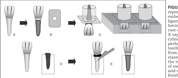
groups (n = 10) for each root embedment method and periodontal ligament simulation. Two types of resin were used: a self-cured acrylic resin (Jet Clássico, São Paulo, SP, Brazil), and a polystyrene resin (Cristal, Piracicaba, SP, Brazil). The teeth were mounted individually in plastic cylinders (Tigre, Rio Claro, SP, Brazil) and the roots were embedded in resin up to 2 mm below the cemento-enamel junction (CEJ). Four methods were used in order to reproduce the periodontal ligament: 1 - tooth embedded directly in the resin cylinder; 2 - tooth embedded using a polyether impression ma-terial (Impregum F, 3M-Espe, Seefeld, Germany); 3 - tooth embedded using a polysulfide impression material (Permlastic, Kerr, Romulus, USA); and 4 - tooth embedded using a polyurethane elastomeric material (Ultra flex, Solplas, Santo André, SP, Bra-zil). Root surfaces were dipped into melted wax (Epoxiglass, Diadema, SP, Brazil) up to 2.0 mm below the CEJ, resulting in a 0.2 to 0.3 mm thick wax layer. An X-ray film (Kodak, New York, USA) with a centralized circular hole with 5 mm in di-ameter was used to stabilize the teeth for the em-bedment procedure, 2.0 mm from the CEJ. This set was positioned downward over a perforated wood plate, and a plastic cylinder with 25.0 mm in diameter was positioned and fixed with wax. The resins were manipulated according to the manu-facturers’ instructions and inserted in the cylinder. After resin polymerization, the teeth were removed from the cylinder, and the wax was removed from the root surface and resin cylinder “alveolus”. The elastomeric materials were placed in the resin cyl-inders, the tooth was re-inserted into the cylinder and the excess elastomeric material was removed with a scalpel blade (Xishan Medical Instrument factory, Xishan, China) (Figure 1).
FIGURE 1 - Schematic
representation of root embedment and periodontal ligament simulation: (A) bovine incisor tooth; (B) tooth root covered with wax and X-ray fi lm; (C) fi lm and plastic cylinder positioned over a perforated wood plate; (D) tooth without wax removed from the resin cylinder and elastomeric material placed in the resin cylinder; (E) removal of excess elastomeric material and embedment procedure fi nished.
A B C
12 13
12 13
The teeth were stored for 24 hours at 37°C in 100% humidity, and then submitted to a tangential load at 0.5 mm/minute crosshead speed, on a test-ing machine (Instron 4411, Canton, MA, USA). The antagonistic metallic tooth (NiCr alloy, Verabond, Cordelha, USA) was fixed to the universal testing
machine and positioned on the incisal third of the lingual surfaces of the teeth (Figure 2A). The load required to fracture the specimens was recorded (kgf) and data were submitted to statistical analy-sis by two-way ANOVA (Table 1) and Tukey’s test (α = 5%), with two factors being studied: embed-ment material and periodontal membrane simula-tion material. The fracture modes were analyzed using the scale shown in Figure 2B.
RESULTS
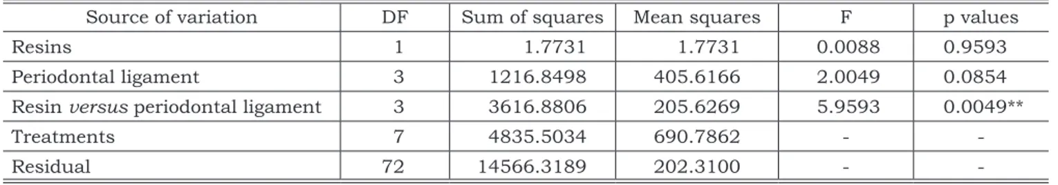
Mean values of fracture resistance are de-scribed in Table 2. Statistical analysis by two-way ANOVA indicated that there were significant differ-ences only for the interaction between periodontal ligament simulation and root embedment mate-rials (Table 1). Tukey’s test revealed statistically significant differences among the groups (Table 2 and Graph 1).
The fracture modes of the specimens are pre-sented in Table 3. The teeth embedded in acrylic resin or polystyrene resin without periodontal
FIGURE 2 - Load application method (A); Classification of the fracture modes (B) – (I) coronal fracture; (II) fracture at
the limit of the resin cylinder; (III) fracture with partial invasion of the cylinder insertion; (IV) root fracture. B. Classification of the fracture modes
A. Antagonist metallic tooth used in fracture test
III IV
II I
TABLE 1 - Two-way analysis of variance.
Source of variation DF Sum of squares Mean squares F p values
Resins 1 1.7731 1.7731 0.0088 0.9593
Periodontal ligament 3 1216.8498 405.6166 2.0049 0.0854
Resin versus periodontal ligament 3 3616.8806 205.6269 5.9593 0.0049**
Treatments 7 4835.5034 690.7862 -
-Residual 72 14566.3189 202.3100 -
-**Statistical significance at the level of 5%. DF: degrees of freedom.
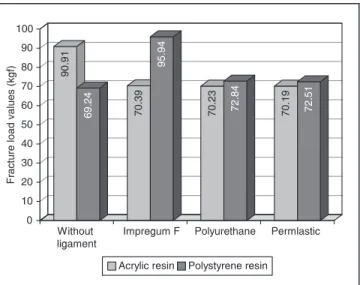
TABLE 2 - Means and standard deviations (SD) of
frac-ture load values in kgf.
Resins Ligament Materials Means ± SD
A
cr
yl
ic
resi
n
Without ligament 90.91 ± 13.59a
Impregum F (polyether) 70.39 ± 13.27b
Ultra flex (polyurethane) 70.23 ±10.71b
Permlastic (polysulfide) 70.19 ± 11.13b
Po
lyst
yr
en
e
resi
n
Impregum F (polyether) 95.94 ± 14.65a
Ultra flex (polyurethane) 72.84 ± 14.32b
Permlastic (polysulfide) 72.51 ± 11.84b
Without ligament 69.24 ± 21.72b
14 15
14 15
ligament simulation tended to fracture on the top of the resin cylinders, while the teeth with peri-odontal ligament simulation tended to fracture in different locations with a greater prevalence at the root portion (types III and IV fracture modes).
DISCUSSION
Teeth can fracture during normal function or traumatic occlusal contacts23. Normal function is
usually related to low load values, whereas trauma can be related to fast and/or high load values. Tooth fracture resistance is analyzed by in vitro studies through mechanical tests4,6,7,8,14,25.
Sever-al studies have used compressive and tangentiSever-al load application over teeth, restorations, posts and prostheses in order to determine their fracture re-sistance. However, a great number of factors may influence the results, especially if some procedures
like mode of load application, crosshead speed, mode of fracture and root embedment method are not standardized. An in vitro test should standard-ize these factors to better represent a clinical situ-ation.
The periodontal ligament is an important structure for the stress distribution generated by load application over teeth18,22. When load is
applied, periodontal fibers are compressed, the tooth dislodges slightly and there is bone distor-tion in the direcdistor-tion of the root movement19. The
initial resistance of the periodontal fibers against tooth displacement is low. However, as the tooth is forced within its alveolus, the periodontal resis-tance is progressively increased17. When the
peri-odontal fibers achieve maximum load resistance, similar to an hydraulic system, the periodontal membrane gets rigid, the load is transferred to the bone support and the stress is distributed to bone in all root surfaces. The mechanical response of a soft tissue to external stress is non-linear and viscous20, which is similar to the characteristics
of elastomeric materials used on impression pro-cedures.
Rees21 (2001), analyzing the importance of
periodontal ligament through a finite element analysis, showed that it is mandatory to include the characteristics of both the periodontal ligament and the alveolar bone in these tests. Isidor et al.12
(1996) reported that the simulation of periodon-tal ligament is essential to determine the stress distributions as similar as possible to the clinical situation, and that a small bar of resin composite at the tip of the embedded roots should be used to create sufficient retention of the mounted roots during load application when a silicone artificial periodontal membrane is used. The tooth remo-tion from the artificial alveolus was not verified in this study. Sirimai et al.24 (1999) reported that it is
TABLE 3 - Fracture modes of the specimens after tangential load tests.
Embedment Material Ligament type Distribution of fracture modes on specimens
I II III IV Total
Polystyrene resin
Without ligament 1 8 1 - 10
Impregum F (polyether) - - 5 5 10
Permlastic (polysulfide) - 5 4 1 10
Ultra flex (polyurethane) - 3 3 4 10
Acrylic resin
Without ligament 2 7 1 - 10
Impregum F (polyether) - 3 4 3 10
Permlastic (polysulfide) 1 4 4 1 10
Ultra flex (polyurethane) - 4 4 2 10
GRAPH 1 - Means of fracture load values in kgf for each
of the four types of simulation of periodontal ligament for the two resins used to simulate bone support.
90
.9
1
69
.2
4
70
.3
9
95
.9
4
70
.2
3
72
.8
4
70
.1
9
72
.5
1
0 10 20 30 40 50 60 70
F
ra
ct
ure
lo
ad
va
lu
es
(kg
f) 80 90 100
Without
ligament Impregum F Polyurethane Permlastic
14 15
14 15
fundamental to simulate not only the periodontal ligament, but also the tooth supporting structures by means of a root embedment method. However, several studies have ignored the simulation of the periodontal ligament, embedding the tooth directly in a non-resilient resin3 because the interposed
layer can contribute to mask the influence of the factor in analysis15. In this study, the simulation
of the periodontal ligament modified considerably the fracture modes, demonstrating that the stress distribution is altered when periodontal ligament is simulated.
There was not a clear difference with regard to the resin type used. Irrespective of the statisti-cal analysis, the means were not greatly different. However, the results demonstrate that more impor-tant than the studies based only on fracture load values, which seem to be different from intraoral values observed when teeth are fractured, is the mode of fracture as the principal parameter on a comparative analysis. This factor is currently used to compare different aspects in restorative pro-cedures6,25. Soares et al.25 (2004) observed higher
fracture resistance of teeth restored with indirect resins than of those restored with feldspathic ce-ramic; however, when the fracture mode was ana-lyzed, they observed that the resin restorations tended to fracture catastrophically, while teeth restored with ceramic tended to show fractures within the restoration.
This study showed a significant difference among the modes of fracture, mainly in relation to the periodontal ligament simulation. When teeth were embedded directly in resin cylinders, stresses seemed to get concentrated around the tooth region localized at the cylinder top. Rigid attachment of the root is not found in nature and may alter the root fracture resistance; this could be clearly seen in the fracture mode analysis. A great number of fractures characterized by failure at the union between the resin cylinder and tooth coronal structure occurred when the periodontal ligament was not simulated. Instead of promoting stress concentration in one particular region, the periodontal ligament transfers the stresses pro-duced by load application over the tooth coronal structure to all root surfaces. This aspect may have a huge importance when the aim, for example, is to analyze the influence of post insertion on tooth fracture resistance, because if the hypotheses that the periodontal ligament can uniformly distribute the stress to all root surfaces is true, this factor can clearly alter the fracture mode produced on in vitro tests. A significant increase in load values was seen in the subgroup of acrylic resin without
periodontal ligament simulation. This fact can be explained by the high resilience of this resin, which allows a deformation great enough to absorb the forces.
Ashby, Jones2 (1986) stated that the degree
of how much cross-linking affects the elastic re-covery of the material is similar to the effect of the periodontal ligament in the oral environment when load is applied to the tooth structure. Thus, the elastomeric material type used to simulate the periodontal membrane appears be a secondary fac-tor in fracture resistance tests. The polysulfide ma-terial tended to show fracture modes more similar to oral situations than in the group without the use of periodontal ligament simulation. Although it is not commonly used, the polyurethane elastomeric material demonstrated that it can be satisfactorily used to simulate periodontal ligament, principally because it is cheaper than impression materials and produces similar results. The polyether im-pression material presented a great number of samples with fractures involving root surfaces. These findings can be explained by the results found by Klooster et al.13 (1991), who analyzed the
physical characteristics of elastomeric impression materials and observed that polysulfide materi-als exhibited the greatest amount of permanent deformation. Polysulfide materials exhibited lower ultimate tensile strength than polyether materials and the greatest amount of elongation at break, with the highest values occurring at the higher strain rates, showing that the stress is less uni-formly transferred to all root surfaces. Based on the facility of usage, consistence, deformation limit and values observed in this study, the polyether material might be the best choice. However, more important than the material used to reproduce the periodontal ligament is the simulation of the periodontal ligament with one of the elastomeric materials.
16 PB
CONCLUSIONS
According to the methodology employed in this study and based on the statistical analysis applied to data, it is possible to conclude that:
• A greater influence of periodontal ligament simulation is noted on the fracture mode rath-er than on the fracture load values.
• The resin type used to embed tooth roots did not have a significant effect on fracture
re-sistance values, but considering the fracture mode, root embedment with polystyrene ma-terials tends to show more homogeneous frac-tures.
ACKNOWLEDGMENTS
This study was supported by the State of Minas Gerais Research Foundation (FAPEMIG), grant 009/2003, and the Coordination for the Improve-ment of Higher Education Personnel (CAPES).
REFERENCES
1. Al-Wahadni A, Gutteridge DL. An in vitro investigation into the effects of retained coronal dentine on the strength of a tooth restored with a cemented post and partial core restoration. Int Endod J 2002;35:913-8.
2. Ashby MF, Jones DRH. Engineering materials 2: an intro-duction to microstructures, processing and design. Oxford: Pergamom Press; 1986.
3. Assif D, Bitenski A, Pilo R, Oren E. Effect of post design on resistance to fracture of endodontically treated teeth with complete crowns. J Prosthet Dent 1993;69:36-40. 4. Ausiello P, De Gee AJ, Rengo S, Davidson CL. Fracture
resistance of endodontically-treated premolars adhesively restored. Am J Dent 1997;10(5):237-41.
5. Behr M, Rosentrit M, Leidrock A, Shneider-Feyrer S, Handel G. In-vitro study of fracture strength and marginal adap-tation of fiber-reinforced adhesive fixed partial inlay den-tures. J Dent 1999;27:163-8.
6. Brunton PA, Cattell P, Burke FJ, Wilson NH. Fracture resistance of teeth restored with onlays of three contem-porary tooth-colored resin-bonded restorative materials. J Prosthet Dent 1999;82:167-71.
7. Burke FJ, Wilson NH, Watts DC. Fracture resistance of teeth restored with indirect composite resins: the ef-fect of alternative luting procedures. Quintessence Int 1994;25:269-75.
8. Burke FJ, Wilson NH, Watts DC. The effect of cavity wall taper on fracture resistance of teeth restored with resin composite inlays. Oper Dent 1993;18:230-6.
9. Dalpino PH, Francischone CE, Ishikiriama A, Franco EB. Fracture resistance of teeth directly and indirectly restored with composite resin and indirectly restored with ceramic materials. Am J Dent 2002;15:389-94.
10. Dias de Souza GM, Pereira GD, Dias CT, Paulillo LA. Fracture resistance of premolars with bonded class II amal-gams. Oper Dent 2002;27:349-53.
11. Dias de Souza GM, Pereira GD, Dias CT, Paulillo LA. Fracture resistance of teeth restored with the bonded amal-gam technique. Oper Dent 2001;26:511-5.
12. Isidor F, Odman P, Brondum K. Intermittent loading of teeth restored using prefabricated carbon fiber posts. Int J Prosthodont 1996;9:131-6.
13. Klooster J, Logan GI, Tjan A. Effects of strain rate on the behavior of elastomeric impression. J Prosthet Dent 1991;66:292-8.
14. Mak M, Qualtrough AJE, Burke FJ. The effect of dif-ferent ceramic materials on the fracture resistance of den-tin-bonded crowns. Quintessence Int 1997;28:197-203.
15. Martinez-Gonzalez A, Amigo-Borras V, Fons-Font A, Selva-Otaolaurruchi E, Labaig-Rueda C. Response of three types of cast posts and cores to static loading. Quintes-sence Int 2001;32:552-60.
16. Mezzomo E, Massa F, Libera SD. Fracture resistance of teeth restored with two different post-and-core designs cemented with two different cements: an in vitro study. Part I. Quintessence Int 2003;34(4):301-6.
17. Parfitt GJ. The physical analysis of the tooth support-ing structures. In: Anderson DJ, Eastoe JE, Melcher AH, Picton DC. The mechanism of tooth support. Philadelphia: Bristol Wright; 1967. p. 154-68.
18. Picton DC. On the part played by the socket in tooth support. Arch Oral Biol 1965;10:945-55.
19. Picton DC, Davies WI. Dimensional changes in the periodontal membrane of monkeys (Macaca irus) due to horizontal thrusts applied to the teeth. Arch Oral Biol 1967;12:1635-43.
20. Pini M, Wiskott HW, Scherrer SS, Botsis J, Belser UC. Mechanical characterization of bovine periodontal liga-ment. J Periodontal Res 2002;37:237-44.
21. Rees JS. An investigation into the importance of the periodontal ligament and alveolar bone as support-ing structures in finite element studies. J Oral Rehabil 2001;28:425-32.
22. Rosentritt M, Furer C, Behr M, Lang R, Handel G. Comparison of in vitro fracture strength of metallic and tooth-coloured posts and cores. J Oral Rehabil 2000;27:595-601.
23. Salis SG, Hood JA, Kirk EE, Stokes AN. Impact-frac-ture energy of human premolar teeth. J Prosthet Dent 1987;58:43-8.
24. Sirimai S, Riis DN, Morgano SM. An in vitro study of the fracture resistance and the incidence of vertical root fracture of pulpless teeth restored with six post-and-core systems. J Prosthet Dent 1999;81:262-9.
25. Soares CJ, Martins LR, Pfeifer JM, Giannini M. Frac-ture resistance of teeth restored with indirect-composite and ceramic inlay sysytems. Quintessence Int 2004;35:281-6.