Manuella Dias Furtado Belém(a) Gláucia Maria Bovi Ambrosano(a) Cínthia Pereira Machado Tabchoury(b)
Rívea Inês Ferreira-Santos(c) Francisco Haiter-Neto(a)
(a)Department of Oral Diagnosis, Faculdade de Odontologia de Piracicaba - FOP, Univ Estadual de Campinas - UNICAMP, Piracicaba, SP, Brazil.
(b)Department of Physiological Science, Faculdade de Odontologia de Piracicaba - FOP, Univ Estadual de Campinas - UNICAMP, Piracicaba, SP, Brazil. (c)Department of Pediatric Dentistry and
Orthodontics, School of Dentistry, Univ Cidade de São Paulo - UNICID, São Paulo, SP, Brazil.
Corresponding Author: Manuella Dias Furtado Belém E-mail: [email protected]
Performance of digital radiography with
enhancement filters for the diagnosis of
proximal caries
Abstract: Enhancement ilters are potentially supposed to improve the diagnostic performance of digital images. Thus, the aim of this study was to compare the performance of digital radiography with and without enhancement ilters for the detection of induced proximal caries lesions. The total sample consisted of 120 sound human teeth (40 premolars, 80 molars). Enamel subsurface demineralization was induced in one of the proximal surfaces of 60 teeth. Standardized radiographs of all teeth were acquired after the demineralization phase using the Digora-Optime
system. Four radiologists examined the digital radiographs and applied
the following ilters provided by the Digorafor Windows 2.6 package:
Negative, Sharpen and both (Negative plus Sharpen). Validation of ra-diographic diagnosis was carried out by Knoop cross-sectional micro-hardness proiling on the proximal surfaces. Intraobserver agreement was estimated using Kappa statistics (k). Sensitivity, speciicity and
over-all accuracy were compared using ANOVA/Tukey test (a = 5%).
Intraob-server agreement ranged from good to very good/optimal (k: 0.65–0.83). Although not statistically signiicant, the highest sensitivity (0.68 ± 0.22)
and accuracy (0.76 ± 0.16) values were observed using the Sharpen
il-ter as opposed to the Negative ilil-ter, which presented the lowest per-formance indices (0.57 ± 0.13 and 0.70 ± 0.10, respectively). Speciicity ranged from 0.84 to 0.85, considering all imaging modalities (p > 0.05). Insofar as the Sharpen ilter had the highest performance indices, it may be considered a useful adjunct for detecting subtle proximal caries le-sions.
Descriptors: Dental Caries; Dental Enamel; Radiographic Image Enhancement; Diagnosis.
Introduction
Radiography is a very suitable method of diagnosing proximal caries lesions, in addition to its relatively good availability and simple technical
demands.1-11 The enhancement of brightness, contrast and edges carries
the potential for increasing the diagnostic value of digital radiographs.12,13
Some authors have stated that ilter enhancement may increase the diag-nostic accuracy for detection of proximal caries lesions.14 On the other
hand, no signiicant differences have been found between enhanced and original images acquired with RVG10 and Vistascan11 systems.
To date, there is but scant information on the performance of the
Declaration of Interests: The authors certify that they have no commercial or associative interest that represents a conflict of interest in connection with the manuscript.
Submitted: Aug 27, 2012
Digora Sharpen ilter for the diagnosis of proximal
caries lesions. In the Digorafor Windows 2.6
pack-age, the Sharpen ilter is a unique tool that allows non-linear iltering, which groups pixels in subma-trix-evidencing high-contrast regions. In addition, the Sharpen ilter may be applied simultaneously to other ilters, such as the Negative ilter. Thus, the aim of this study was to compare the performance of digital radiographic images without ilter en-hancement and with enen-hancement by the Digora
Sharpen, Negative, and combination of Negative and Sharpen ilters, for the detection of subsurface proximal enamel demineralization. The null hy-pothesis stated no differences between iltered and non-iltered images.
Methodology
This study was approved by the Institutional Re-view Board (protocol #148/2009) and is in agree-ment with the ethical principles for research with humans.
Specimen preparation
Sound human premolars (N = 40) and third molars (N = 80) were selected after extraction for orthodontic and surgical reasons. The root por-tion of each tooth was embedded in a rectangular block of utility wax. The crowns were coated with a fast-drying acid-resistant red varnish (Colorama Express, Colorama/CEIL, São Paulo, Brazil),
leav-ing a 7 mm²circular window of exposed enamel on
one of the proximal surfaces. Specimens were num-bered and then randomly assigned to two groups of 60 specimens (1 - no demineralization, 2 - deminer-alization).
Demineralization phase
A buffer solution, 50% saturated in relation to dental enamel, was tested in pilot studies and used to induce subsurface demineralization.7 This
demin-eralizing solution contained 0.05 M acetate buffer, pH 4.8, 1.12 mM calcium, 0.77 mM phosphate,
and 0.03 ppm luoride.15 The ratio recommended for
use is 2 mL of demineralizing solution to 1 mm²of
exposed enamel. Since the exposed enamel area was 7 mm², the teeth in group 2 were kept individually
immersed in 14 mL of the demineralizing solution
and incubated at 37°C for 120 days. After 60 days
of immersion, the demineralizing solution was re-placed to avoid supersaturation.7
To test the possibility of demineralizing solu-tion penetrasolu-tion and ionic exchange after coating the tooth crown with the fast-drying acid-resistant red varnish, the specimens in group 1 were sepa-rated into two subgroups. Thirty specimens were kept individually in plastic recipients on a pellet of cotton moistened with distilled and deionized wa-ter, at 37°C for 120 days. The other 30 specimens were coated totally on their crown with the red nail varnish, and were kept immersed in demineralizing solution for 120 days at 37°C. No signiicant dif-ference was found between mean Knoop microhard-ness numbers (KHN) obtained for both subgroups (p > 0.05). Hence, these subgroups were considered the control group, for statistical purposes.
Image acquisition
Standardized radiographs of all the specimens
were taken after 120 days using the Digora-Optime
system (Orion Corp.; Soredex, Helsinki, Finland).
The specimens were radiographed using a GE 1000
X-ray unit (General Electric Co., Milwaukee, USA), operating at 65 kVp, 10 mA, 2.5 mm total alumi-num iltration, and at a 32 cm focus-receptor dis-tance. The exposure time selected was 0.16 s. An acrylic device was manufactured to hold the speci-men, the X-ray beam indicator device and the im-age receptor in a reproducible relationship (Figure 1). A constant specimen-receptor distance of 2 cm was maintained in this acrylic device, and the X-ray tube vertical and horizontal angulations were set at 0° and 90°, respectively. A 1.5 cm thick acrylic plate was positioned in front of the specimens to simulate soft tissues. The image receptors were scanned at a standard resolution of 397 dpi.
Digital radiographs were then exported in TIFF (tagged image ile format), 8 bits, to Digorafor
Windows 2.6 (Soredex, Tuusula, Finland). The Neg-ative ilter was applied and the modiied images were recoded and stored in TIFF (8 bits). The matrix sizes
of the original and the enhanced images were 480 ×
tion session, the observers were instructed to ana-lyze each image and score the proximal surfaces (1 = absence of subsurface demineralization, 2 = presence of subsurface demineralization). Images were presented in random order on a 19-inch liq-uid crystal display monitor screen (W1952TQ, LG Electronics, Taubaté, Brazil), with a resolution of
1440 × 900 pixels, in true color(32 bits). All view-ing was performed under uniform subdued lightview-ing in a quiet and secluded room.8 The analog
bright-ness and contrast controls on the monitor were kept constant during the assessments. The observers were positioned from 50 to 70 cm away from the monitor during image analysis. The duration of the ilter was applied and the enhanced images were
re-coded and stored in TIFF (8 bits) with the same ma-trix size. Both the Negative and the Sharpen ilters were applied altogether, and these enhanced images were also recoded and stored in TIFF (8 bits). Four subsamples of images were generated: the Digora
original images and the images with ilter enhance-ment: Negative, Sharpen and Negative plus Sharpen (Figure 2).
Image assessment
Four blinded and experienced dental radiologists assessed the digital radiographs using the Digora
for Windows 2.6 package. Following each
calibra-Figure 1 - Acrylic device holding the X-ray beam indicator, specimen and image receptor in a standardized position.
interpretation sessions was not preset, but the ob-servers were instructed to examine no more than 20 radiographs in each session to avoid visual fatigue.
Reproducibility
To estimate reproducibility, intraobserver agree-ment was analyzed using Kappa (k) statistics. Fol-lowing the calibration session, the observers were asked to examine 20 images of the specimens twice. A ten-day interval was established between the irst and second assessments. Intraobserver agreement ascertained k coeficients ranging from 0.65 to 0.83, indicating good or very good/optimal reliability.
Validation
The enamel test areas were submitted to Knoop cross-sectional microhardness proiling,7 insofar as
this method provides both microscopic visualization and measurement of mechanical resilience of the demineralized tissue, which in turn allows a quan-titative assessment of the demineralization.15,16 This
procedure was carried out using the FM Series
digital microhardness tester (Future-Tech Corp.,
To-kyo, Japan) connected to the FM-ARS 7000
soft-ware (Sun-Tec Corp., Novi, USA), which automati-cally calculated the KHN.17 One independent and
well-trained operator carried out the microhardness measurements.
Statistical analyses
Cross-sectional microhardness proiling data were used as a reference for the true presence/ab-sence of enamel subsurface demineralization. For this reason, absolute performance numbers (true positive, false positive, true negative and false nega-tive) and indices—i.e. sensitivity (true positive ratio), speciicity (true negative ratio) and overall accuracy (number of true positives + number of true negatives / all recordings)—were estimated for each imaging modality. In addition, receiver operating character-istic (ROC) analysis was used to assess the observ-ers’ performance in detecting proximal enamel sub-surface demineralization. The areas under the ROC curves, designated as Az, represent the eficiency of
the diagnostic imaging modality. ANOVA/Tukey test (a = 0.05) were used to compare the performance indices between the imaging modalities studied. All the analyses were carried out using the STATA 7.0
package (StataCorp. LP, College Station, USA).
Results
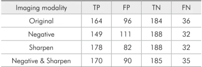
As summarized in Table 1, observers using the Sharpen ilter made the highest number of true posi-tive diagnoses, and also the smallest number of false positive observations.
Although not statistically signiicant, average sensitivity (0.68 ± 0.22) was higher for images en-hanced with the Sharpen ilter (Table 2). Mean overall accuracy was also higher for this imaging modality (0.76 ± 0.16), though not signiicant from a statistical standpoint. Conversely, the use of the Negative ilter was associated to the lowest means of sensitivity and overall accuracy. Speciicity ranged
Imaging modality Sensitivity Specificity Accuracy Original 0.62 ± 0.14 (A) 0.84 ± 0.07 (A) 0.73 ± 0.08 (A) Negative 0.57 ± 0.13 (A) 0.85 ± 0.02 (A) 0.70 ± 0.10 (A) Sharpen 0.68 ± 0.22 (A) 0.85 ± 0.06 (A) 0.76 ± 0.16 (A) Negative & Sharpen 0.65 ± 0.17 (A) 0.84 ± 0.03 (A) 0.74 ± 0.14 (A) Means followed by the same letter in the same column do not significantly differ according to the Tukey test at a 95% confidence interval
Table 1 - Overall true positive (TP), false positive (FP), true negative (TN), and false negative (FN) numbers for the de-tection of enamel subsurface demineralization by imaging modality.
Imaging modality TP FP TN FN
Original 164 96 184 36
Negative 149 111 188 32
Sharpen 178 82 188 32
Negative & Sharpen 170 90 185 35
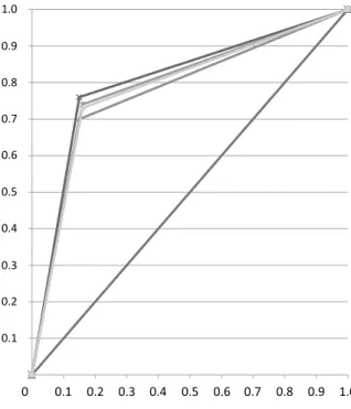
from 0.84 to 0.85, considering all imaging modali-ties (p > 0.05). It may be assumed, however, that the speciicity mean values were generally high without affecting sensitivity (0.62–0.68) for subsurface de-mineralization, except for the images with the Neg-ative ilter. The Az was greater for the Sharpen ilter,
corroborating its higher overall accuracy (Figure 3).
Discussion
Proximal caries lesion depths are generally un-derestimated, but the measurements in this study
were more accurate using the Digora
(photostimu-lable storage phosphor) and Dixi (CCD-based)
sys-tems in comparison to other two digital intraoral systems.4 The Digora-Optimesystem has more
lex-ible plates, which are presumably more comfortable for the patients, and a reduced scanning time (from approximately 30 seconds to about 8 seconds) in re-lation to its predecessor Digora FMX. However, an in vitro study on diagnostic performance for detect-ing non-cavitated proximal caries lesions reported
that there was no statistically signiicant difference
between the Digora FMX and the Digora-Optime
(an older and a relatively newer version, respective-ly), in regard to overall accuracy.9 From a practical
point of view, it may be suggested that both system versions are suitable for caries diagnosis.
Some studies mentioned that image enhancement with ilters improved the detection of proximal car-ies lesions13,14 or reduced the observer variability.10
However, other authors suggested that ilter en-hancement did not provide a remarkable improve-ment in caries diagnosis accuracy in relation to the original digital and conventional radiographs.18
Because no statistically signiicant differences were found between the imaging modalities for the di-agnosis of proximal subsurface demineralization (Table 2), the null hypothesis had to be accepted. This inding corroborates previous studies.10,18 At
irst glance, one should not indicate the use of il-ter enhancement for the diagnosis of subtle proxi-mal demineralization. In fact, another experimental study also did not report any signiicant differences between the original images acquired with photo-stimulable storage phosphor plates and those pro-cessed with task-speciic ilters (Caries 1 and Caries 2).11 However, the use of the Fine enhancement ilter
was recommended whenever a dentist searches for a shallow caries lesion, even though this procedure did not provide signiicant statistical improvement in comparison with the original radiographs.11
The main objective of the enhancement tech-niques is to modify the physical attributes of an im-age to make it more suitable for a given task and a speciic observer.18 Filters are tools derived from
image processing algorithms (mathematical equa-tions) included in imaging analyzing software ap-plications.14 An enhancement ilter could modify
the input image (original image) to compensate for losses in image quality caused by underexposure or noise,11 rendering an output image (iltered
im-age). The Sharpen ilter included in the Digorafor
Windows 2.6 package was developed with an algo-rithm that enables computer-assisted manipulation of the input image data array or matrix, grouping high-contrast pixels together in a submatrix, thus evidencing subtle changes in the output image.19
0,0 0,1 0,2 0,3 0,4 0,5 0,6 0,7 0,8 0,9 1,0
Negaive Filter Sharpen Filter
Negaive plus Sharpen Filter Original
1.0
0.9
0.8
0.7
0.6
0.5
0.4
0.3
0.2
0.1
Figure 3 - Receiver operating characteristic curves for the imaging modalities. Original (Az = 0.73), Negative filter
(Az = 0.70), Sharpen filter (Az = 0.76) and Negative plus
In general, radiographic examination provides low sensitivity and high speciicity in the detection of non-cavitated caries lesions.20 This inding is in
agreement with those presented in Table 2, albeit with the sensitivity and speciicity observed for the Sharpen ilter. As shown in Table 2, the Digora-Optime radiographs enhanced with the Sharpen
ilter exhibited the most favorable relationship be-tween sensitivity (0.68) and speciicity (0.85), fol-lowed by the images with both Negative and Sharp-en ilters, and the original images (sSharp-ensitivity: 0.62, speciicity: 0.84). A plausible explanation for the su-perior performance of observers using the Sharpen ilter may be related to the two-dimensional nature of the radiographic examination, which more fre-quently evidences the radiolucency of advanced le-sions extending to the dentine. However, because the Sharpen ilter can highlight contrast regions, proximal enamel subsurface demineralization that could have been overlooked in the original images may have been depicted in the iltered images.
The most unbalanced relationship was registered for the images with the Negative ilter. Interestingly, the radiographs enhanced with both Negative and Sharpen ilters had the second best performance in-dices (Table 2). Considering absolute numbers (Ta-ble 1) and indices (Ta(Ta-ble 2) for the Sharpen iltered images, in relation to the original and the Negative ilter images, it may be assumed that the Sharpen ilter actually improved the diagnostic performance of these two imaging modalities. Not surprisingly, the Negative ilter yielded the lowest performance
indices for detecting proximal subsurface deminer-alization (Table 2). This result may be related to the radiographic interpretation method, since dentists are taught and trained to diagnose caries lesions as radiolucent images.
As in many other studies on the performance of imaging modalities for the detection of caries le-sions,2,6,13,21,22 dental radiologists were selected as
observers. These professionals are well-trained for imaging diagnosis; therefore, many enhancement tools may have little effect on their performance, as opposed to what would be expected from a general practitioner. Nevertheless, considering that caries lesion depth is often underestimated in radiographic images4 and that proximal subsurface
demineraliza-tion is dificult to be detected,2,6,11 a diagnostic
ad-junct that could add any improvement in the radio-graphic image with no additional cost or signiicant time-consuming techniques should be recommended for and welcomed by dental radiologists and gen-eral practitioners. Therefore, the applicability of the Sharpen ilter to Digora-Optime radiographs
should be considered as a suitable adjunct for en-hancing the diagnosis of proximal surface deminer-alization such as caries lesion.
Conclusion
The Digora-Optimeimages with the Sharpen
il-ter had the best performance-related indices. There-fore, the use of the Sharpen ilter is advisable to improve the diagnosis of incipient proximal caries lesions.
References
1. Zhang ZL, Qu XM, Li G, Zhang ZY, Ma XC. The detection accuracies for proximal caries by cone-beam computerized tomography, film, and phosphor plates. Oral Surg Oral Med Oral Pathol Oral Radiol Endod. 2011 Jan;111(1):103-8. 2. Haiter-Neto F, Ferreira RI, Tabchoury CPM, Bóscolo FN.
Linear and logarithmic subtraction for detecting enamel subsurface demineralization. Dentomaxillofac Radiol. 2005 May;34(3):133-9.
3. Seneadza V, Koob A, Kaltschmitt J, Staehle HJ, Duwenhoeg-ger J, Eickholz P. Digital enhancement of radiographs for assessment of interproximal dental caries. Dentomaxillofac Radiol. 2008 Mar;37(3):142-8.
4. Jacobsen JH, Hansen B, Wenzel A, Hintze H. Relationship between histological and radiographic caries lesion depth measured in images from four digital radiography systems. Caries Res. 2004 Jan-Feb;38(1):34-8.
5. Haiter-Neto F, Wenzel A, Gotfredsen E. Diagnostic accuracy of cone beam computed tomography scans compared with intraoral image modalities for detection of caries lesions. Den-tomaxillofac Radiol. 2008 Jan;37(1):18-22.
7. Ferreira RI, Haiter-Neto F, Tabchoury CPM, Bóscolo FN. In vitro induction of enamel subsurface demineralization for evaluation of diagnostic imaging methods. J Appl Oral Sci. 2007 Oct;15(5):392-8.
8. Wenzel A, Haiter-Neto F, Gotfredsen E. Influence of spatial resolution and bit depth on detection of small caries lesions with digital receptors. Oral Surg Oral Med Oral Pathol Oral Radiol Endod. 2007 Mar;103(3):418-22.
9. Haiter-Neto F, Pontual AA, Frydenberg M, Wenzel A. A comparison of older and newer versions of intraoral digital radiography systems: diagnosing noncavitated proximal cari-ous lesions. J Am Dent Assoc. 2007 Oct;138(10):1353-9. 10. Haiter-Neto F, Pontual AA, Frydenberg M, Wenzel A.
De-tection of non-cavitated approximal caries lesions in digital images from seven solid-state receptors with particular focus on task-specific enhancement filters. An ex vivo study in hu-man teeth. Clin Oral Investig. 2008 Sep;12(3):217-23. 11. Haiter-Neto F, Casanova MS, Frydenberg M, Wenzel A.
Task-specific enhancement filters in storage phosphor images from the Vistascan system for detection of proximal caries lesions of known size. Oral Surg Oral Med Oral Pathol Oral Radiol Endod. 2009 Jan;107(1):116-21.
12. Kamburoğlu K, Murat S, Yüksel SP, Cebeci AR, Paksoy CS. Occlusal caries detection by using a cone-beam CT with different voxel resolutions and a digital intraoral sensor. Oral Surg Oral Med Oral Pathol Oral Radiol Endod. 2010 May;109(5):e63-9.
13. M∅ystad A, Svanæs DB, van der Stelt PF, Gröndahl H-G, Wenzel A, van Ginkel FC, et al. Comparison of standard and task-specific enhancement of Digora storage phosphor images for approximal caries diagnosis. Dentomaxillofac Radiol. 2003 Nov;32(6):390-6.
14. Lehmann TM, Troeltsch E, Spitzer K. Image processing and enhancement provided by commercial dental software pro-grams. Dentomaxillofac Radiol. 2002 Jul;31(4):264-72. 15. Argenta R, Tabchoury C, Cury JA. A modified pH-cycling
model to evaluate fluoride effect on enamel demineralization. Pesqui Odontol Bras. 2003 Jul-Sep;17(3):241-6.
16. Featherstone JDB, ten Cate JM, Shariati M, Arends J. Com-parison of artificial caries-like lesions by quantitative mi-croradiography and microhardness profiles. Caries Res. 1983;17(5):385-91.
17. Arends J, ten Bosch JJ. Demineralization and remineralization evaluation techniques. J Dent Res. 1992 Apr;71 Spec No:924-8. 18. Eickholz P, Kolb I, Lenhard M, Hassfeld S, Staehle H. Digital
radiography of interproximal caries: effect of different filters. Caries Res. 1999 May-Jun;33(3):234-41.
19. Analoui M. Radiographic image enhancement. Part I: spatial domain techniques. Dentomaxillofac Radiol. 2001 Jan;30(1):1-9.
20. Bader JD, Shugars DA, Bonito AJ. A systematic review of the performance of methods for identifying carious lesions. J Public Health Dent. 2002 Fall;62(4):201-13.
21. Şenel B, Kamburoğlu K, Üçok Ö, Yüksel SP, Özen T, Avsever H. Diagnostic accuracy of different imaging modalities in detection of proximal caries. Dentomaxillofac Radiol. 2010 Dec;39(8):501-11.