327 Contribution of radiology to the forensic activity
Radiol Bras 2007;40(5):327–330 Original Article
CAN CHANGES ASSOCIATED WITH AGING HINDER
THE IDENTIFICATION OF INDIVIDUALS SUBMITTED TO LUMBAR
SPINE RADIOGRAPHY? A POTENTIAL CONTRIBUTION
OF RADIOLOGY TO THE FORENSIC ACTIVITY*
Sílvia Falcão de Oliveira1
, Glória Maria Martins Gomes1
, Lucio Ronaldo Cardoso2
, Hilton Augusto Koch3
, Edson Marchiori4
, Bianca Gutfilen5
OBJECTIVE: The present study was aimed at evaluating the possibility of a radiological study of the lumbar spine determining the correct identification of an individual despite the changes associated with aging. MATERIALS AND METHODS: The study sample included 60 pairs of lumbar spine radiographic images of both male and female, adult patients of different ages, acquired at different times, at three-year minimum intervals. The pairs of images were mixed up so two experienced radiologists could put them back together. The vertebrae of each pair were compared for similarities and differences in anatomical details. Pairing cri-teria adopted were finding a specific anatomical variation or detail, or finding two or more similarities among anatomical details, with no point of divergence. RESULTS: Correct pairing of radiographs of the whole sample was achieved by both observers, who presented countless coincidence points in their analyses. The statis-tical analysis demonstrated a good-to-perfect interobserver agreement. CONCLUSION: Comparison between radiographic images of lumbar spine can determine a correct identification of individuals, despite changes associated with aging. Therefore, radiography represents a potential tool to be utilized in forensic identifica-tion studies.
Keywords: Radiographic comparison; Identification; Forensic anthropology.
Alterações decorrentes do envelhecimento podem impedir a identificação de indivíduos submetidos a radio-grafias da coluna lombar? Potencial contribuição da avaliação radiológica para a atividade forense. OBJETIVO: O objetivo deste trabalho foi avaliar a possibilidade de o exame radiológico da coluna lombar determinar a identificação correta dos indivíduos, apesar das alterações evolutivas do envelhecimento. MA-TERIAIS E MÉTODOS: Foi constituída amostra com 60 pares de radiografias de coluna lombar, feitas em épocas distintas, com intervalo mínimo de três anos, de pacientes de ambos os sexos, adultos e com idades diversas. Os pares foram misturados para que dois experientes radiologistas os reconstituíssem. As vérte-bras de cada par foram comparadas em relação a semelhanças e diferenças de detalhes anatômicos, sendo estabelecido, como critério de pareamento, o encontro de uma variação anatômica ou de uma particularidade específica, ou o encontro de duas ou mais igualdades entre os detalhes anatômicos, sem pontos de diver-gência. RESULTADOS: O correto pareamento de todas as radiografias foi alcançado por ambos os observa-dores, os quais apresentaram inúmeros pontos de coincidência em suas análises. O estudo estatístico de-monstrou que a concordância entre os dois observadores foi considerada de boa a perfeita. CONCLUSÃO: A comparação radiográfica da coluna lombar é capaz de determinar a correta identificação dos indivíduos, apesar das alterações evolutivas do envelhecimento. Dessa forma, as radiografias representam potencial ins-trumento para uso em perícias de identificação forense.
Unitermos: Comparação radiográfica; Identificação; Antropologia forense. Abstract
Resumo
* Study developed in the Department of Radiology at Facul-dade de Medicina da UniversiFacul-dade Federal do Rio de Janeiro (UFRJ), Rio de Janeiro, RJ, Brazil.
1. Masters in Radiology by Universidade Federal do Rio de Janeiro (UFRJ), Experts Medical Examiners at Instituto Médico-Legal Afrânio Peixoto (IMLAP), Rio de Janeiro, RJ, Brazil.
2. Associate Professor of Medical Practice at Faculdade de Medicina da Universidade Federal do Rio de Janeiro (UFRJ), Rio de Janeiro, RJ, Brazil.
3. Full Professor, Department of Radiology – Faculdade de Medicina da Universidade Federal do Rio de Janeiro (UFRJ), Rio de Janeiro, RJ, Brazil.
4. Full Professor of Radiology at Universidade Federal Fluminense (UFF), Niterói, RJ, Adjunct Coordinator for the Course of Post-graduation in Radiology – Universidade Federal do Rio de Janeiro (UFRJ), Rio de Janeiro, RJ, Brazil.
5. Associate Professor, Department of Radiology – Universi-dade Federal do Rio de Janeiro (UFRJ), Rio de Janeiro, RJ, Brazil.
INTRODUCTION
The pioneering utilization of radiologi-cal resources to elucidate problems related to forensic identification dates back to 1927. Culbert & Law wrote an interesting
article where they report a case of identifi-cation based on the comparison between nasal accessory sinuses and mastoid pro-cesses on preoperative radiographs and on radiographs of a deceased individual, where they could enumerate 20 coinciden-tal items(1).
From that time on, the comparative analysis of ante- and post-mortem radio-graphs in association with studies of finger-prints and dental arcades, and currently with DNA study, started constituting the Mailing address: Dra. Sílvia Falcão de Oliveira. Academia de
Polícia Civil do Estado do Rio de Janeiro, Centro de Estudos de Assuntos Policiais. Rua Frei Caneca, 162, Centro. Rio de Janeiro, RJ, Brazil, 20211-040. E-mail: [email protected]
328
Oliveira SF et al.
Radiol Bras 2007;40(5):327–330
routine protocol in forensic identification studies(2).
A study performed in 2002 by Ameri-can scientists identified the poor quality of images on ante- and post-mortem radio-graphs as the major limitation for the use of this method(3)
, but nevertheless such limitations usually do not use to affect the performance of experienced professionals, provided protocols are observed. There-fore, a methodology should be adopted in the process of forensic identification in order to facilitate the procedures: radiolo-gists should classify the bone morphologi-cal features into categories such as typimorphologi-cal morphological differences, anatomical variants, aging degenerative processes, traumatisms and congenital malforma-tions(4).
The present study is aimed at evaluat-ing the possibility of a radiological study of the lumbar spine determining the correct identification of an individual despite changes associated with aging, to demon-strate the potential role of lumbar spine radiography in the forensic activity.
MATERIALS AND METHODS
A survey aiming at finding adult pa-tients with at least two radiographs of their lumbar spine was performed in the logical Archives of the Service of Radio-diagnosis at Hospital Universitário Cle-mentino Fraga Filho. The inclusion crite-rion was patients with two radiographs of the lumbar spine acquired at three-year minimum interval, independently from the degree of degeneration observed or the presence of other osteoarticular diseases. Sixty pairs of studies performed at intervals ranging between three and thirty years (mean 4.8 years and median 4.0 years) were selected. The youngest patient was 20 years old, and the oldest, 88 years old (mean age 60.82 years and standard deviation, 16.14) (Tables 1 and 2).
Once the pairs of films had been se-lected, they had their register numbers hid-den and were mixed up before being ana-lyzed by two experienced radiologists. The comparative study included the analysis of morphological variations of vertebral bod-ies, spinous processes, transverse pro-cesses, pedicles and intervertebral spaces,
degenerative aging processes, and also any particular findings that could be of help in the process of images pairing.
The pairing criterion adopted was a single finding of an anatomical variant or specific detail, or the finding of two or more similarities among anatomical details, with no point of divergence
Besides being evaluated at the nega-toscope, the radiographic images were pho-tographed and submitted to an images pro-cessing software (Adobe Photoshop), to enhance or reduce their contrast and/or brightness, or for segmentation aiming at optimizing the comparison process.
RESULTS
Both observers could pair off all of the 60 studies. The process of putting the im-age pairs back together was developed through a careful observation of anatomi-cal details. The first pairs of images were based on findings of anatomical variants or specific details — on the very spinal col-umn or not — capable, by themselves, to
Table 2 Distribution of men and women according their age range.
Age range 20–30 years 31–40 years 41–50 years 51–60 years 61–70 years 71–80 years
81 or more years
Total Men 1 6 3 2 3 2 4 21 Women 0 3 3 10 11 9 3 39
allow the identification. The reading of the analyses performed by both observers dem-onstrated 20 coincidence points, with high-lights on the following findings: “partial vertebra collapse”, “spondylolysis”, “bi-zarre transverse process”, “very short trans-verse process”, “elongated left transtrans-verse process”, “upward transverse process”, “transverse mega-apophysis” and “gross calcifications in the pelvic excavation – calcified myoma?”.
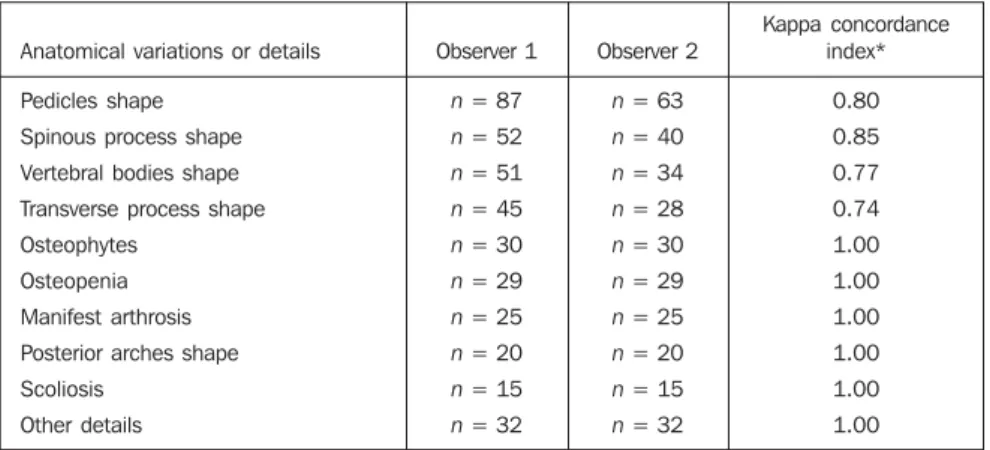
The other anatomical details were con-sidered as a whole for establishing a posi-tive identification. Correct pairing of radio-graphs of the whole sample was achieved by both observers, who presented countless coincidence points in their analyses, and the statistical analysis demonstrated a good-to-perfect interobserver agreement (Table 3).
Signs of traumatisms and/or bone sur-geries or congenital malformations, consid-ered as a comparison criterion, have not been found among the radiographic images included in the sample.
Figure 1 shows a pair of images corre-sponding to the case with the longest inter-val between the two radiographs (30 years).
DISCUSSION
The 60 pairs of radiographic images were correctly identified by both observers, with a concordance index specifically cal-culated for each anatomical detail or patho-logical alteration analyzed. These indices ranged between 0.74 and 1.00, with the statistical analysis demonstrating a good-to-perfect interobserver agreement.
The observers’ confidence to define the images pairing was due to the finding of similarly shaped anatomical details — in-vestigated and evaluated separately or in conjunction — and the absence of discrep-ancies amongst images. Findings of either pathological alteration on the very spinal column, such as osteophytes and other evi-dences of arthrosis, or occasional abnormal findings, such as “gross calcification in the pelvic excavation – calcified myoma?”, or atypical anatomical details, such as “up-ward transverse process”, have allowed some pairings. Essentially, the perfect pair-ing was based on morphological param-eters.
Table 1 Sampling for radiographic comparisons.
329 Contribution of radiology to the forensic activity
Radiol Bras 2007;40(5):327–330
The large number of degenerative fea-tures found on the majority of radiographic images is attributed to the high age range of the patients included in the present study; however, osteopenia and osteophy-tes were pointed out as contributory factors in the pairing process. Manifest arthrosis does represent a relevant factor, as well as do alterations capable of considerably af-fecting anatomical patterns.
Some anthropologists have been insist-ing that the interval between ante- and
post-mortem radiographies should be no
longer than few years, in cases where such radiographic images are suggested as a support to human remains identification(5).
However, the results of the present study go against this idea: among the 60 cases in-cluded in the present study, six corre-sponded to individuals with intervals of more than tem years between studies. It is important to mention a case described in the literature where the ante-mortem radio-graph had been acquired 13 years before the disappearance of an individual whose cadaver could be identified(6).
The trabecular pattern can be utilized in comparative studies(7), but only in cases
where the images are concerned with stud-ies performed in not very distant times, considering that the trabecular structure is susceptible to reabsorption, even in early stages of osteoporosis, particularly in women, and may lead to erroneous inter-pretation(4).
Reference to forensic authors demon-strates that anatomical details should be meticulously observed despite the absence of a mandatory minimum number of items to be compared for determining the identi-fication of human remains. Usually, one to four coincidental characteristics and no discordance are considered as sufficient parameters(8).
Detailed anatomical structures with natural variations, especially the skull, lumbar-sacral column and chondrosternal joints, constitute the best elements for hu-man remains identification(9). However, not
only these structures can be of help in com-parative studies. The literature suggests the mastoid apophysis(10), sella turcica of the
sphenoid bone(10), hand and wrist(11), and
clavicle(12).
At global level, comparative analysis of
ante- and post-mortem radiographs has rep-resented a potential resource both in necro-scopic studies of bone specimens and analy-ses of carbonized or putrefied cadavers, or even recent cadavers whose physiognomic lines individualization is still feasible(13).
It is evident that the major contribution of this method is associated with recent cadavers, considering that, nearly always in such cases, relatives show up to claim the cadaver
The procedure could be routinely adopted in forensic identification pro-cesses, and it is a responsibility of coroners providing the family relatives with guid-ance in the search of ante mortem radio-graphs of the deceased.
Considering that the comparison be-tween radiographic images of lumbar spine can determine a correct identification of individuals, despite changes associated with aging, it may be concluded that radi-ography represents a potential tool to be utilized in forensic identification studies, comparable in utility to fingerprints and dental evidences.
Table 3 Interobserver findings of anatomical variations or details considered abnormalities or similari-ties between both radiographic images.
Anatomical variations or details
Pedicles shape
Spinous process shape
Vertebral bodies shape
Transverse process shape
Osteophytes
Osteopenia
Manifest arthrosis
Posterior arches shape
Scoliosis
Other details
Observer 1
n = 87
n = 52
n = 51
n = 45
n = 30
n = 29
n = 25
n = 20
n = 15
n = 32
Observer 2
n = 63
n = 40
n = 34
n = 28
n = 30
n = 29
n = 25
n = 20
n = 15
n = 32
Kappa concordance index*
0.80
0.85
0.77
0.74
1.00
1.00
1.00
1.00
1.00
1.00
* Kappa standard error = 0,05; p < 0.000.
330
Oliveira SF et al.
Radiol Bras 2007;40(5):327–330
Acknowledgement
Professor Mário Newton Leitão Azeve-do, Head for the Service of Rheumatology, Faculty of Medicine at Universidade Fede-ral do Rio de Janeiro, for his collaboration in the survey for the sampling of the pre-sent study.
REFERENCES
1. Culbert WL, Law FM. Identification by compari-son of roentgenograms of nasal accessory sinuses and mastoid process. JAMA J Am Med Assoc 1927;88:1634–1636.
2. Iscan MY. Rise of forensic anthropology. Yrbk Phys Anthropol 1988;31:203–230.
3. Kuehn CM, Taylor KM, Mann FA, Wilson AJ,
Harruff RC. Validation of chest X-ray compari-sons for unknown decedent identification. J Fo-rensic Sci 2002;47:725–729.
4. Kahana T, Goldin L, Hiss J. Personal identifica-tion based on radiographic vertebral features. Am J Forensic Med Pathol 2002;23:36–41. 5. Angyal M, Dérczy K. Personal identification on
the basis of antemortem and postmortem radio-graphs. J Forensic Sci 1998;43:1089–1093. 6. Valenzuela A. Radiographic comparison of the
lumbar spine for positive identification of human remains. Am J Forensic Med Pathol 1997;18: 215–217.
7. Mann RW. Use of bone trabeculae to establish positive identification. Forensic Sci Int 1998;98: 91–99.
8. Kahana T, Hiss J. Forensic radiology. Br J Radiol 1999;72:129–133.
9. Quatrehomme G, Fronty P, Sapanet M, Grévin G,
Bailet P, Ollier A. Identification by frontal sinus pattern in forensic anthropology. Forensic Sci Int 1996;83:147–153.
10. Voluter G. The V-test. Radiol Clin 1959;Supl 28: 5–7.
11. Greulich WW. Skeletal feature: visible on the roentgenogram of hand and wrist which can be used for establishing individual identification. Am J Roentgenol Radium Ther Nucl Med 1960; 83:756–764.
12. Sanders I, Woesner ME, Ferguson RA, Noguchi TT. A new application of forensic radiology: iden-tification of deceased from a single clavicle. Am J Roentgenol Radium Ther Nucl Med 1972;115: 619–622.