Leishmania
(
Leishmania
)
major
-infected Rhesus Macaques
(
Macaca mulatta
) Develop Varying Levels of Resistance
against Homologous Re-infections
VF Amaral, A Teva, R Porrozzi*, AJ Silva, MS Pereira, MP Oliveira-Neto**,
G Grimaldi Jr/
+Departamento de Imunologia *Departamento de Ultraestrutura e Biologia Celular, Instituto Oswaldo Cruz-Fiocruz **Hospital Evandro Chagas-Cruz-Fiocruz, Av. Brasil 4365, 21045-900 Rio de Janeiro, RJ, Brasil
Sevenrhesus macaques were infected intradermally with 107 promastigotes of Leishmania
(Leish-mania) major. All monkeys developed a localized, ulcerative, self-healing nodular skin lesion at the site of inoculation of the parasite. Non-specific chronic inflammation and/or tuberculoid-type granuloma-tous reaction were the main histopathological manifestations of the disease. Serum Leishmania-specific antibodies (IgG and IgG1) were detected by ELISA in all infected animals; immunoblot analyses indi-cated that numerous antigens were recognized. A very high degree of variability was observed in the parasite-specific cell-mediated immune responses [as detected by measuring delayed-type hypersensi-tivity (DTH) reaction, in vitro lymphocyte proliferation, and gamma interferon (IFN-γ) production] for individuals over time post challenge. From all the recovered monkeys (which showed resolution of the lesions after 11 weeks of infection), 57.2% (4/7) and 28.6% (2/7) animals remained susceptible to secondary and tertiary infections, respectively, but the disease severity was altered (i.e. lesion size was smaller and healed faster than in the primary infection). The remaining monkeys exhibited complete resistance (i.e. no lesion) to each rechallenge. Despite the inability to consistently detect correlates of cell-mediated immunity to Leishmania or correlation between resistance to challenge and DTH, lym-phocyte transformation or IFN-γ production, partial or complete acquired resistance was conferred by experimental infection. This primate model should be useful for measuring vaccine effectiveness against the human disease.
Key words: rhesus macaques - Macaca mulatta nonhuman primates experimental leishmaniasis -Leishmania (L.) major - immune responses - histopathology
Protozoan parasites of the genus Leishmania
are associated with a broad spectrum of diseases, ranging from mild cutaneous to lethal visceral forms. The outcome of leishmanial infection in humans depends largely on the immune respon-siveness of the host and the virulence of the infect-ing parasite strain. When infected with Leishma-nia, especially species causing the more benign
This work was supported by grants from Fiocruz and Faperj, and the UNDP/World Bank/WHO Special Pro-gram for Research and Training in Tropical Diseases. The data presented in this paper have been submitted by VFA as part of a thesis for a PhD degree in Cellular and Molecular Biology at Fiocruz (Rio de Janeiro, Brazil). Present address: Departamento de Imunobiologia, Universidade Federal Fluminense, Outeiro de São João Batista s/no, 24210-000 Niteroi, RJ, Brasil
+Corresponding author. Fax: +55-21-2280.1589. E-mail: grimaldi@gene.dbbm.fiocruz.br
Received 14 November 2000 Accepted 18 April 2001
self-healing cutaneous leishmaniasis (CL) forms, most humans develop an effective cell-mediated immunity that resolves the infection and usually confers clinical protection against reinfection (Grimaldi & Tesh 1993). Both Leishmania
-reac-tive CD4+ and CD8+ T cells are associated with cure of human CL (Carvalho et al. 1985, Da-Cruz et al. 1994). However, T lymphocytes reactive to leishmanial antigens may also participate in the development of chronic and destructive mucosal lesions (Conceição-Silva et al. 1990, Pirmez et al. 1993).
The use of murine models for the study of im-munology of CL has greatly improved our under-standing of the regulation of the immune response and of the cellular immune mechanisms involved in host resistance and susceptibility to leishmanial infection. Inoculation of genetically resistant in-bred strains of mice with L. (L.) major results in
pro-gressive and fatal infections that develop in more susceptible mouse strains are correlated with acti-vation of the Th2 cytokines IL-4, IL-5, and IL-10, that promote humoral immunity (Reiner & Locksley 1995, Sjölander et al. 1998).
Current research employing mouse models is also providing the foundation for studies designed to identify leishmanial protective immunogens and the immune mechanisms responsible for immunity in vaccinated mice (Reed & Scott 1993). However, small experimental animals may not be good pre-dictors of human responses. Simian species most closely related to humans should be more attrac-tive as animal models to evaluate the safety and immunogenicity of vaccines. The hominoid pri-mates (chimpanzees, orangutans, gorillas, and gib-bons), which diverged from humans over 5 mil-lion years ago, are the most likely to mimic accuratly the disease state and the immunological response to infection (Kennedy et al. 1997a). How-ever, cost considerations and other factors (great apes are endangered species) for using hominoid primate species in biomedical studies represent serious limitations. Next in evolutionary distance are Old World monkeys (macaques, baboons, man-drills, and mangageys), which diverged from the human line between 15 and 20 million years ago. Most distantly related to humans (over 30 million years apart) are the New World monkeys (aotus, owl, cebus monkeys, and marmosets) (Kennedy et al. 1997b). Genetic studies have shown that class I and class II major histocompatibility complex (MHC) products in great apes and nonhominoid Old World monkeys much resemble their human counterparts; in contrast, New World monkeys have a condensed or smaller MHC as compared to humans (reviewed in Kennedy et al. 1997a). There-fore, vaccine trials in Old World primates will prob-ably not be hindered due to divergence of MHC molecules (Prilliman et al. 1996).
A variety of studies have examined the suscep-tibility of Old and New World monkeys to infec-tion and subsequent disease inducinfec-tion by various
Leishmania species (Dennis et al. 1986, Lujan et
al. 1986, Pung & Kuhn 1987, Olobo et al. 1992, Gicheru et al. 1995). Nonhuman primate models have also provided valuable data on the efficacy of putative vaccine candidates against leishmania-sis (Olobo et al. 1995, Kenney et al. 1999, Gicheru et al. 2001). We have previously examined the Asian rhesus macaques as an experimental model for study of CL caused by L. (L.) amazonensis
infection (Amaral et al. 1996, 2000). In the present study, M. mulatta monkeys were infected with L.
(L.) major and the patterns of primary and
chal-lenge infections and immune responses were fol-lowed. An important aspect of this animal system
in experimental CL lies in the reproduction of clini-cal events that are common in L. (L.) major
-in-fected patients, namely, the self-healing disease and resistance to homologous challenges, indicating development of an acquired immunity.
MATERIALS AND METHODS
Animals - A total of ten laboratory-bred and
-reared young adult (3- to 8-year-old, weighting between 4,600 and 10,560 g) male rhesus macaques (M. mulatta) obtained from the Fiocruz rhesus
colony were used. Experimental animals were housed indoors in individual steel squeeze-back cages in a temperature (25oC) – and humidity (60 ± 5%) – controlled environment. Water was pro-vided ad libitumvia an automatic watering sys-tem, and High Protein Monkey Diet (NUVILAB; Ministério da Agricultura e Reforma Agrária, Brasil), supplemented with eggs, fruits and veg-etables, was fed twice daily. The primate facilities
are maintained according to the guidelines of the Committee on the Care and Use of Laboratory Animals of the Institute of Laboratory Animal Resources, National Research Council and Health and Human Services (NIH: MD, USA).
The experiments were conducted using a pro-tocol approved by the Institutional Committee of the Center for Biological Evaluation and Care of Research Animals (CEUA-Fiocruz). The monkeys were aclimatized to the laboratory conditions for at least two weeks before the experimental proce-dures were begun. Animals were anesthetized be-fore infection and prior to each sampling or test-ing procedure. Monkeys were initially restrained in their cages, and subsequently they were given, intramuscularly, Ketamine (Ketalar: Ketamine hydrochloride; Parke Davis; 10-40 mg/kg body weight) for anesthesia.
Parasites and experimental infections - The
strain LV39 (MRHO/SU/59/P) of L. (L.) major
Seven rhesus macaques were each infected by intradermal inoculation of 1 x 10⁷ stationary phase promastigotes in 0.1 ml. The various challenge and rechallenge experiments were performed as fol-lows. The animals were initially infected by in-jecting the promastigote suspension in the area below the brow (orbit) of the right eye and, 26 and 40 weeks later, they were challenge-infected in the opposite orbit (secondary infection) and in the left forearm (tertiary infection) respectively with identical numbers of parasites. Three healthy mon-keys, designated as control, were inoculated with PBS.
Pathology - The size and appearance of
leish-manial lesions (lesion development) was followed sequentially. Lesion area was calculated using the formula πr1r2, as described by Wilson et al. (1979).
Skin biopsies were removed from the border of cutaneous lesions during active stages (between three and six weeks post-inoculation) using a 4 mm punch and fixed in 10% buffered formalin. Paraf-fin sections were prepared from central and pe-ripheral zones of the lesion and stained with he-matoxylin-eosin.
Leishmanin skin test (LST) - The Leishmania
-derived antigens or leishmanin [consisting of pooled heat-killed L. (L.) major, L. (L.) ama-zonensis, and L. (Viannia) guyanensis
pro-mastigotes suspended in PBS with 0.5% phenol] used for skin testing of the primates was prepared at the Fiocruz (Biomanguinhos Unit), Brazil. A volume 0.1 ml containing 5 x 10⁶ parasites was injected into the left forearm. Delayed-type hyper-sensitivity (DTH) reactions were measured as skin indurations at the site of injection (Fig. 2D) after 72 h using the ballpoint pen method (Agwale et al. 1998). An average induration diameter of equal or more than 5 mm was considered as a positive LST.
Lymphocyte blastogenesis assay - The
meth-ods followed for peripheral blood leukocytes (PBL) preparation and proliferation assays were as de-scribed (Amaral et al. 1996). Purified PBL were cultured at 2 x 10⁶ viable cells ml-1 in the pres-ence of optimal culture concentration of mitogen (Phytohemagglutinin PHA-P at 12.5 µg ml-1; Sigma) or parasite antigen (10 µg protein/well). The soluble leishmanial antigens (SLA) for in vitro blast transformation assays were prepared from L.
(L.) major (LV39) promastigotes, according to the
method described by Dennis et al. (1986). Cul-tures were incubated at 37oC in a humidified at-mosphere containing 5% CO2 for three days in the case of mitogen or for four days in the case of an-tigens. The cells were pulsed with [3H]thymidine (Amersham, Co., U.K.; 1 µCi/well; 5 µCi/mM) over
the last 18 h and harvested onto glass fiber filter
mats (Titertek, FlowLab). Radioactive incorpora-tion into DNA was determined by liquid scintilla-tion spectrometry. Results are expressed as the stimulation index (SI, mean cpm stimulated cul-tures/mean cpm unstimulated cultures). To deter-mine significance, data from the lymphocyte pro-liferative responses (LPR) of infected monkeys were compared with those of healthy animals (con-trol group) using the unpaired Student’s t-test.
Concordance between the LST and the in vitroLPR was assessed by the ratio: number of monkeys posi-tive by both tests + number of those negaposi-tive by both tests : total number of individual tested (Sassi et al. 1999).
The staining and cytofluographic analysis of stimulated PBL was as described previously (Amaral et al. 1996). For the analyses of pheno-type of responding T cell subsets of infected ani-mals, the following commercial monoclonal anti-bodies were employed: CD4 helper/inducer T cell subset (OKT4 antibody; Ortho Diagnostic Systems, Raritan, NJ), CD8 suppressor/cytotoxic T cell sub-set (Leu 2a antibody; Becton-Dickinson, San Jose, CA), and anti-CD25 (for IL-2 receptor; Becton-Dickinson).
Interferon-γ production by stimulated cells and measurements of IFN-γ- Purified PBL were
ad-justed to 2 x 10⁶ cells ml-1 in medium and stimu-lated with either PHA-P or SLA. Cell culture me-dium were pooled from duplicate wells after 72 h of stimulation. A rhesus monkey IFN-γ ELISA immunoassay kit obtained commercially (Bio-source International, Camarillo, CA) was used for the in vitro quantitative determination of IFN-γ in the supernatants. Streptavidine-Peroxidase (HRP) conjugate and stabilized chromogen, Tetra-methylbenzidine (TMB) substrate were used for detection, as per the manufacturer’s instructions. A standard curve (obtained for the various stan-dards over the range of 0 to 1000 pg ml-1 IFN-γ) was run with each assay. IFN-γ concentrations for unknown samples and controls were read from the standard curve plotted.
Serologic assays - Antigen-specific serum
antibodies were detected employing cross-reactive (Shearer et al. 1999) anti-human IgG1(clone 8c/ 6-39), IgG2(clone HP-6014), and IgG4 (clone HP 6025) biotinylated monoclonal antibodies obtained commercially (Sigma). The reaction was revealed with biotin-avidin peroxidase system. The mono-clonal antibodies were diluted 1:800 and avidin-peroxidase was diluted 1:1,000. The substrate con-sisted of 0.04% o-phenylenediamine (OPD) dihidrochloride and 0.012% hydrogen peroxidase in phosphate-citrate buffer, pH 5. All sera were tested in duplicate and those yielding positive re-sults were re-tested at least once. The lower limit of positivity (cut off) was determined by mean of negative control + 2 s.d. (Richardson et al. 1983). Moreover, detection of serum antibodies to leishmanial (LV39) antigens was performed by Western blot analysis. Briefly, the SLA extracts were resolved by sodium dodecyl sulphate-poly-acrylamide gel electrophoresis (SDS-PAGE) us-ing 12% slab gels in non-reducus-ing conditions, and electrophoretically transferred to nitrocellulose paper (Schleider and Schuell, Keene, NH). The nitrocellulose strips were incubated overnight at 4oC with either immune sera obtained from infected primates or human CL patients (positive controls) or sera from healthy macaques (negative controls). Subsequently after washing, strips were incubated with either rabbit anti-monkey IgG-peroxidase conjugate or rabbit anti-human IgG (Sigma), or anti-human IgG1(clone 8c/6-39) biotinylated monoclonal antibody. After rinsing, strips were developed in a satured solution of 3,3’-diaminobenzidine in Tris buffer (pH 7.4) contain-ing 0.01% H2O2. The techniques have been de-scribed in detail previously (Leon et al. 1992).
RESULTS
Establishment of infection and lesion develop-ment - As illustrated (Figs 1, 2), all L. (L.) major
-infected rhesus monkeys inoculated (Experiment 1) in the orbit of the right eye with 107 virulent promastigotes developed nodular skin lesions at the site of inoculation. An erythematous papule was first visible at 1-2 weeks postinoculation (wk p.i.); lesion development progressed rapidly (nodules varying between approximately 20 to 160 mm2), peaking at 3 to 8 wk p.i., and all lesions ulcerated (after 3 wk p.i.) and subsequently regressed and healed (macroscopically, primary lesions had dis-appeared from infected animals by 11 wk p.i.). In contrast, the convalescent monkeys (which had recovered from primary skin lesions) developed significant levels of resistance to subsequent chal-lenges using the same parasite strain/dose. As shown (Fig. 1) some of the recovered monkeys remained susceptible to secondary infection (4/7
animals; 57.2%) and/or to tertiary infection (2/7 animals; 28.6%), but in general the lesions were smaller and healed faster than in the initial infec-tion. The remaining monkeys developed complete clinical protection in that no skin lesion developed after each rechallenge.
Pathology - A firm elastic nodule developed
(after 3 wk p.i.) at the site of parasite inoculation. The chronic nodular lesions (Fig. 2A-B) diminished in size after spontaneous ulceration and healing of the CL. At the biopsy animals showed histological evidence of infection. In early phases (3-4 wk p.i.) of the developing primary ulcerated skin lesions (data not shown), the histopathological findings in the dermis showed a non-specific chronic mono-nuclear infiltrate, containing indistinctly delimited differentiated macrophage accumulations and neu-trophils (apparently associated with lysis of few parasitized macrophages). In late stages (6-8 wk p.i.), differentiated macrophages, interspersed with more or less numerous lymphocytes and plasma cells, represented the principal feature in this in-flammatory process, which evolved to the forma-tion of tuberculoid-type granulomatous nodules (Fig. 2C). The granulomas were surrounded by a diffuse mononuclear infiltrate containing giant cells. The histological picture of lesions (biopsy samples obtained at 2-3 wk p.i.) developed in ani-mals after secondary and/or tertiary infections was not the exudative cellular reaction (commonly found at this stage following primary infection), but an earlier formation of mature granulomatous nodules.
Cell-mediated immune responses - DTH
reac-tion to LST was used as in vivo correlate of
cellu-0 20 40 60 80 100 120 140 160 180
0 3 5 8 11 26 27 31 37 40 45 46 47 WEEKS POST INFECTION
L
ESI
O
N
A
R
EA
( m
m
2 )
N25 N5 N15 O35 O19 O41 O25
Fig 1: time course of skin lesion development in Leishmania
(Leishmania) major-infected rhesus macaques. The animals
were initially inoculated in the orbit of the right eye with 1 x 10⁷ L. (L.) major promastigotes. After recovery from the
lar immunity. No cell-mediated reaction to skin test parasite antigen developed in monkeys prior to infection, but LST reactivity (range 3-10 mm) was detected as early as 3 wk p.i. and continued up to 63 wk p.i. (Fig. 3). In spite of a transient immu-nounresponsiviness to the LST observed in indi-viduals over time post challenge, the pooled leishmanin identified all the monkeys with active or healed CL. The reaction sizes found among posi-tive animals did not correlate however with lesion development.
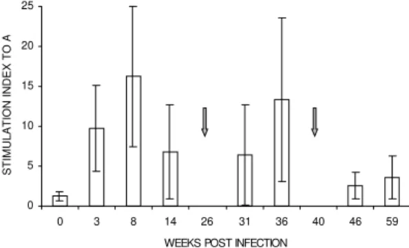
In spite of wide variations from one infected animal to another (as shown by some large SD values), the findings indicate that a proliferative response in vitro to SLA of blood lymphocytes develops during primary and challenge infections (Fig. 4). Parasite-specific LPR were negative (SI<
2.5) in all animals at the initiation of infection, and PBL of animals were comparably responsive to control mitogen PHA-P prior to and throughout infection (data not shown). A positive reaction developed as early as 3 wk p.i. (mean ± SD of SI 9.7 ± 5.4) and subsequently continued to increase, peaking at 8 wk p.i. (mean ± SD of SI 16.3 ± 8.8), but SI mean values (3.6 ± 2.7 at 59 wk p.i.) were significantly lower after the tertiary challenge than during the primary or secondary infection. No positive correlation (r = 0.36, p> 0.05) was
ob-served between the overall lymphocyte blastogen-esis positivity and LST induration size values.
detected in these animals during the initial infec-tion when compared with non-infected controls. In fact only two individuals, after challenge, pro-duced IFN-γ (30-40 pg/ml) above the level ob-served at time zero (10 pg/ml). The sensitivity of the assay was evidently low since supernatants from PHA stimulated PBL cell cultures produced only marginal levels of IFN-γ prior to and throughout primary infection.
Antigen-stimulated PBL cells from all pri-mates were examined (during 45-60 wk p.i.) for phenotype analysis of cell surface antigens. Re-sults showed variation (but not significantly; P > 0.05) in the ratio of CD4+ to CD8+ responding (CD25+) T cells in experimental animals
(CD4:CD8 mean ratio, 1.65; range, 0.8-2.6) as compared to uninfected controls (CD4:CD8 mean ratio, 1.95; range, 1.1-3.5).
Antibody responses - Anti-L. (L.) major
anti-body responsesdetected by ELISA in experimen-tal animals varied during infection (Fig. 5). IgM response was variable (absorbance values ranging from 0.41 ± 0.17 to 0.74 ± 0.25 at 1:50 serum dilution), but was not significantly above the ab-sorbance values (0.48 ± 0.20) of the uninfected animals. In contrast, both total IgG and IgG1 lev-els were detectable 3 wk p.i. and subsequently con-tinued to increase, peaking at 8 wk p.i. after which levels declined in animals with healing lesion. Fol-lowing re-challenge, IgM levels did not increase significantly in comparison to primary infection (data not shown), but the IgG and IgG1 titers in-creased and continued up to 46 wk p.i. No detect-able IgG2 or IgG4antibody responses to Leishma-nia were observed in control (uninfected) or
in-fected animals.
Western blots of promastigote homogenates were performed employing immune sera (1:100 was taken as the optimal titer, lowest dilution at which control animals gave no signal on the anti-gen profile) from monkeys at various times post-0
2 4 6 8 10 12
0 3 8 14 26 31 36 40 46 63
WEEKS POST INFECTION
INDURATION SIZE ( m
m
Fig 3: delayed-typed hypersensitivity reaction to skin test para-site antigen was measured to assess levels of cell-mediated immunity in vivodeveloped in Leishmania (Leishmania) ma-jor-infected rhesus macaques. A volume of 0.1 ml containing 5 x 10⁶ heat-killed promastigotes was injected into the shaven
area of the right forearm. Skin induration was read after 72 h and results are expressed as the diameter of skin induration in millimetres. Data are mean ± SD of 7 infected monkeys. Ani-mals were rechallenged at different time points as indicated (arrows).
0 5 10 15 20 25
0 3 8 14 26 31 36 40 46 59
WEEKS POST INFECTION
STIMULATION INDEX TO
A
Fig 4: parasite-specific proliferative responses of blood lym-phocytes from rhesus monkeys are detected during primary and challenge infections. Cell suspensions of purified periph-eral blood leukocytes were restimulated in vitroin the pres-ence of soluble leishmanial antigens prepared from Leishma-nia (Leishmania) major. Cell proliferation was assessed by measuring [3H]thymidine incorporation. Results are expressed
as the stimulation index (SI, mean cpm stimulated cultures/ mean cpm unstimulated cultures). Data are mean ± SD of 7 infected monkeys. Animals were rechallenged at different time points (arrows).
0,0 0,5 1,0 1,5 2,0 2,5
0 3 8 14 26 31 40 46 59
Cut off(0,73)
0,0 0,2 0,4 0,6 0,8 1,0
0 3 8 14 26 31 40 46 59
WEEKS POST IN FEC TION
O
D
(
45
0 nm )
Cut off (0,04)
WEEKS POST INFECTION A
B
Fig 5: evolution of serum levels of Leishmania-specific
infection (data not shown). The antibodies (both IgG and IgG1 isotypes) produced by the animals recognized multiple bands ranging from 35 to 210 kDa, but there was variation in the number of anti-gen components and/or in the intensities of sig-nals. Animals giving high ELISA antibody titers gave stronger reactions and recognized more anti-gens in western blots.
DISCUSSION
Localized CL caused by L. (L.) major is an
endemic parasitic zoonosis in North Africa and the Middle East (WHO/CID/Leish/98.9 Add.1). The disease is generally benign, in that most cases heal spontaneously within three months (Kemp et al. 1994). The experiments recorded here show that
M. mulatta is susceptible to L. (L.) major infection
and confirm the potential for this to be used as a nonhuman primate model of the human disease. In spite of a variation observed in the clinical course of infection, each of the experimental animals developed a simple cutaneous lesion which pro-gressed spontaneously to ulceration and complete resolution within about three months. The study shows that both the incubation period and self-cure in L. (L.) major-infected rhesus macaques compare
well with those observed in humans (Beach et al. 1984). The pathologic features in this model were also very similar to human CL (Ridley & Ridley 1983), in that the exudative cellular inflammatory process evolved to a granulomatous reaction in developing ulcerated skin lesion. The fact that the tuberculoid-type granulomatous reaction took place much earlier in reinfected animals (as com-pared with those with primary infection) indicates that histological changes reflect host immune sta-tus in CL.
A more marked variation in the clinical course of infection [as compared to L. (L.) major] was
observed when rhesus monkeys were examined for susceptibility to L. (L.) amazonensis (Amaral et al.
1996). These observations suggest that the out-come of leishmanial infection in M. mulatta may
depends on the biological behaviour (virulence) of the infecting parasite strain. Similarly, the patho-logic analyses in L. (L.) amazonensis-infected
rhesus monkeys indicated that lesions contained amastigotes with a mononucluear infiltrate of mac-rophages, lymphocytes, and plasma cells, and for-mation of tuberculoid-type granulomas (Amaral et al. 1996). The granulomas showed a mixture of T-cell subpopulations with the ratio of CD4:CD8 phenotypes less than one. The percentage of cells in the granulomas expressing the MHC class II antigens (HLA-DR+) in active lesions (95 ± 7.1%) was significantly higher (P < 0.005)from the
heal-ing lesions (42 ± 12.7%) (Amaral et al. 2000).
Re-sults similar to ours were previously reported, studying the infiltrate in the skin of humans with CL (Pirmez et al. 1990, Lima et al. 1994).
Self-cure CL in humans is generally thought to give rise to a long-lasting immunity to reinfection (Alvarado et al. 1989, Kemp et al. 1994). Some observations suggest, however, that immunity con-ferred by prior self-resolving L. braziliensis sp.
infection may not always be complete (Saravia et al. 1990). Aditionally, partial protection was re-ported in a patient who had a previous infection of
L. (L.) major and became reinfected one year later
with the same parasite strain (Killick-Kendrick et al. 1985). In this study, monkeys which had re-covered from skin lesions were challenged using an identical inoculum of the same parasite strain. The animals developed distinct levels of clinical resistance (as reflected by either an absence of skin lesion or a smaller size and faster resolution of the lesions as compared to the inicial infection) to each rechallenge, indicating that acquired protective immunity against the parasite occurred. Similar results have been demonstrated in näive (Amaral et al. 1996) or vacinnated (Kenney et al. 1999) L. (L.) amazonensis or L. (L.) tropica (Wolf 1976)
infected rhesus macaques, as well as in other pri-mates species (Dennis et al. 1986, Githure et al. 1987, Gicheru et al. 2001).
Humans with CL due to L. (L.) major develop
DTH to intradermal injection of killed pro-mastigotes (LST), and their peripheral mono-nuclear cells proliferate in response to Leishmania
antigens (Kemp et al. 1994, Sassi et al. 1999). Both in vitro lymphocyte proliferative and IFN-γ re-sponses tend to increase during healing (Gaafar et al. 1999). Therefore, healing of the skin lesions and immunity in zoonotic CL are believed to be mediated by cell-mediated immune response. In contrast, the DTH response reported in vaccinees as in vivocorrelates of cellular-mediated immu-nity may not indicate resistance to infection (Antunes et al. 1986, Castes et al. 1994). This study shows that the L. (L.) major-infected rhesus
macaques were equally able to develop parasite-specific cell-mediated immune responses. Despite the inability to consistently detect correlates of cell-mediated immunity to L. (L.) major, complete or
partial resistance was conferred by experimental infection. Correlates of protection were also sought, but they were not consistently found in other non-human primate models of non-human leishmaniasis (Dennis et al. 1986, Lujan et al. 1986, Olobo et al. 1992, Gicheru et al. 1995).
Although marked variation in the magnitude of immune response to Leishmania has been
agreement with the immune profile of patients with healing CL due to L. (L.) major. Our results showed
a week correlation (r = 0.36) between the DTH
reaction assessed by LST and the in vitroLPR of PBL from L. (L.) major-infected rhesus macaques,
as compared to values (98% concordance) de-scribed for patients with active or healed CL caused by the same parasite species (Sassi et al. 1999). However, a positive correlation (r = 0.88) was
found between the size of skin induration and lym-phocyte proliferation in animals with healing le-sion at 14 wk p.i. (data not shown).
The fact that SI mean values obtained were sig-nificantly lower after the tertiary challenge than during the primary or secondary infection was not expected at all. The biological significance of this decline is not clear, although adult rhesus mon-keys may exhibit stress response [such as when removed from their social group to a novel envi-ronment (Gust et al. 1993) and/or submitted to excess of manipulation] that might justify the lower response. Alternatively, those animals completely recovered and parasite-free would develop lower or even negative response as well.
Unlike the typical response in humans, little or no IFN-γ was detected in supernatants after pri-mary infection (data not shown), which may indi-cate a limit of the model. Nevertheless, it is very possible that this could in fact represent a false-negative result, either because the time point to measure the cytokine was not the optimal one (IFN-γ was measured after up to 72 h of stimulation), or because the kit was not sensitive enough to detect this production. Although a Th1-like activity re-sponse (as revealed by an increased production of IFN-γ by the responding T cells) was associated with CL in vervet monkeys (Cercopithecus aethiops) following secondary rechallenge with L. (L.) major (Olobo et al. 1992), recent data
sug-gest that protection against CL in either vaccinated rhesus macaques (Kenney et al. 1999) or vervets (Gicheru et al. 2001) may require more than the activation of Leishmania-specific IFN-γ
-produc-ing T cells.
In late stages of infection, no significant varia-tion (P > 0.05) in the ratio of CD4+ to CD8+ re-sponding (CD25+) T cells was found in experi-mental animals as compared to uninfected controls. However, in the original model description (Amaral et al. 1996), the circulating T cell subpopulations from the L. (L.) amazonensis-infected monkeys
vary throughout the course of infection. In early phases of infection blood CD4+ T cells appear to predominate (CD4:CD8 mean ration, 5.0; range 2.5-7.1), but subsequently, an increase in CD8+ T cells was observed (CD4:CD8 mean ration, 1.05; range 0.37-2.1). Moreover, in this model, there was
apparently no correlation between lesional T cell subsets (T-cell ratios favor CD8+ cells in both ac-tive and healing lesions) (Amaral et al. 2000) and those found in blood [initially during infection CD4:CD8 mean ratio was 5.0; subsequently, an increase in CD8+ T cells was observed (CD4:CD8 mean ration, 1.05)] (Amaral et al 1996).
In this study, immune recognition was con-firmed by detection of an antigen specific antibody response. Immunoblot analyses showed that in-fected monkeys produced antibodies (both IgG and IgG1 isotypes) which bound to a number of Leish-mania antigen components, consistent with human
data (Leon et al. 1992). A differential decline in leishmanial antigen-specific IgG, IgM, IgE, and IgG subclasses was shown in visceral leishmania-sis patients after chemotherapy (Anam et al. 1999), in particular with IgG1 and IgG3 levels being sig-nificantly reduced following treatment (Elassad et al. 1994, Ghosh et al. 1995). The decline observed in the IgG and IgG1 responses in monkeys with healing lesion is probably due to similar regula-tory function of T cells as described for the murine and human models (Abbas et al. 1996). However, little information is available on the IgG subclasses expressed in the sera of non-human primate spe-cies. The data suggest that baboon sera, like hu-man sera, contain four IgG subtypes, whereas macaque sera exhibit only three of the human sub-class analogous (Shearer et al. 1999). Southern blot experiments with genomic DNA samples of rhesus monkeys and human C gamma-specific probes also indicated that IgG3 gene is not expressed in macaques (Calvas et al. 1999).
In conclusion, this primate model showed that experimental infection confers partial or complete immunity to challenge in a variable proportion of individuals as happens in humans. This difference in protection (contrary to the susceptibility of BALB/c mice) provides outcomes that should al-low correlates of complete protection to be identi-fied. Furthermore, the ability of candidate vaccines to equal or exceed the rate of protection conferred by experimental infection provides a basis of evalu-ation of efficacy. Because the incubevalu-ation period and the disease tempo (i.e., the progression and resolution of skin lesions) were similar to those seen in human CL, the endpoint for prophylaxis and vaccine trials can be easily determined in the rhesus model.
ACKNOWLEDGMENTS
REFERENCES
Abbas AK, Murphy KM, Sher A 1996. Functional di-versity of helper T lymphocytes. Nature383: 787-793.
Agwale SM, Duhlinska DD, Grimaldi Jr G 1998. Re-sponse to heterologous leishmanins in cutaneous leishmaniasis in Nigeria - Discovery of a new fo-cus. Mem Inst Oswaldo Cruz 93: 23-27.
Almeida RP, Barral-Netto M, De Jesus AMR, De Freitas LAR, Carvalho EM, Barral A 1996. Biological be-havior of Leishmania amazonensis isolated from humans with cutaneous, mucosal, or visceral leish-maniasis in BALB/c mice. Am J Trop Med Hyg54: 178-184.
Alvarado R, Enk C, Jaber K, Schur L, Frankenburg S 1989. Delayed-type hypersensitivity and lymphocyte proliferation in response to Leishmania major in-fection in group of children in Jericho. Trans R Soc Trop Med Hyg83: 189-192.
Amaral VF, Pirmez C, Gonçalves AJS, Ferreira V, Grimaldi Jr G 2000. Cell populations in lesions of cutaneous leishmaniasis of Leishmania (L.) amazonensis-infected rhesus macaques, Macaca mulatta. Mem Inst Oswaldo Cruz 95: 209-216. Amaral VF, Ransatto VAO, Conceição-Silva F,
Molinaro E, Ferreira V, Coutinho SG, McMahon-Pratt D, Grimaldi Jr G 1996. The Asian rhesus macaques (Macacamulatta) as an experimental model for study of cutaneous leishmaniasis. Exp Parasitol 82: 4-44.
Anam K, Afrin F, Baneriee D, Pramanik N, Guha SK, Goswami RP, Sahaq SK, Ali N 1999. Differential decline in Leishmania membrane antigen-specific immunoglobulin G (IgG), IgM, IgE, and IgG sub-class antibodies in Indian kala-azar patients after chemotherapy. Infect Immun67: 6663-6669. Antunes CM, Mayrink W, Magalhães PA, Costa CA,
Melo MN, Dias M, Michalick MS, Williams P, Lima AO, Vieira LB, Schettini APM 1986. Controlled field trials of a vaccine against New World cutane-ous leishmaniasis. Int J Epidemiol 15: 572-579. Beach R, Kilu G, Hendricks L, Oster C, Leeuwenberg J
1984. Cutaneous leishmaniasis in Kenya: Transmis-sion of L. major to man by the bite of Phlebotomus duboscqi. Trans R Soc Trop Med Hyg 78: 747-751. Calvas P, Apoil P, Fortenfant F, Roubinet F, Andris J, Capra D, Blancher A 1999. Characterization of the three immunoglobulin G subclasses of macaques. Scand J Immunol49: 595-610.
Carvalho EM, Johnson Jr WR, Barreto E, Marsden PD, Costa JLM, Reed S, Rocha H 1985. Cell mediated immunity in American cutaneous and mucocutane-ous leishmaniasis. J Immunol 135: 4144-4148. Castes M, Blackwell J, Trujillo D, Formica S, Cabrera
M, Zorrilla G, Rodas A, Castellanos PL, Convit J 1994. Immune response in healthy volunteers vac-cinated with killed leishmanial promastigotes plus BCG. I. Skin-test reactivity, T-cell proliferation and interferon-γ production. Vaccine 12: 1041-1051. Conceição-Silva F, Dórea RC, Pirmez C, Schubach A,
Coutinho SG 1990. Quantitative study of Leishma-nia braziliensis braziliensis reactive T cells in
pe-ripheral blood and in the lesions of patients with American mucocutaneous leishmaniasis. Clin Exp Immunol79: 221-226.
Da-Cruz AM, Conceição-Silva F, Bertho AL, Coutinho SG 1994. Leishmania-reative CD4 and CD8 T cells associated with cure of human cutaneous leishma-niasis. Infect Immun 62: 2614-2618.
Dennis VA, Lujan R, Chapman Jr WL, Hanson WL 1986. Leishmania donovani: cellular and humoral immune responses after primary and challenge in-fections in squirrel monkeys, Saimiri sciureus. Exp Parasitol 61: 319-334.
Elassad AMS, Younis SA, Siddig M, Gravson J, Petersen E, Ghalib HW 1994. The significance of blood lev-els of IgM, IgA, IgG and IgG subclasses in Sudanese visceral leishmaniasis patients. Clin Exp Immunol 95: 294-299.
Gaafar A, Veress B, Permin H, Kharazmi A, Theander TG, El Hassan AM 1999. Characterization of the local and systemic immune responses in patients with cutaneous leishmaniasis due to Leishmania major. Clin Immunol 91: 314-320.
Ghosh MK, Dasgupta S, Ghose AC 1995. Immunoglo-bulin G sub-class-specific antileishmanial antibody responses in Indian kala-azar and post kala-azar der-mal leishmaniasis. Clin Diagn Lab Immunol2: 291-296.
Gicheru MM, Olobo JO, Anjili CO, Orago AS, Modabber F, Scott P 2001. Vervet monkeys vacci-nated with killed Leishmania major parasites and interleukin-12 develop a type 1 immune response but are not protected against challenge infection. InfectImmun 69: 245-251.
Gicheru MM, Olobo JO, Kariuki TM, Adhiambo C 1995. Visceral leishmaniasis in vervet monkeys: Immu-nological responses during asymptomatic infections. Scand J Immunol41: 202-208.
Githure JI, Reid GD, Binhazim AA, Anjili CO, Shatry AM, Hendricks LD 1987. Leishmania major: the suitability of East African nonhuman primates as animal models for cutaneous leishmaniasis. Exp Parasitol 64: 438-447.
Grimaldi Jr G, Tesh RB 1993. Leishmaniasis of the New World: current concepts and implications for future research. Clin Microbiol Rev6: 230-250.
Grimaldi Jr G, Momen H, Naiff RD, McMahon-Pratt D, Barrett TV 1991. Characterization and classifica-tion of leishmanial parasites from humans, wild animals, and sand flies in the Amazon region of Bra-zil. Am J Trop Med Hyg 44: 645-661.
Gust DA, Gordon TP, Hambright MK 1993. Response to removal from and return to a social group in adult male rhesus monkeys. Physiol Behav53: 599-602. Kemp M, Hey AS, Kurtzhals JA, Christensen CB, Gaafar A, Mustafa MD, Kordofani AA, Ismail A, Kharzmi A, Theader TG 1994. Dichotomy of the human T cell response to Leishmania antigens. I. Th1-like activity response to Leishmania major promastigote antigens in individuals recovered from cutaneous leishmaniasis. Clin Exp Immunol 96: 410-415. Kennedy RC, Shearer MH, Hildebrand WH 1997a.
and immunogenicity. Vaccine15: 903-908. Kennedy RC, Shearer MH, Hildebrand WH, Simmonds
RS 1997b. Nonhuman primates and their use in im-munologically based investigations. The Immunolo-gist5/5: 150-156.
Kenney RT, Sacks DL, Sypek JP, Vilela L, Gam AA, Evans-Davis K 1999. Protective immunity using recombinant human IL-12 and alum as adjuvants in a primate model of cutaneous leishmaniasis. J Immunol 163: 4481-4488.
Killick-Kendrick R, Bryceson ADM, Peter W, Evans DA, Leaney AJ, Rioux JÁ 1985. Zoonotic cutane-ous leishmaniasis in Saudi Arabia: Lesions healing naturally in man followed by a second infection with the same zymodeme of Leishmania major. Trans R Soc Trop Med Hyg 79: 363-365.
Leon LL, Barral A, Machado GMC, Grimaldi Jr G 1992. Antigenic differences among Leishmania amazonensis isolates and their relationships with distinct clinical forms of the disease. Mem Inst Oswaldo Cruz 87: 229-234.
Lima HC, Vasconcelos AW, David JR, Lerner EA 1994. American cutaneous leishmaniasis: in situ charac-terization of the cellular immune response with time. Am J Trop Med Hyg50: 743-747.
Lujan R, Dennis VA, Chapman Jr WL, Hanson WL 1986. Blastogenic responses of peripheral blood leu-kocytes from owl monkeys experimentally infected with Leishmania braziliensis panamensis. Am J Trop Med Hyg 35: 1103-1109.
Olobo JO, Anjili CO, Gicheru MM, Mbati PA, Kariuki TM, Githure JI, Koech DK, McMaster WR 1995. Vaccination of vervet monkeys against cutaneous leishmaniasis using recombinant Leishmania ‘ma-jor surface glycoprotein’ (gp63). Vet Parasitol 60: 199-212.
Olobo JO, Reid GD, Githure JI, Anjili CO 1992. IFN-gamma and delayed-type hypersensitivity are asso-ciated with cutaneous leishmaniasis in vervet mon-keys following secondary rechallenge with Leish-mania major. Scand J Immunol (Suppl.) 11: 48-52. Pirmez C, Cooper C, Paes-Oliveira M, Schubach A, Torigian VK, Modlin RL 1990. Immunologic re-sponsiveness in American cutaneous leishmaniasis lesions. J Immunol145: 3100-3104.
Pirmez C, Yamamura M, Uyemura K, Paes-Oliveira M, Conceição-Silva F, Modlin RL 1993. Cytokine
pat-terns in the pathogenesis of human leishmaniasis. J Clin Investig91: 1390-1395.
Prilliman K, Lawlor D, Ellexson M, McElwee N, Confer D, Cooper DKC, Kennedy RC, Hildebrand W 1996. Characterization of baboon class I major histocom-patibility molecules. Transplantation 61: 989-996. Pung OJ, Kuhn RE 1987. Experimental leishmaniasis
in the Brazilian squirrel monkey (Saimiri sciureus): lesions, hematology, cellular, and humoral immune responses. J Med Primatol16: 165-174.
Reed SG, Scott P 1993. T-cell and cytokine responses in leishmaniasis. Curr Opin Immunol 5: 524-531. Reiner SL, Locksley RM 1995. The regulation of
im-munity to Leishmania major. Annu Ver Immunol13: 151-177.
Richardson MD, Turner A, Warnock DW, Llewellyn PA 1983. Computer-assisted rapid enzyme-linked immunosorbent assay (ELISA) in the serological diagnosis of aspergillosis. J Immunol Methods 56: 201-207.
Ridley DS, Ridley MJ 1983. The evolution of the lesion in cutaneous leishmaniasis. J Pathol 141: 83-96. Saravia NG, Weigle K, Segura I, Giannini SH, Pacheco
R, Labrada LA, Gonçalves A 1990. Recurrent le-sions in humans Leishmania braziliensis infection -Reactivation or reinfection? Lancet 336: 398-402. Sassi A, Louzir H, Bem Salah A, Mokni M, Bem Osman
A, Dellagi K 1999. Leishmanin skin test, lym-phoproliferative responses and cytokine production after symptomatic or asymptomatic Leishmania major infection in Tunisia. Clin Exp Immunol116: 127-132.
Shearer MH, Dark RD, Chodosh J, Kennedy RC 1999. Comparison and caracterization of immunoglobu-lin G subclasses among primate species. Clin Diagn Lab Immunol6: 953-958.
Sjölander A, Baldwin TM, Curtis JM, Handman E 1998. Induction of a Th1 immune response and simulta-neous lack of activation of a Th2 response are re-quired for generation of immunity to leishmaniasis. J Immunol160: 3949-3957.
Wilson HR, Dickman BS, Childs GE 1979. Leishma-nia braziliensis and L. mexicana mexicana: experi-mental cutaneous infections in golden hamsters. Exp Parasitol 47: 270-83.