209 209 209 209 209 Mem Inst Oswaldo Cruz, Rio de Janeiro, Vol. 95(2): 209-216, Mar./Apr. 2000
Cell Populations in Lesions of Cutaneous Leishmaniasis of
Leishmania
(
L.
)
amazonensis-
infected Rhesus Macaques,
Macaca mulatta
VF Amaral, C Pirmez*, AJS Gonçalves*, V Ferreira**, G Grimaldi Jr/
+Departamento de Imunologia *Departamento de Bioquímica e Biologia Celular e Molecular, Instituto Oswaldo Cruz **Departamento de Primatologia-Fiocruz, Av. Brasil 4365, 21045-900 Rio de Janeiro, RJ, Brasil
The cellular nature of the infiltrate in cutaneous lesion of rhesus monkeys experimentally infected with Leishmania (L.) amazonensis was characterized by immunohistochemistry.
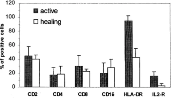
Skin biopsies from infected animals with active or healing lesions were compared to non-infected controls (three of each type) to quantitate inflammatory cell types. Inflammatory cells (composed of a mixture of T lymphocyte subpopulations, macrophages and a small number of natural killer cells and granulocytes) were more numerous in active lesions than in healing ones. T-cells accounted for 44.7 ±
13.1% of the infiltrate in active lesions (versus CD2+ = 40.3 ± 5.7% in healing lesions) and T-cell ratios favor CD8+ cells in both lesion types. The percentage of cells expressing class II antigen (HLA-DR+) in active lesions (95 ± 7.1%) was significantly higher (P < 0.005) from the healing lesions (42.7 ±
12.7%). Moreover, the expression of the activation molecules CD25 (≅ 16%), the receptor for interleukin-2, suggests that many T cells are primed and proliferating in active lesions. Distinct histopathological patterns were observed in lesions at biopsy, but healing lesions contained more organized epithelioid granulomas and activated macrophages, followed by fibrotic substitution. The progression and resolu-tion of skin lesions appears to be very similar to that observed in humans, confirming the potential for this to be used as a viable model to study the immune response in human cutaneous leishmaniasis.
Key words: Leishmania (L.) amazonensis - Macaca mulatta - experimental leishmaniasis - systemic and local cellular immunity
The outcome of leishmanial infection in hu-mans depends largely on the immune responsive-ness of the host and the virulence of the infecting parasite strain. The protozoa in this genus are ca-pable of producing a broad spectrum of disease in humans, ranging from asymptomatic infection to disfiguring forms of mucosal leishmaniasis (ML) or the potentially fatal visceral form (kala-azar).
The data presented in this paper have been submitted by VF Amaral as part of a thesis for a PhD degree in Cellu-lar and MolecuCellu-lar Biology at the Oswaldo Cruz Insti-tute/Fiocruz, Rio de Janeiro, RJ, Brazil. Present address: Departamento de Imunobiologia, Universidade Federal Fluminense, Outeiro de São João Batista s/n, Niteroi, RJ, Brasil.
This work was supported in part by grants from the UNDP/World Bank/WHO Special Program for Research and Training in Tropical Diseases, Faperj and CNPq/ RHAE Program (Brazil).
+Corresponding author. Fax: + 55-21-280-1589. E-mail:
grimaldi@gene.dbbm.fiocruz.br Received 2 August 1999 Accepted 6 December 1999
The more benign self-healing cutaneous leishma-niasis (CL) forms usually result in protection against reinfection, and cell-mediated immunity is involved in this protection (Grimaldi & Tesh 1993).
The use of murine models for the identifica-tion of T-cell subsets secreting defined patterns of cytokines that lead to strikingly different effector functions has greatly improved our understanding of the regulation of the immune response involved in host resistance and susceptibility to Leishmania
infection (Mossman & Moore 1991). The innate or acquired capacities of macrophages for killing intracellular amastigotes appear ultimately to elimi-nate or control leishmanial infection. The current hypothesis is that T-cell-mediated macrophage activation is the major protective mechanism; re-sistance to infection is associated with a TH1 cytokine profile, characterized principally by in-terferon-γ (IFN-γ), interleukin (IL) 2, and IL-12 production, whereas susceptibility is dependent of TH2 cytokines, with secretion of IL-4, IL-5, and IL-10 (Heinzel et al. 1995).
210 210 210 210
210 Immune Response in Rhesus Macaques VF Amaral et al.
of CL (Barral et al. 1987, Castes et al. 1989, Martinez-Arends et al. 1991, Esterre et al. 1992, Lima et al. 1994, Isaza et al. 1996). Differential expression of cytokines parallels the variation ob-served on the nature of the cellular inflammatory infiltrate (Modlin et al. 1985, Nilsen & Mshana 1987, Pirmez et al. 1990a). T lymphocytes reac-tive to leishmanial antigens contribute to protec-tive immunity, but may also participate in the de-velopment of chronic and destructive mucosal le-sions (Conceição-Silva et al. 1990, Pirmez et al. 1990a, 1993). The pathogenesis of CL may be explained by the balance of CD4+ type 1 and CD8+ type 2 T cells, which have distinct T cell reper-toires and may recognize distinct sets of antigens (Uyemura et al. 1993, Russo et al. 1993). How-ever, the mechanisms leading to preferential induc-tion and/or expansion of distinct TH cell subsets are still not well understood.
Non-human primates of several species, al-though to a lesser extent than inbred strains of mice, have also been used for studying immune response to Leishmania (Lainson & Bray 1966, Dennis et al. 1986, Lujan et al. 1986, 1987, Pung & Kuhn 1987, Olobo et al. 1992, Curry et al. 1994, Amaral et al. 1996). The availability of these experimental models is desirable, since they will provide valu-able data on the efficacy of newly developed vac-cines and the immunotherapeutic agents against leishmaniasis. We have previously examined (i) the susceptibility of Macaca mulatta to L. (L.)
amazonensis infection and the clinico-pathologic changes of the disease and (ii) the immunological profile of the host occurring during primary and challenge infections (Amaral et al. 1996). The purpose of this study was to further characterize the local T-cell responses associated with cure of cutaneous lesions in this experimental host.
MATERIALS AND METHODS
Animals A total of nine laboratorybred and -reared young adult (5 to 7 years old) rhesus macaques (M. mulatta) of both sexes, obtained from the Oswaldo Cruz Foundation Primate Re-search Center (Rio de Janeiro, Brazil), were used. The animal facility is maintained according to the guidelines of the Committee on the Care and Use of Laboratory Animals of the Institute of Labora-tory Animal Resources, National Research Coun-cil, and the HHS (MD, USA). Animals were housed indoors in individual steel squeeze-back cages in a temperature (25oC) – and humidity (60±5%) – controlled environment. The monkeys were acclimatized to the laboratory conditions for at least two weeks before the experimental proce-dures begun. Animals were anesthetized before infection and prior to each sampling or testing
pro-cedure. Monkeys were initially restrained in their cages, and subsequently were given, intramuscu-larly, Ketamine (Ketalar: Ketamine hydrochloride; Parke Davis) for anesthesia.
Experimental infection - Animals (n = 6) were infected by intradermal inoculation of 108 viru-lent promastigotes of L. (L.) amazonensis (strain MHOM/BR/77/LTB0016) in the upper eyelid. Le-sion area was calculated using the formula πr1r2, as described by Wilson et al. (1979).
Leishmanin skin test (LST) and serological studies - The evaluation of delayed type hypersen-sitivity (DTH) responses to L. (L.) amazonensis -derived leishmanin was done as described (Agwale et al. 1998). The tests (reactions measured as skin indurations at the site of injection) were read after 48 or 72 h using the ballpoint pen method. Class-specific (IgM and IgG) antibodies against Leish-mania were detected using indirect immunofluo-rescence (IFA) as described (Grimaldi et al. 1980). Each individual serum was tested in serial twofold dilutions [1:10 to 1:640] in PBS. Pooled normal monkey serum and pooled human cutaneous leish-maniasis serum were included in each analysis as negative and positive control, respectively.
Lymphocyte blastogenesis assay - The meth-ods described (Lujan et al. 1986, Amaral et al. 1996) were followed for peripheral blood leuko-cytes (PBL) preparation and proliferation assays. Purified PBL were cultured at 2 x 10⁶ viable cells ml-1 in the presence of optimal culture concentra-tion of mitogen (PHA-P at 12.5 µg ml-1; Sigma) or parasite antigen (10 µg protein/well). The soluble antigens for in vitro blast transformation assays were prepared from L. amazonensis promastigotes, according to the method described by Dennis et al. (1986). Cultures were incubated at 37oC in a hu-midified atmosphere containing 5% CO2 for three days in the case of mitogen or for four days in the case of antigens. The cells were pulsed with [3H]thymidine (Amersham, Co., U.K.; 1 µCi/well; 5 µCi/mM) over the last 18 h and harvested onto glass fiber filter mats (Titertek, FlowLab, U.S.A.). Radioactive incorporation into DNA was deter-mined by liquid scintillation spectrometry. Results were expressed as stimulation index (SI), which was obtained by dividing the proliferations of test cultures by proliferation of control cultures. The data from the lymphocyte proliferative responses (LPR) were compared with those of healthy ani-mals (control group) using the unpaired Student’s
t-test.
211 211211 211211 Mem Inst Oswaldo Cruz, Rio de Janeiro, Vol. 95(2), Mar./Apr. 2000
Naperville, IL). Naive animals (n = 3) were stud-ied for characterizing the distribution and immunophenotype of lymphocyte subpopulations in normal rhesus skin. The samples were snap fro-zen and stored in liquid nitrogen until processed. Portions of the biopsies were used for routine his-topathologic studies.
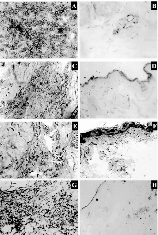
Immunoperoxidase staining - Cryostat sections 4-5 µm thick were air-dried and fixed in acetone. After rehydratation with phosphate buffered saline (PBS), serial sections of each specimen were stained with a biotin-avidin immunoperoxidase technique using the following monoclonal antibod-ies (mAbs): CD2 (T11 pan T cell antibody; Coulter), CD4 (IOR helper/inducer T cell subset antibody; CIMAB, Havana, Cuba), CD8 (Leu 2a suppressor/cytotoxic T cell subset antibody; Becton-Dickinson, San Jose, CA), CD23 (Leu 20 antibody for B cells, Becton-Dickinson), and CD16 (Leu-11 antibody for NK cells, granulocytes, acti-vated monocyte, imature fetal B/T cells and mac-rophages; Becton Dickinson). Also used were mAbs anti-HLA-DR (which identifies B cells, monocytes, macrophages and activated T cells; Becton Dickinson) and anti-CD25 (for IL-2 recep-tor; Becton Dickinson). Whenever possible, a sec-ond mAb of a similar specificity but from differ-ent commercial sources was used in a cross-check-ing strategy. The sections were blocked with nor-mal goat serum, then incubated with the primary murine mAb (at a concentration predetermined by checkerboard titration), followed by biotin-labeled anti-mouse antibody. After incubation with streptavidin-peroxidase complex (Amersham, UK), the sections were developed with the chromogen 3,9 amino-ethyl-carbazole (AEC, Sigma) plus 0.03% hydrogen peroxide in 0.01 M acetate buffer pH 5.0. Slides were counterstained with Mayer’s hematoxilin. Controls included untreated sections. The percentage of cells reacting with each mAb in relation to the total number of cells was determined as described by Esterre et al. (1992). Depending on the number of cells within the infiltrate, the whole section or 10 microscopic fields (magnifi-cation x 250) were evaluated blind by two inde-pendent observers. To normalize the results, the total number of cells and the number of positive cells were calculated per square millimetre of in-flammatory infiltrate (Sobel et al. 1984, Ridel et al. 1988) and the result given as a mean percent-age ± SD of positive cells within the granuloma. The data were analyzed using Student’s t-test.
RESULTS
Immunologic features - The immunologic re-sponses during infection were determined by LST, LPR, and measurements of antibodies. Individual
cell-mediated immune responses to leishmanial antigens varied in the course of CL in L. (L.)
amazonensis-infected rhesus monkeys, but specific DTH reactivity (mean ± SD of LST size at 14 wk p.i; 8.0 ± 3.0) and LPR of PBL (mean ± SD of SI at 14 wk p.i; 9.0 ± 3.9) were detected in all ani-mals, and both immune responses persisted beyond apparent self-cure (PBL of animals were compa-rably responsive to control mitogen PHA prior to and throughout infection). Both IgM and IgG re-sponses to promastigote antigens rose during in-fection and then declined. IgM titers peaked (1:40) at 2 wk p.i.; IgG levels were detectable 2 wk p.i. and subsequently continued to increased, peaking (1:130) at 10 wk p.i. after which levels declined (1:30) in animals with healing lesion.
Lesion development - All L. (L.) amazonensis -infected animals developed skin lesions at the site of inoculation. Lesion development progressed as follows: the skin lesions (of an erythematous-papu-lar nature) were first visible at 1-2 wk p.i., then the developing nodular lesion progressed rapidly (nod-ules varying between approximately 20 to 120 mm2), peaking at 8 to 15 weeks p.i. All lesions ulcerated (after 6-8 wk p.i.) and were subsequently followed by regression and healing.
Pathology - Biopsies were obtained from in-fected animals with active (n = 3) or healing (n = 3) lesions and skin sections were stained with ei-ther hematoxylin and eosin stains or specific mAbs for cell typing and quantitate the relative numbers of cells in the inflammatory infiltrate. In early phases of developing ulcerated skin lesions, indis-tinctly delimited and more or less differentiated macrophage accumulations were found (which encircled a central area of vacuolated macrophages, with or without intracellular parasites), and then evolved to the formation of granulomatous nod-ules. The granulomas were surrounded by a dif-fuse mononuclear infiltrate containing giant cells. In late stages, fibroblasts proliferated at the periph-ery of the granulomas and finally invaded them with fibrotic substitution.
The relationship between lesion type and the size/profile of the cellular infiltrate was examined. The mean ± SD area of skin inflammatory infil-trates (composed of a mixture of T lymphocyte subpopulations, macrophages and a small number of natural killer cells and granulocytes) in devel-oping nodular lesions (2.4 ± 0.9 mm) was greater (p < 0.05) than that obtained in lesions (0.4 ± 0.1 mm) during the healing process.
212 212 212 212
212 Immune Response in Rhesus Macaques VF Amaral et al.
(CD2+) accounted for 44.7 ± 13.1% (with a local CD4 (17.5 ± 10.6%)/CD8 (30.0 ± 15.3%) ratio of 0.7 ± 0.6] of the infiltrate in active lesions [versus CD2+= 40.3 ± 5.7%; with a local CD4 (18.7 ± 10.9%)/CD8 (23.0 ± 3.0%) ratio of 0.8 ± 3.7 in healing lesions]. The percentage of cells express-ing HLA-DR antigen in the active lesion group (95 ± 7.1%) was significantly higher (p < 0.005) from the healing lesion group (42.7 ± 12.7%). In com-parison to the expression of class II antigens, the percentage of T cells bearing IL-2 receptors (CD25+ phenotype) present in the inflammatory infiltrate was lower in either active (16.0 ± 5.7%) or healing (2.0 ± 3.5) lesions. We observed a vari-able but moderate percentage of cells with the NK phenotype (CD16+ cells = 20.0 ± 15.0% and 28.3 ± 12.0% in active and scarring lesions, respectively) and only few B lymphocytes (CD20+ cells ≅ 2%) in the infiltrate.
Moreover, skin sections from naive animals were studied as controls. Small numbers of CD2+ cells were found in the epidermis and/or dermis, clustered mainly around postcapillary venules of the papillary vascular plexus or adjacent to cuta-neous appendages. Most of these cells were CD8+ and presented an activated phenotype (HLA-DR+), but they did not express CD25 antigen on their surface.
DISCUSSION
A variety of Old World and New World mon-keys are susceptible to infection with various hu-man pathogens, but the issue of pathological con-sequence and mimicry of the human disease state is not clear cut in these animals as it is with the great apes (Kennedy et al. 1997). Non-human pri-mate model is an animal system closer in term of immunological responses to humans (Letvin et al. 1983, Klein et al. 1993, Kennedy et al. 1997). The closer phylogenetically the Old World monkey
species is to humans, the better the cross-reactiv-ity with human reagents. The majorcross-reactiv-ity of anti-human CD antigen reagents cross-react with Old World monkey species, although differences among species are observed (Letvin et al. 1983, Klein et al. 1993).
The characteristic clinical and pathologic fea-tures of the self-healing CL in humans were ob-served in M. mulatta experimentally infected with
L. (L.) amazonensis, confirming the potential for this to be used as a primate model to study the im-mune response in human disease. The findings indicate that systemic immune responses (both humoral and cell-mediated) developed in infected animals correspond to those for human CL. In the original model description (Amaral et al. 1996), the results also show that the circulating T cell sub-populations from the infected monkeys vary throughout the course of infection. In early phases of infection blood CD4+ T cells appear to predomi-nate (6-8 wk p.i.; CD4:CD8 mean ration, 5.0; range 2.5-7.1), but subsequently, an increase in CD8+ T cells was observed (15-21 wk p.i.; CD4:CD8 mean ration, 1.05; range 0.37-2.1).
Here we have characterized the cellular nature of the infiltrate in the skin lesion of rhesus macaques infected with the same parasite. Frozen skin biopsies from animals with active or healing lesions and non-infected controls were stained with a panel of mAbs to quantitate the cell types. Dis-tinct histopathological patterns were observed in different lesions at biopsy, but there was predomi-nance of lymphocytes and/or macrophages in the inflammatory infiltrate; granulomas showed a mix-ture of T-cell subpopulations with the ratio of helper:suppressor phenotypes less than one. The percentage of cells in the granulomas expressing the major histocompatibility complex (MHC) class II antigens in active lesions was significantly higher from the healing lesions. Results similar to ours were previously reported, studying the infiltrate in the skin of humans with CL (Pirmez et al. 1990b, Lima et al. 1994). Moreover, approximately 16% of the intralesional lymphocytes expressed IL-2 receptors, indicating that these activated cells were expanded when in contact with the parasite anti-gens. In spite of wide variations from one infected animal to another (as shown by some large SD values), the data show that the cell types and per-centages of each cell type change uniformly dur-ing the infection, suggestdur-ing that the same compo-nents of the immune response are working in uni-son, at least at the level of the listed cell markers. In humans, the intralesional immune response is heterogenous during the course of CL. Differ-ential expression of cytokines parallels the varia-tion observed on the nature of the cellular
213 213213 213213 Mem Inst Oswaldo Cruz, Rio de Janeiro, Vol. 95(2), Mar./Apr. 2000
214 214 214 214
214 Immune Response in Rhesus Macaques VF Amaral et al.
matory infiltrate, with particular reference to the T cell phenotypes (Modlin et al. 1985, Nilsen & Mshana 1987, Pirmez et al. 1990a). T-cells bear-ing γδ antigen receptors responding to infection (Modlin et al. 1989) are specifically selected by a limited set of antigens (Russo et al. 1993) and ex-panded within microanatomic region (Uyemura et al. 1992).
Most reports concerning characterization of cells in active skin lesions of patients with CL have shown that T cells constituted 37-75% of the in-flammatory cells and the ratio of CD4+ T cells to CD8+ T cells ranged from 0.8 to 1.8 (Barral et al. 1987, Castes et al. 1989, Martinez-Arends et al. 1991, Esterre et al. 1992, Lima et al. 1994, Isaza et al. 1996). The numbers noted above are consis-tent with those reported here, indicating that the cellular populations found in active or healed le-sions of L. (L.) amazonensis-infected rhesus macaques are similar to what has been described in human CL. In this model, there is apparently no correlation between lesional T cell subsets and those found in blood (Amaral et al. 1996). Results similar to ours were reported in human CL studies (Modlin et al. 1985, Conceição-Silva et al. 1990, Ghosh et al. 1995).
The activation of CD4+ T and/or CD8+ T cells may be essential for the healing that was subse-quently observed in these monkeys as has been found in human studies. These T-cell populations proliferating in response to leishmanial antigen stimulation (Uyemura et al. 1993) might be in-volved in protection from and healing of CL (Maasho et al. 1998). NK cells were relatively abundant in the inflammatory infiltrate as well, and they could play a role in the local control of para-site dissemination (Ridel et al. 1988). In humans, the immunological effectiveness of granulomas appear to be related less to the numbers and loca-tion of T cell phenotypes than the funcloca-tional as-pects of these cells, particularly the ability to gen-erate lymphokines (Modlin et al. 1985, Pirmez et al. 1993). The immunity to the parasite and/or pathogenesis of CL may be explained by the bal-ance of CD4+ type 1 and CD8+ type 2 T cells, which probably recognize distinct sets of antigen (Uyemura et al. 1993).
The skin immune system plays a critical role in the T-cell response to Leishmania infection (Modlin et al. 1989, Conceição-Silva et al. 1990, Pirmez et al. 1993). The majority (90%) of lym-phocytes present in normal human skin is activated, and approximately evenly distributed between CD4+ inducer and CD8+ suppressor-cytotoxic T-cell subsets (Maasho et al. 1998). Our results indi-cate that the preferential perivascular localization of activated T lymphocytes (with a predominance
of CD8+ T cells) observed in humans (Bos et al. 1987) is also the characteristic of normal monkey skin.
Prospects for human immunoprophylaxis with a new generation of safe and effective subunit vac-cines is now within our reach (Grimaldi 1995). For a vaccine to elicit a cellular immune response, the class I and class II products of the MHC must present peptide fragments of the vaccine to T-cells. While the repertoire of MHC alleles present among Old World primates may differ, the organization and expression of MHC alleles is similar to that in humans; in contrast, New World monkeys have a smaller MHC when compared to humans (Klein et al. 1993, Kennedy et al. 1997). As the rhesus monkey is an animal system closer phylogeneti-cally to human than the New World primates (Kennedy et al. 1997), the value of this primate model to the researcher lies in its abilitity to pro-duce physiological and immunological responses to leishmanial infection, which are quantitatively similar to those of humans infected with the same agent. The rhesus model should provide an indi-cation of the potential success and/or limitations for candidate vaccines employing similar delivery systems in humans.
REFERENCES
Agwale SM, Duhlinska DD, Grimaldi Jr G 1998. Re-sponse to heterologous leishmanins in cutaneous leishmaniasis in Nigeria - Discovery of a new fo-cus. Mem Inst Oswaldo Cruz 93: 23-27.
Amaral VF, Ransatto VAO, Conceição-Silva F, Molinaro E, Ferreira V, Coutinho SG, McMahon-Pratt D, Grimaldi Jr G 1996. The Asian Rhesus macaques (Macacamulatta) as an experimental model for study of cutaneous leishmaniasis. Exp Parasitol 82: 34-44. Barral A, Jesus AR, Almeida RP, Carvalho EM, Barral-Netto M, Costa JM, Badaró R, Rocha H, Johnson WD 1987. Evaluation of T-cell subsets in the lesion infiltrates of human cutaneous and mucocutaneous leishmaniasis. Parasite Immunol 9: 487-497. Bos JD, Zonneveld I, Das PK, Krieg SR, van der Loos
CM, Kapsenberg ML 1987. The skin immune sys-tem (SIS): distribution and immunophenotype of lymphocyte subpopulations in normal human skin.
J Invest Dermatol 88: 569-573.
Castes M, Moros Z, Martinez A, Trujillo D, Castellanos PL, Rondon AJ, Convit J 1989. Cell-mediated im-munity in localized cutaneous leishmaniasis patients before and after treatment with immunotherapy or chemotherapy. Parasite Immunol 11: 211-222. Conceição-Silva F, Dórea RC, Pirmez C, Schubach A,
Coutinho SG 1990. Quantitative study of Leishma-nia braziliensis braziliensis reactive T cells in pe-ripheral blood and in the lesions of patients with American mucocutaneous leishmaniasis. Clin Exp Immunol 79: 221-226.
215 215215 215215 Mem Inst Oswaldo Cruz, Rio de Janeiro, Vol. 95(2), Mar./Apr. 2000
to defined Leishmania T-cell epitopes. Infect Immun 62: 1733-1741.
Dennis VA, Lujan R, Chapman Jr WL, Hanson WL 1986.
Leishmania donovani: cellular and humoral immune responses after primary and challenge infections in squirrel monkeys, Saimiri sciureus. Exp Parasitol 61: 319-334.
Esterre P, Dedet JP, Frenay C, Chevallier M, Grimaud J-A 1992. Cell populations in the lesion of human cutaneous leishmaniasis: a light microscopical, im-munohistochemical and ultrastructural study.
Virchows Arch A Pathol Anat Histopathol 421: 239-247.
Ghosh MK, Nandy A, Addy M, Maitra TK, Ghose AC 1995. Subpopulations of T lymphocytes in the pe-ripheral blood, dermal lesions and lymph nodes of post kala-azar dermal leishmaniasis patients. Scand J Immunol 41: 11-17.
Grimaldi Jr G 1995. Meeting on vaccine studies towards the control of leishmaniasis. Mem Inst Oswaldo Cruz 90: 553-556.
Grimaldi Jr G, Tesh RB 1993. Leishmaniasis of the New World: current concepts and implications for future research. Clin Microbiol Review 6: 230-250. Grimaldi Jr G, Moriearty PL, Hoff R 1980. Leishmania
mexicana: immunology and histopathology in C3H mice. Exp Parasitol 50: 45-56.
Heinzel FP, Rerko RM, Ahmed F, Pearlman E 1995. Endogenous IL-12 is required for control of of Th2 cytokine responses capable of exacerbating leish-maniasis in normally resistant mice. J Immunol 155: 730-739.
Isaza DM, Restrepo M, Restrepo R, Caceres-Dittmar G, Tapia FJ 1996. Immunocytochemical and histopatho-logic characterization of lesions from patientes with localized cutaneous leishmaniasis caused by Leish-mania panamensis. Am J Trop Med Hyg 55: 365-369.
Kennedy RC, Shearer MH, Hildebrand WH, Simmonds RS 1997. Nonhuman primates and their use in im-munologically based investigations. The Immunolo-gist 5/5: 150-156.
Klein J, Satta Y, O’Huigin C, Takahata N 1993. The molecular descent of the major histocompatibility complex. Immunol Reviews 11: 213-244. Lainson R, Bray RS 1966. Studies on the immunology
and serology of leishmaniasis. II. Cross-immunity experiments among different forms of American cutaneous leishmaniasis in monkeys. Trans R Soc Trop Med Hyg 60: 526-532.
Letvin NL, Kim NW, Reinhertz EL, Hunt RD, Lane JH, Schlossman SF 1983. T lymphocyte surface an-tigens in primates. Eur J Immunol 13: 345-347. Lima HC, Vasconcelos AW, David JR, Lerner EA 1994.
American cutaneous leishmaniasis: in situ charac-terization of the cellular immune response with time.
Am J Trop Med Hyg 50: 743-747.
Lujan R, Dennis VA, Chapman Jr WL, Hanson WL 1986. Blastogenic responses of peripheral blood leukocytes from owl monkeys experimentally infected with
Leishmania braziliensis panamensis. Am J Trop Med Hyg 35: 1103-1109.
Lujan R, Hanson WL, Chapman Jr WL, Dennis VA 1987. Antibody responses, as measured by the enzyme-linked immunosorbent assay (ELISA), in owl mon-keys experimentally infected with Leishmania braziliensis panamensis. J Parasitol 73: 430-432. Maasho K, Sanchez F, Schurr E, Hailu A, Akuffo H 1998.
Indications of the protective role of natural killer cells in human cutaneous leishmaniasis in an area of en-demicity. Infect Immun 66: 2698-2704.
Martinez-Arends A, Tapia FJ, Caceres-Dittmar G, Mosca W, Valecillos L, Convit J 1991. Immunocytochemi-cal characterization of immune cells in lesions of American cutaneous leishmaniasis using novel T cell markers. Acta Trop 49: 271-280.
Modlin RL, Pirmez C, Hofman FM, Torigian V, Uyemura K, Rea TH, Bloom BR, Brenner MB 1989. Lym-phocytes bearing antigen-specific γδ T-cell recep-tors accumulate in human infectious disease lesions.
Nature 339: 544-548.
Modlin RL, Tapia FJ, Bloom BR, Gallinoto ME, Castes M, Rondon AJ, Rea TH, Convit J 1985. In situ char-acterization of the cellular immune response in American cutaneous leishmaniasis. Clin Exp Immunol 60: 241-248.
Mossman TR, Moore KW 1991. The role of IL-10 in cross-regulation of TH1 and TH2 responses.
Immunol Today 12: A49-53.
Nilsen R, Mshana RN 1987. In situ characterization of the cutaneous immune response in Ethiopian cuta-neous leishmaniasis. Scand J Immunol 26: 503-512. Olobo JO, Reid GD, Githure JI, Anjili CO 1992. IFN-gamma and delayed-type hypersensitivity are asso-ciated with cutaneous leishmaniasis in vervet mon-keys following secondary rechallenge with Leish-mania major. Scand J Immunol 1 (Suppl. I): 48-52. Pirmez C, Cooper C, Paes-Oliveira M, Schubach A, Torigian VK, Modlin RL 1990a. Immunologic re-sponsiveness in American cutaneous leishmaniasis lesions. J Immunol 145: 3100-3104.
Pirmez C, Oliveira-Neto MP, Grimaldi G Jr, Savino W 1990b. Immunopathology of American cutaneous leishmaniasis. Modulation of MHC class II gene products by keratinocytes before and after Glucantime therapy. Mem Inst Oswaldo Cruz 85: 203-209.
Pirmez C, Yamamura M, Uyemura K, Paes-Oliveira M, Conceição-Silva F, Modlin RL 1993. Cytokine pat-terns in the pathogenesis of human leishmaniasis. J Clin Investig 91: 1390-1395.
Pung OJ, Kuhn RE 1987. Experimental leishmaniasis in the Brazilian squirrel monkey (Saimiri sciureus): lesions, hematology, cellular, and humoral immune responses. J Med Primatol 16: 165-174.
Ridel PR, Esterre P, Dedet JP, Pradinaud R, Santoro F, Capron A 1988. Killer cells in human cutaneous leishmaniasis. Trans R Soc Trop Med Hyg 82: 223-226.
Russo DM, Armitage RJ, Barral-Netto M, Barral A, Grabstein KH, Reed SG 1993. Antigen-reactive gamma delta T cells in human leishmaniasis. J Immunol 151: 3712-3718.
216 216 216 216
216 Immune Response in Rhesus Macaques VF Amaral et al.
The immunopathology of experimental allergic en-cephalomyelitis. Quantitative analysis of inflamma-tory cells in situ. J Immunol 132: 2393-2401. Uyemura K, Klotz J, Pirmez C, Ohmen J, Wang XH, Ho
C, Hoffman WL, Modlin RL 1992. Microanatomic clonality of γδ T cells in human leishmaniasis le-sions. J Immunol 148: 1205-1211.
Uyemura K, Pirmez C, Sieling PA, Kiene K,
Paes-Oliveira M, Modlin RL 1993. CD4+ type 1 and CD8+ type 2 T cell subsets in human leishmaniasis have distinct T cell receptor repertoires. J Immunol 151: 7095-7104.