193
Dorsolateral head muscles of the catfish families Nematogenyidae and
Trichomycteridae (Siluriformes: Loricarioidei): comparative
anatomy and phylogenetic analysis
Aléssio Datovo and Flávio Alicino Bockmann
The skeletal muscles of the dorsolateral region of the head of the Nematogenyidae and representatives of the all major clades of the Trichomycteridae are described and illustrated. A hypothesis on the phylogenetic relationships among these taxa exclusively based on the surveyed musculature is presented. The single most parsimonious cladogram obtained from the phylogenetic analysis of the 36 myological characters gathered and 35 terminal-taxa mostly agrees with the previous hypotheses of trichomycterid intrarelationships. The Copionodontinae and Trichogeninae form a monophyletic lineage that is the sister-group to all remaining trichomycterids. The monophyly of the clades formed by Glanapteryginae plus Sarcoglanidinae; Stegophilinae plus Tridentinae plus Vandelliinae; and the assemblage comprising all of these five subfamilies (TSVSG clade) is corroborated. Two of our findings are, however, discordant with the previous prevailing hypotheses: the sister-group relationship among Tridentinae and Stegophilinae and the monophyly of the Trichomycterinae lato sensu, i.e., including the genera
Scleronema and Ituglanis. In addition, the previously proposed osteological synapomorphies supporting the close affinities of Scleronema and Ituglanis with the TSVSG clade were revised, revealing that they are either invalid or ambiguous. Most of the synapomorphies herein proposed are homoplasy-free, with some of them corroborating the monophyly of weakly-supported groups, such as Stegophilinae.
Os músculos esqueléticos da região dorsolateral da cabeça de Nematogenyidae e de representantes de todos os maiores clados de Trichomycteridae são descritos e ilustrados. Uma hipótese das relações filogenéticas entre estes táxons, baseada exclusivamente na musculatura estudada, é apresentada. O único cladograma mais parcimonioso obtido da análise filogenética dos 36 caracteres miológicos levantados e 35 táxons-terminais concorda com a maior parte das hipóteses anteriores de relações entre os tricomicterídeos. Copionodontinae e Trichogeninae formam uma linhagem monofilética que é grupo-irmã de todos os demais tricomicterídeos. O monofiletismo dos clados formados por Glanapteryginae mais Sarcoglanidinae; Stegophilinae mais Tridentinae mais Vandelliinae; e do agrupamento incluindo estas cinco subfamílias (clado TSVSG) é corroborado. Duas de nossas descobertas são, entretanto, discordantes com relação às hipóteses anteriores mais aceitas: a relação de grupo-irmão entre Tridentinae e Stegophilinae e o monofiletismo de Trichomycterinae lato sensu, i.e., incluindo os gêneros Scleronema e Ituglanis. Além disso, as sinapomorfias osteológicas previamente propostas suportando as relações de afinidade de Scleronema
e Ituglanis com o clado TSVSG foram revisadas, revelando serem inválidas ou ambíguas. A maior parte das sinapomorfias aqui
propostas são livres de homoplasias, com algumas delas corroborando o monofiletismo de grupos fracamente suportados, tais como o da subfamília Stegophilinae.
Key words: Myology, Ostariophysi, Teleostei.
Laboratório de Ictiologia de Ribeirão Preto, Universidade de São Paulo, FFCLRP, Departamento de Biologia, Programa de Pós-Graduação em Biologia Comparada. Av. dos Bandeirantes, 3900, 14040-901 Ribeirão Preto, SP, Brazil. adatovo@gmail.com, fabockmann@ffclrp.usp.br
Introduction
The Siluriformes is a worldwide, megadiverse group of primarily freshwater fishes with 3370 valid species described to date (Eschmeyer & Fong, 2010). Historically, most of the anatomical studies on catfishes, either in descriptive approaches or as sources of information for cladistic analyses, relied heavily upon their skeleton, due its known utility, easy access and extensive literature. Alternative body systems and
remains poorly explored by systematists. This is likely due to the obstacles intrinsic to the investigation of this body system: dissections are laborious, time-consuming, and, many times, destructive (preventing future verification of conditions in the same material); observations are difficult, and a standardized comparative framework is deficient. In the catfish literature, exceptions are found in the recent contributions by Diogo and collaborators (e.g. Diogo, 2007; Diogo &
Vandewalle, 2003; Diogo et al., 2004, 2006).
In the present study we carried out an investigation on the dorsolateral head myology of two Neotropical catfish lineages that are currently thought to be sister-groups, the Nematogenyidae and Trichomycteridae (de Pinna, 1992, 1998; Diogo, 2005). These taxa are of special interest due to their key positions in the phylogeny of Siluriformes. First of all, they comprise the basal most lineage of the suborder Loricarioidei (sensu Sullivan et al., 2006; interchangeably treated as
superfamily Loricarioidea by Schaefer & Lauder, 1986; de Pinna, 1998), a group which also includes the families Callichthyidae, Scoloplacidae, Astroblepidae, and Loricariidae. Moreover, the Loricarioidei is a group that apparently diverged relatively early in the catfish evolution (cf. Arratia, 1992; de Pinna, 1998; Diogo,
2005), being recently regarded as the basal most group in the Siluriformes (Sullivan et al., 2006; Lundberg et al., 2007).
Accordingly, morphological studies in Nematogenyidae and Trichomycteridae, especially of the former (de Pinna, 1998), may be important for an understanding of the evolution of traits in the Loricarioidei in particular and the Siluriformes as a whole.
The Nematogenyidae is endemic to the upper reaches of central Chile (de Pinna, 1998). The single living species of the family, Nematogenys inermis, inhabits the plane sectors with
slow-running waters of rivers and streams, being associated with aquatic vegetation as juveniles and occupying deeper areas (pools) when adults (Arratia, 1983). Nematogenys is of
singular interest because it is thought to retain several primitive character states within the suborder Loricarioidei (Arratia & Huaquín, 1995; de Pinna, 1998). It is presently accepted as the sister-group of Trichomycteridae, and this hypothesis is based on several anatomical features (Mo, 1991; de Pinna, 1992; Diogo, 2005; Diogo et al., 2006).
External cheek and opercular muscles of Nematogenys inermis were briefly described by Howes (1983a, 1983b) and
illustrated by Arratia (1992: fig. 43b) and de Pinna (1998: fig. 7), whereas Schaefer & Lauder (1986) briefly commented on the adductor mandibulae muscle-complex and hyoid
musculature of N. inermis. The most complete descriptive
treatment of the myology for the Nematogenyidae was provided by Diogo et al. (2006). However, examination of
material of N. inermis revealed various problematic
interpretations of the muscular structures proposed by Diogo (2005) and Diogo et al. (2006), revealing the need of
an in depth re-analysis of the myology of this species. The Trichomycteridae, also a Neotropical group, is one of the most specious catfish families, currently encompassing 241 valid species (Eschmeyer & Fong, 2010). This family is widely distributed throughout the major river drainages from
Costa Rica to Patagonia (de Pinna & Wosiacki, 2003). The monophyly of Trichomycteridae is very well supported, with the most conspicuous characters involving its highly specialized opercular-interopercular apparatus (Baskin, 1973; de Pinna, 1992, 1998). This system allows a generalized trichomycterine to climb a steep rocky substrate with fast-flowing waters (pers. obs.; de Pinna, 1998) and a parasite vandelliine to anchor in the branchial chambers of its host (Zuanon & Sazima, 2004a, 2004c; Adriaens et al., 2010).
Trichomycterids encompass one of the greatest ranges of feeding modes and habitats in a single fish family. Adults of Copionodontinae and Trichogeninae explore the water column, with the former resting on the bottom but making constant incursions to the upper water levels, while the latter is markedly nektonic, and both feed mostly on insects (FAB, pers. obs.; Sazima, 2004). Trichomycterines are primarily invertebrate-eaters and dwell on rocky bottoms, where they have remarkable skills to resist and ascend against strong currents (pers. obs.; de Pinna, 1998). Glanapterygines and sarcoglanidines are mostly insectivorous, with the former living inside the leaf litter (FAB, pers. obs.; Nico & de Pinna, 1996) and the latter inhabiting the most superficial layers of clear loose sand and fine gravel (pers. obs.; Zuanon & Sazima, 2004b; Zuanon et al., 2006). The glanapterygines Pygidianops
and Typhlobelus are also psamophilous (Schaefer et al., 2005),
but they occupy the deeper interstices of the sandy habitats (FAB, pers. obs.). Members of the Stegophilinae are usually reported to be scale- or mucus-eaters (Baskin et al., 1980;
Winemiller & Yan, 1989; de Pinna, 1998; Fernández & Schaefer, 2009), but also include carnivores and even scavengers (pers. obs.; Goulding, 1979, 1980; Lüling, 1984). The Trichomycteridae also includes the vandelliines, whose infamous hematophagous members obtain blood from the gills of other fishes (Kelley & Atz, 1964; Machado & Sazima, 1983; de Pinna, 1998). Vandelliines and most stegophilines – the so-called “candirus”, “caneros”, or “carneros” – rest buried in sand bars, fine gravel, and muddy substrate (pers. obs.; Haseman, 1911; Devincenzi & Teague, 1942; Roberts, 1972; Baskin et al., 1980; Winemiller & Yan, 1989; Spotte, 2002). The
stegophilines Pareiodon and Pseudostegophilus and the
Tridentinae are presumably nektonic (FAB, pers. obs.; cf.
Roberts, 1972; Ferraris, 1991), although members of the latter subfamily are also said to hide in sand bottoms (Burgess, 1989). Such a stunning diversity of feeding specializations, behaviors, and wide range of habitats are obviously mirrored in their morphology.
The Trichomycteridae is recognized as one of the best known siluriform groups in phylogenetic terms (de Pinna, 1998; de Pinna & Wosiacki, 2003). Members are currently grouped in eight subfamilies: Copionodontinae, Trichogeninae, Trichomycterinae, Glanapteryginae, Sarcoglanidinae, Tridentinae, Stegophilinae, and Vandelliinae. The genera Ituglanis and Scleronema were proposed as
Wosiacki (2003) who maintained these genera within the Trichomycterinae by default. Monophyly of all subfamilies of the Trichomycteridae is well-corroborated (Baskin, 1973; de Pinna, 1989a, 1989b, 1992, 1998; de Pinna & Britski, 1991; Costa & Bockmann, 1994), with the exception of the Trichomycterinae, whose phyletic status has been object of numerous discussions over recent decades (de Pinna, 1989a; Arratia, 1990, 1998; Costa & Bockmann, 1993; Bockmann et al., 2004;
see “Discussion”). Elucidation of the relationships of the members of the Trichomycterinae is critical to an understanding of the evolution of the family as a whole inasmuch as they form a large gray zone containing about 57% of the diversity of the group at a basal region of the Trichomycteridae cladogram (cf. de Pinna, 1998).
In spite of the remarkable morphological diversity in the Trichomycteridae, its musculature is one of the less known among all catfish families, both in phylogenetic and descriptive terms. Howes (1983b) briefly described the superficial cheek and opercular muscles of the Trichomycteridae on the basis of observations done on one vandelliine (Branchioica = Paravandellia; cf. de Pinna & Wosiacki, 2003) and three
trichomycterines (Eremophilus and two species of Trichomycterus). Schaefer & Lauder (1986) provided
commentaries on the adductor mandibulae complex and hyoid
musculature of the Trichomycteridae based on single species of both Henonemus and Trichomycterus. The most important
contribution to the knowledge of the myology of the Trichomycteridae published so far is the study of Adriaens et al. (2010) in which the cranial musculature of Trichomycterus guianensis is described in greater detail. Phylogenetic
relationships among the major groups of the family were established on the basis of 72 characters (de Pinna, 1998), most of them from the skeleton (83.3%). A single myological synapomorphy emerged as informative in that analysis, the presence of the protractor operculi muscle, an element first
identified as the “preopercular muscle” by Howes (1983b: 329 and fig. 12b), and which was treated as a synapomorphy for non-Copionodontinae, non-Trichogeninae trichomycterids (de Pinna, 1992, 1998). Diogo (2005) proposed several new putative synapomorphies for the Trichomycteridae, including two derived from observations in the dilatator operculi
(however, see Discussion) based on examination of Hatcheria macraei and three species of Trichomycterus. For most
trichomycterid subfamilies, virtually nothing is known about their myology. In this scenario, myology may potentially offer new lines of evidence for elucidating these relationships, by testing previous hypotheses and highlighting new ones.
In light of the above situation, the purposes of this study were: (1) to provide phylogenetically-oriented descriptions of the dorsolateral head musculature for the Nematogenyidae and each key group of the Trichomycteridae, in order to standardize the future myological comparisons involving these families; (2) evaluate the relationships between the Nematogenyidae and Trichomycteridae and postulate a phylogenetic hypothesis on the relationships within the latter family, based on characters from the investigated musculature; and (3) compare the obtained
hypothesis of relationships with others from the literature and assess the congruency among the different phylogenetic sources of information.
Material and Methods Anatomical nomenclature
Myological nomenclature follows Winterbottom (1974a) except for the dorsal branchial muscles which follows Springer & Johnson (2004). Based mainly on the discoveries of Gosline (1989), Diogo & Chardon (2000) proposed a new terminology for the sections of the adductor mandibulae in Ostariophysi. However, several problems with this nomenclature were detected (see “Discussion: Nematogenyidae” and “Discussion: Trichomycterinae”; Datovo & Vari, in prep.). For that reason, for purpose of the present study, we adopt the traditional nomenclature for the adductor mandibulae
(Vetter, 1878; Allis, 1897; Winterbottom, 1974a), identifying the outer portion plesiomorphically inserted onto the lower jaw as the “A2” and the inner portion as “A3”. The name
protractor operculi, applied to a trichomycterid muscle described by de Pinna (1992) is used. Nomenclature for new muscles or muscle sections is explained throughout the text. Osteological terminology follows Bockmann et al. (2004), except as noted below. The so-called “tendon-bone supraorbital” [= “antorbital” of Britski & Ortega (1983) and de Pinna (1989a); “fronto-lacrimal tendon bone”of Baskin (1973) and de Pinna (1989a); “nasal” of Eigenmann (1918)] seems to be an ossification associated with a ligament rather than with a tendon (pers. obs., see below; Adriaens et al., 2010). Hence, this bone is herein designated “sesamoid supraorbital” according to Adriaens et al. (2010). Arratia & Schultze (1990) demonstrated that the so-called “urohyal” in catfishes does not entirely correspond to the urohyal of other teleosts. For this reason, these authors proposed the name “parurohyal” for that bone in catfishes, a terminology herein followed. The commonly used term “pleural rib” is replaced by “rib” only. “Pleural rib” or “ventral rib” has been traditionally used in opposition to the “dorsal ribs” in gnathostomes (Britz & Bartsch, 2003). Since Britz & Bartsch (2003) have demonstrated that gnathostomes have only one type of rib (the “pleural” or “ventral” one), the use of the adjective “pleural” is dispensable (e.g. Britz et al., 2009; Britz & Johnson, 2010). Furthermore, since fishes lack a true pleural cavity, the use of the term “pleural” may be somewhat confusing (Adriaens, pers. comm.).
and lower jaws, and forms a membrane over the roof of the anterolateral part of buccal cavity.
Anatomical preparations
Specimens dissected for musculature were double-stained for cartilage and bone. For this purpose, they underwent the steps 2 and 4 of the protocol of Taylor & van Dyke (1985) with cartilage stained using their “alternate” Alcian Blue 8GX solution (with 80 parts of 95% ethanol and 20 parts of glacial acetic acid; Dingerkus & Uhler, 1977). In order to prevent further demineralization, specimens were then transferred to a sodium borate-based neutralization solution, according to the step 5 of Taylor & van Dyke (1985), for a shorter period of time of 2-8 hours. Subsequently, they were placed into an ethanol-based solution of Alizarin Red-S for 3-6 hours, in order to stain the bones (Springer & Johnson, 2000). Potassium hydroxide-based solutions for neutralization and bone stain of Taylor & van Dyke (1985) were not used because the KOH seems to macerate tendons and ligaments (Springer & Johnson, 2000). Specimens were maintained in 60% to 95% ethanol, according to the desired hardness of the muscles to facilitate dissections. Typically, more concentrated ethanol solution works better for very small sized specimens, since their muscles become more rigid due the higher dehydration level, making dissections easier. In larger sized specimens, on the other hand, intense dehydration can makes muscle ablation difficult and may damage fine surgical instruments. Cleared and double-stained skeletal specimens were prepared following Taylor & van Dyke (1985). Histological sections were stained according to Masson’s trichrome staining protocol.
Illustrations
Drawings were sketched by hand (AD) using a digital pen tablet, based on photographs and direct observation of specimens under stereomicroscope. Colors of structures on drawings are merely illustrative, rather than corresponding of the true coloration of fixed or living specimens. Colors and patterns used to represent each tissue in the drawings are presented in Table 1.
Photographs were taken by a Leica DFC420 digital image capture device attached to a Leica MZ16 stereomicroscope. The photographs herein presented are multifocal montages made with several individual photographs taken from different focal planes of the anatomical structures. This final fully focused montage was made on CombineZP (Hadley, 2009).
Phylogenetic analysis
Character matrix was assembled in Mesquite 2.7 (Maddison & Maddison, 2009). Multistate characters were ordered whenever a clear morphoclinal gradient between the more extreme states was identifiable (Maslin, 1952). Ordering states according to morphoclines minimizes the amount of morphological changes between character states thus choosing the most parsimonious hypothesis of transformation (Maslin, 1952; Wilkinson, 1992; de Pinna, 1992; Slowinski, 1993; Wiens, 2001: 693-694). Multistate characters which morphoclines were
not detected were unordered. Maximum parsimony analysis was performed under TNT 1.1 (Goloboff et al., 2008). A “traditional search” was run with the following settings: 1000 replicates; up to 10000 trees retained in memory; remaining settings follows program defaults (which includes TBR-branch swapping). Obtained trees were rooted in Nematogenys inermis
(outgroup), the sole extant member of the Nematogenyidae and the sister-group of Trichomycteridae (de Pinna, 1992, 1998). Bremer support for the clades were calculated through the script bremer.run of TNT with the following settings: swap existing trees up to 10 steps longer; do 10 ratchet iterations in constrained searches; remaining settings follows script defaults. Numbers of steps, consistency indexes, and retention indexes were obtained from Mesquite. The most parsimonious reconstruction for characters with ambiguous optimization (as ACCTRAN or DELTRAN; Swofford & Maddison, 1987) was not a priori assumed, but rather evaluated case by case (Amorim, 1997; Agnarsson & Miller, 2008), with the decisions in favor of one or another reconstruction justified into the textual comments of each character.
Material examined
listed below, radiographs of the types of all trichomycterids available at the “All Catfish Species Inventory” (Morris et al., 2006) and “Primary Types Imagebase of California
Academy of Sciences” (California Academy of Sciences, 2007) websites were also examined.
Albuliformes: Albulidae: Albula vulpes; LIRP 7427, 1: 1ms. Amiiformes: Amiidae: Amia calva; USNM 64338, 4: 1ms (174). Characiformes: Acestrorhynchidae: Acestrorhynchus falcatus; LIRP 5115, 5: 2cs (128.6-135.2), 1ms (147.3). Alestidae: Chalceus erythrurus; LIRP 5955, 2: 1cs (167.4), 1ms (192.0). Phenacogrammus interruptus; LIRP 7441, 4: 2cs (65.0-68.9), 2ms (67.4-69.2). Anostomidae: Leporellus vittatus; LIRP 231, 56: 2ms. Characidae: Agoniatinae: Agoniates halecinus; MZUSP 34333, 2: 1cs (152.1); MZUSP 53827, 1: 1ms (196.9). Aphyocharacinae: Aphyocharax cf. pusillus; LIRP 4559, 84: 2cs (39.6-42.7), 2ms (44.8-45.4). Bryconinae: Brycon nattereri; LIRP 3558, 3: 1ms. Brycon orbignyanus; LIRP 6003, 32: 2cs (17.4-24.1), 5ms (22.4-56.7). Characinae: Roeboides cf. prognathus; LIRP 700, 16: 2cs (80.7-88-9), 2ms (71.9-84.9). Cheirodontinae: Odontostilbe pequira; LIRP 5417, 63: 2ms (36.58-37.08). Glandulocaudinae: Lophiobrycon weitzmani; LIRP 4337, 8: 2cs (25.8-27.0); LIRP 4338, 31: 2ms (27.1-28.4). Mimagoniates microlepis; LIRP 5942, 20: 2cs (44.3-53.5), 2ms (47.2-49.4). Iguanodectinae: Piabucus melanostomus; LIRP 5936, 19: 2cs (77.3-78.0), 2ms (77.3-87.5). Serrasalminae: Metynnis mola; LIRP 5937, 21: 2cs (44.1-54.0), 2ms (48.3-58.8). Serrasalmus spilopleura; LIRP 4066, 32: 2ms (128.8-130.7). Stethaprioninae: Brachychalcinus cf. copei; LIRP 5939, 14: 2cs (31.5-34.1), 1ms (36.0). Poptella paraguayensis; LIRP 5940, 21: 2cs (38.2-39.8), 2ms (35.5-38.2). Stevardiinae: Planaltina myersi; LIRP 3356, 27: 2cs (30.5-31.4), 2ms (27.9-31.4). Tetragonopterinae: Tetragonopterus argenteus; LIRP 5941, 10: 2cs (63.3-81.7), 2ms (60.6-66.9). Characidae Incertae sedis: Astyanax fasciatus; LIRP 132, 54: 3cs (72.0-75.5), 2ms (10.2-11.1). A. altiparanae; LIRP 2040, 42: 1cs, 2ms. Bryconops alburnoides; LIRP 5116, 7: 2cs (94.0-99.8), 2ms (104.4-106.7). Creagrutus seductus; LIRP 4417, 16: 2ms (38.90-41.63). Hollandichthys multifasciatus; LIRP 5744, 85: 2cs (79.4-86.5), 2ms (76.4-77.6). Salminus hilari; LIRP 670, 3: 1ms. Triportheus paranaensis; LIRP 5935, 44: 2cs (69.2-75.5), 2ms (72.3-86.9). Citharinidae: Citharinus citharus; USNM 72797, 7: 1ms (192). Crenuchidae: Characidium fasciatum; LIRP 220, 107: 2ms (53.16-54.20). Ctenoluciidae: Boulengerella maculata; LIRP 5974, 3: 2cs (199.3-200.5), 1ms (266.7). Cynodontidae: Hydrolycus scomberoides; LIRP 5943, 4: 2cs (125.5-139.4), 1ms (143.5). Curimatidae: Curimatopsis sp.; LBP 412, 6: 2cs (58.7-60.4), 2ms (61.6-67.1). Distichodontidae: Xenocharax spilurus; AMNH 230302, 3 of 79: 2ms (95.22-104.29). Erythrinidae: Hoplias malabaricus; LIRP 6032, 5: 2ms (84.48-215); LIRP 5594, 5: 2cs (64.4-66.4). Gasteropelecidae: Thoracocharax cf. stellatus; LIRP 715, 16: 2cs (40.9-44.4), 2ms (42.1-42.5). Hepsetidae: Hepsetus odoe; USNM 309529, 6: 1ms (168). Lebiasinidae: Pyrrhulina australis; LIRP 6049, 416: 2cs (32.7-41.6), 2ms (37.22-38.74). Parodontidae: Parodon hilarii; LIRP 638, 38: 2ms (135.06-138.43). Clupeiformes: Engraulidae: Setipinna taty; USNM 265905, 7: 1ms (103.53). Cypriniformes: Cyprinidae: Barilius senegalensis; USNM 271201, 12: 1ms (92.97). Rasbora cephalotaenia; USNM 330848, 146: 1ms (81.20). Elopiformes: Elopidae: Elops saurus; USNM 289648, 9: 1ms (202). Megalopidae: Megalops cyprinoides; USNM 102685, 12: 1ms (106.10). Esociformes: Esocidae: Esox americanus; USNM 237253, 12: 1ms (158). Gonorynchiformes: Chanidae: Chanos chanos; USNM 173572, 5: 1ms (147).
paulensis; LISDEBE 1312, 3: 2ms (46.5-49.3). *Pareiodon microps; LIRP 7422, 3: 1cs (104.5), 2ms (108.1-112.87). *Pseudostegophilus nemurus; LIRP 7428, 1: 1ms (96.3). Trichogeninae: *Trichogenes longipinnis; LIRP 1023, 3; LIRP 1058, 4; LIRP 1059, 12: 1cs (47.4), 2ms (60.8-75.5). Trichomycterinae: *Bullockia maldonadoi; LBP 3112, 39: 1ms (50.45); LIRP 5947: 3: 1cs (45.83). *Hatcheria macraei; ILPLA 1807, 3 of 8: 1ms (181); LBP 451, 1: 1ms (63.97); MLP 5622, 1 of 3; MLP 5668, 1 of 3; MLP 7903, 1 of 2; MZUSP 35687, 2 of TNU: 2cs (42.30-43.98). Ituglanis bambui; MZUSP 79860, holotype; MZUSP 79862; 4 paratypes: 1cs; MZUSP 79864; 4 paratypes: 2cs. I. cf. amazonicus; LIRP 7424, 3: 1ms (57.2); MZUSP 86820, 1. I. eichorniarum; MCP 36243, 1; MCP 36244, 1; MNRJ 780; 2 paralectotypes. I. epikarsticus; MZUSP 79869, holotype; MZUSP 79870, 1 paratype; MZUSP 79871, 1 paratype: 1cs; MZUSP 79872, 1 paratype: 1cs; *I. cf. gracilior; MCP 36257, 13: 1cs (38.2); MCP 36258, 17; MZUSP 86821, 12: 1cs (39.0), 1ms (53.19). I. herberti; MNRJ 1429, 3 paralectotypes; MNRJ 28466, 1 paralectotype; NUP 2238; 1; NUP 2241, 3: 1cs; NUP 2242, 2. I. macunaima; LIRP 5642, 6 paratypes: 1cs (29.1); MZUSP 86237, 7 paratypes: 1cs (25.5); MZUSP 86251, 2 paratypes: 1cs (25.5); MZUSP 86272, 1 paratype; MZUSP 88452; holotype. I. nebulosus; MZUSP 69574; 1 paratype: 1cs (35.1). I. parahybae; MCP 7784, 1; MCP 18026, 1; MCP 18032, 1; MZUSP 71852, 3; MZUSP 79810, 1. I. parkoi; MCP 36240, 1; MCP 36248, 1; MNRJ 3849, holotype. I. passensis; MCP 27436, 3; MZUSP 80097, 3. *I. proops; MNRJ 781, 3 paralectotypes; MZUSP 36502, 7; MZUSP 60255, 20 of 95: 2cs (57.1-77.0), 1ms (70.2). I. ramiroi; MZUSP 79865, holotype; MZUSP 79867, 3 paratypes: 3cs (25.4-31.3); MZUSP 79868, 1 paratype. I. sp. 1 (Tocantins/upper Paraná); MCP 15930, 37: 2cs (34.7-53.3); MCP 23073, 5: 1cs (56.2); NUP 1103, 4. *Scleronema angustirostre; MCP 21436, 17: 1cs (35.2), 1ms (42.4). S. cf. minutum; MCP 19108, 2. Trichomycterus albinotatus; LIRP 4333, 4 (23.9-36.1): 1cs (31.7). T. alternatus; LIRP 266, 1; LIRP 28, 1. T. alterus; MLP 250: 3 of 21. *T. areolatus; LBP 997, 17: 3cs (45.21-91.46), 2ms (60.5-63.5). T. auroguttatus; LIRP 4334, 4 (37.9-92.3): 1cs (59.3). *T. brasiliensis; LIRP 1968, 15: 2cs (68.0-83.8), 2ms (76.4-95.6). T. aff. brasiliensis; LIRP 818, 21: 2cs (40.1-57.0), 1ms (43.6). *T. candidus; LIRP 815, 2; LIRP 1098, 1; LIRP 7425, 12: 1ms (54.63); LIRP 7431, 1; LIRP 7435, 5. *T. chiltoni; LBP 1001, 10: 2ms (83.38-104.25); LBP 1003, 13: 2cs (47.06-50.32). *T. corduvensis; MLP 8221, 1 of 3; MLP 8222, 3 of 20: 1ms (53.53); *T. davisi; LIRP 2798, 18: 2cs (56.7-65.2), 1ms (79.3); LIRP 2799, 34: 3cs (61.0-75.2), 1 hs (63.8), 2ms (17.1-82.2). T. diabolus; LIRP 1128, 21 paratypes; LIRP 3456, 9 paratypes: 2cs. *T. cf. iheringi; LIRP 1027, 1; LIRP 1055, 5: 1cs (77.1); LIRP 3182, 8: 1ms (72.2); LIRP 3183, 2; LIRP 3185, 1. *T. immaculatus; LIRP 285, 28: 2cs (76.2-74.0), 1ms (83.7). T. cf. itatiayae; LIRP 5086, 1: 1 cs (43.3); LIRP 5087, 3. *T. maracaya; LIRP 4381, 4 paratypes (26.7-51.3): 1cs (43.9), 1ms (40.0). T. mirissumba; LIRP 4335, 2 (55.4-56.3): 1cs (56.3). T. paolence; LIRP 5713, 1. T. pauciradiatus; MNRJ 17057, 17: 3cs (27.38-45.90). T. reinhardti; LIRP 1062, 3: 1cs (30.5). T. sp. 1(São Francisco); LIRP 645, 34: 2 cs (48.0-60.0), 1ms (72.2). *T. stawiarski; LIRP 5088, 10 (33.2-60.2): 1cs (50.1), 1ms (45.0). *T. zonatus; LIRP 596, 8: 1ms (28.7); Tridentinae: *Tridentopsis pearsoni; MZUSP 38671, 3 (16.6-18.9): 1cs (16.6), 1ms (18.3). Vandelliinae: *Paracanthopoma parva; LIRP 7399, 16: 2cs (9.60-16.43), 2ms (18.91-22.72); LIRP 7410, 3; LIRP 7411, 9; LIRP 7413, 1. *Paravandellia oxyptera; LIRP 4557, 8: 1ms (22.8). P. sp. 1; LIRP 736, 1; LIRP 5407, 1. Vandellia cf. sanguinea; LIRP 7408, 7: 2cs (23.28-28.00). *V. cirrhosa; LIRP 7401, 6: 1cs (47.06), 1ms (49.94). *V. sanguinea; LIRP 7409, 3; LIRP 7414,
52: 3cs (30.89-68.53), 1ms (77.14). V. sp. 1; LIRP 598, 12: 1cs (60.0), 1ms (64.4). Vandellia sp. 2; LIRP 7391, 7: 2cs (71.42-86.90). Trichomycteridae Incertae sedis: “Trichomycterus” hasemani; LIRP 7392, 2; LIRP 7406, 8; LIRP 7395, 16: 3cs (12.38-13.60); LIRP 7405, 5; LIRP 7404, 3; LIRP 7393, 30: 3cs (13.38-13.63); LIRP 7407, 4; LIRP 7397, 49: 4cs (12.94-13.78), 3ms (14.00-15.27); LIRP 7403, 16; LIRP 814, 1; MCP 23070, 5: 1cs (11.7).
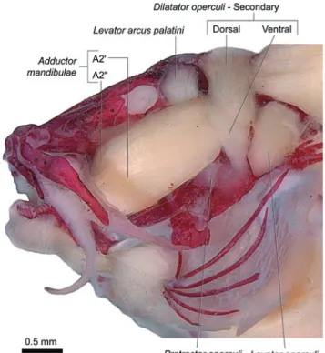
Anatomical descriptions of the cheek muscles Nematogenyidae
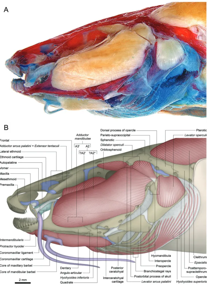
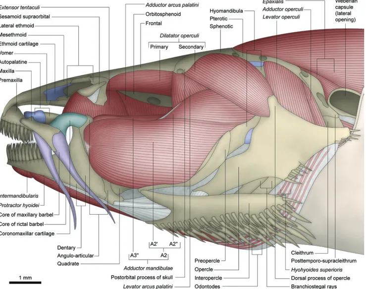
Adductor mandibulae with two main facial subdivisions:
one lateral - A2 - and another medial - A3 (Figs. 1, 2). Separation between these sections is complete only along their posterodorsal origin, where the levator arcus palatini lies
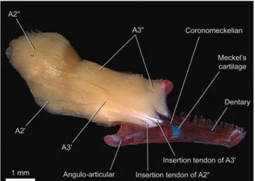
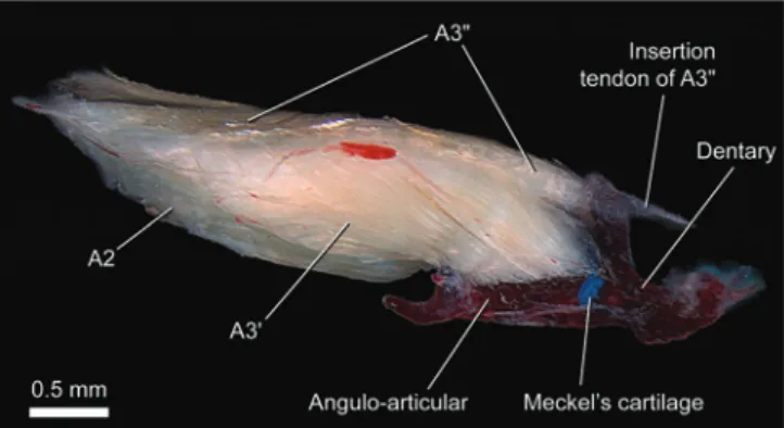
between the dorsal portions of the A2 and A3. Anteriorly, these two main subdivisions are largely continuous with each other (Fig. 2). The A2 section originates primarily from the posteroventral region of the suspensorium on the hyomandibula and preopercle. A few fibers may also attach tendinously to the postorbital process of the skull and suprapreopercle. Subdivisions A2' (ventral) and A2" (dorsal) are barely distinguishable from each other near their insertion (this distinction is more evident in the larger specimen). The A2' has a primarily musculous insertion onto the posterolateral region of the coronoid process of the lower jaw on both angulo-articular and dentary (Fig. 1). The fibers of the A2" deflect abruptly ventrally near to the coronoid process to insert predominantly onto the medial face of the lower jaw, mainly via a strong tendon that attaches to the posterior rim of the coronoid process of the dentary (Fig. 2). Sections A2' and A2" seem superficially distinguishable due to the different orientation of their fibers (Fig. 1). Nonetheless, no effective separation is possible between these two groups of fibers since they intermix more deeply within the muscle. Therefore, a direct correlation between these two superficially distinct groups of fibers and the sections A2' and A2" is problematic. The A3 section is also separable into ventral (A3') and dorsal (A3") portions along their origins (Fig. 2). The A3' is more deeply associated with the A2, originating anterolaterally to the levator arcus palatini from the
hyomandibula, metapterygoid, and quadrate. The lateral most fibers of this portion are exposed on the lateral surface of the
adductor mandibulae just below the A2, and insert onto the lateral aspects of the dentary and angulo-articular. The A3" originates medial to the levator arcus palatini from the hyomandibula and sphenotic. Anteriorly, A3" and A3' fuse to each other and insert mainly onto the medial face of the angulo-articular and coronomeckelian bone (Fig. 2). Section Aω is absent (Fig. 2).
The levator arcus palatini is nearly conically-shaped and positioned between the dorsal portions of the sections A2 and A3of the adductor mandibulae. It arises from ventral region of postorbital process of skull (Fig. 1) and inserts onto the central region of the lateral face of the hyomandibula.
The extensor tentaculi is undifferentiated from the adductor arcus palatini (Fig. 1; see “Discussion:
Fig. 2. Medial view of left adductor mandibulae and attached
lower jaw of Nematogenys inermis (Nematogenyidae), LBP
1002 (76.4 mm SL). tendinous along the anterior most portions but musculous
posteriorly. The fibers arise from the ventrolateral region of the neurocranium on the pterosphenoid, parasphenoid, orbitosphenoid, lateral ethmoid, and vomer. Posteriorly, this muscle inserts only on the medial face of the suspensorium (anterior region of the hyomandibula and posterodorsal region of the metapterygoid). Anteriorly the muscle gradually insert onto the lateral surface of the suspensorium (anterodorsal region of metapterygoid, endopterygoid) and autopalatine (posterior portion).
The adductor hyomandibulae and protractor operculi
are absent (Fig. 1).
Dilatator operculi with one single section (Fig. 1)
originating from the dorsolateral region of the orbitosphenoid and lateral margin of the frontal. The muscle passes medial to the levator arcus palatini and suprapreopercle, but lateral to
the adductor mandibulae. Insertion is tendinous onto the tip
of dorsal process of the opercle.
The levator operculi originates from the posterolateral
region of the hyomandibula (Fig. 1) and lateroventral region of the pterotic. Insertion is onto the posterodorsal region of the lateral face of the opercle (see also pertinent comments about this muscle under “Discussion”).
The adductor operculi is fully covered laterally by the levator operculi. The origin is from the ventral region of the
pterotic with an insertion onto the dorsal face of the medial crest of the opercle.
Trichomycteridae Copionodontinae
Adductor mandibulae with sections A2 and A3 well
separated from each other posteriorly, especially by the
levator arcus palatini (Figs. 3, 4). The A2 section is
undivided with its origin from the posteroventral region of the suspensorium on the hyomandibula, quadrate, and preopercle (Fig. 3). The A3 is fully divided into A3' and A3" (Figs. 4, 5). The A3' originates medial to the levator arcus palatini, from the anterodorsal portion of the suspensorium,
specifically the hyomandibula, quadrate, and metapterygoid. Somewhat anterior to the levator arcus palatini,the A2 and
A3' sections fuse such that they are not separable in the region of insertion. This common insertion occurs solely onto the large posterior face of coronoid process of angulo-articular. The A3" from the anterodorsal part of the hyomandibula and the ligamentous tissue sheet which unites the bones of the suspensorium. It inserts primarily onto the distal part of the maxilla (Figs. 3, 4) with some deeper fibers loosely attaching to the tip of the coronoid process. Because of this insertion onto the maxilla, the A3" could be considered a retractor tentaculi muscle (see Character 7). Section Aω is absent (Fig. 5).
Levator arcus palatini (Fig. 3) comparable to that in the Nematogenyidae.
Adductor arcus palatini situated fully medial to the suspensorium and separated from the extensor tentaculi
(Figs. 3, 6). Origin is from the ventrolateral region of the
neurocranium on the orbitosphenoid and pterosphenoid. Insertion is onto the dorsal regions of the hyomandibula, metapterygoid, and connective tissue sheet which joins the skeletal elements of the suspensorium. The anterior part of this muscle, which inserts mainly onto the metapterygoid, is slightly laterally deflected, originating more dorsally over the orbitosphenoid (Fig. 3).
The extensor tentaculi originates mainly from the
ventrolateral region of the lateral ethmoid (Fig. 3) with adjacent parts of orbitosphenoid sometimes involved. Insertion is onto the dorsal face of the posterolateral region of the autopalatine.
Adductor hyomandibulae present with its origin on the
deep ventral fossa of the pterotic and insertion on the posterior region of the medial face of the hyomandibula (Fig. 6).
The protractor operculi is absent (Fig. 3).
Dilatator operculi with two main recognizable sections:
a primary (basically corresponding to the single section of the dilatator operculi of the Nematogenyidae and other
Siluriformes; see Character 25) and a secondary section found only in trichomycterids (see Characters 30, 32; Fig. 3). These sections are somewhat continuous with each other in the area of their insertion but are separate at their origins. The primary section passes medial to the levator arcus palatini
and lateral to the adductor mandibulae. It originates from the
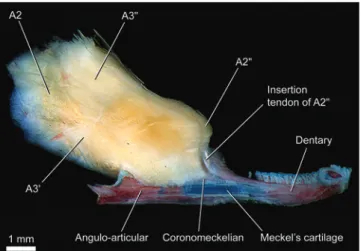
onto the posterolateral region of the coronoid process of the angulo-articular. Fibers of the A2" originate from the posterodorsal region of the hyomandibula and converge onto a long tendon that inserts onto the medial face of the angulo-articular (Fig. 8). The entire A3lies anterior to the levator arcus palatini, and is also distinctly subdivided into two almost fully
separated portions (Fig. 8). The A3' originates from the anteroventral portion of the suspensorium on hyomandibula, quadrate, and metapterygoid. Most of its fibers attach to a conspicuous tendon that inserts onto the coronomeckelian bone, albeit with some of its ventrolateral most fibers attaching to the insertion tendon of the A2" (Fig. 8). The A3" has its origin on the hyomandibula and metapterygoid and its insertion across the posteromedial rim of the coronoid process of the dentary (Fig. 8). Section Aω is absent (Fig. 8).
The levator arcus palatini (Fig. 7) is as in the
Nematogenyidae and Copionodontinae, except for being positioned posterior to the A3 section of the adductor mandibulae, rather than lateral to some part of that section.
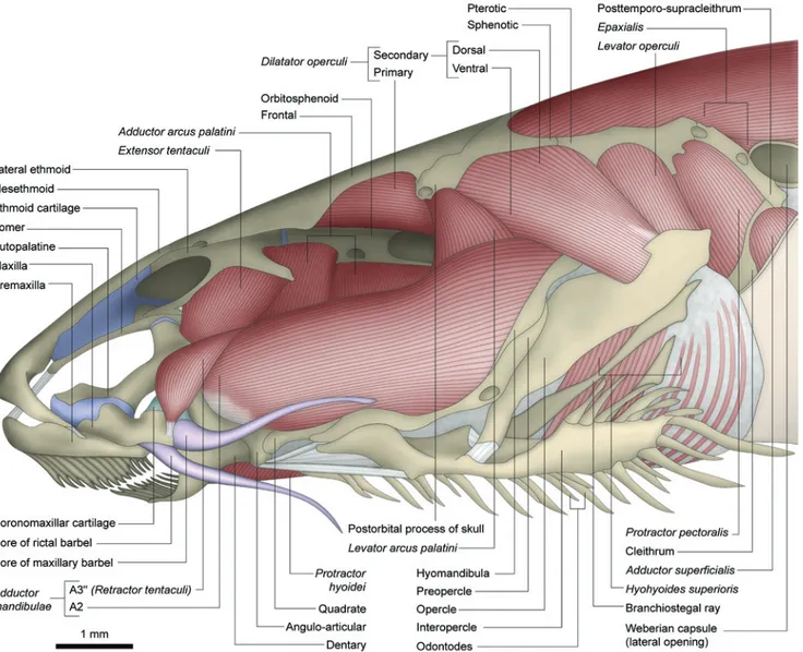
Fig. 3. Left lateral view of head of Copionodon pecten (Copionodontinae), LIRP 1012 (49.5 mm SL). Antorbital and core of nasal barbels removed.
The levator operculi originates from the lateral region
of the pterotic and posterodorsal part of the hyomandibula (Fig. 3). Insertion is onto the dorsomedial region of the opercle.
The adductor operculi is located fully medial to the levator operculi. Origin is from the ventral region of the pterotic
between the origins of the levator operculi (lateral) and adductor hyomandibulae (medial) (Fig. 6). Insertion is onto
the dorsal face of the medial crest of the opercle.
Trichogeninae
Adductor mandibulae with the two main facial
The adductor arcus palatini is fully separated from the extensor tentaculi (Fig. 7). Its origin is from the ventrolateral
region of the neurocranium including the lateral ethmoid, orbitosphenoid, pterosphenoid, and parasphenoid. The insertion is on the dorsal portions of the medial face of the hyomandibula and metapterygoid with a small group of fibers also attaching on the lateral face of the metapterygoid.
Extensor tentaculi with origin mainly from the ventral face
of the lateral process of the lateral ethmoid (Fig. 7). Insertion of the muscle is on the posterolateral region of the autopalatine and anterior part of the metapterygoid.
Adductor hyomandibulae with same condition as in the
Copionodontinae.
The protractor operculi is absent (Fig. 7).
Dilatator operculi with primary and secondary sections
fully separated at their origins but partially continuous with each other in the area of their insertions (Fig. 7). The primary section occupies the same position as in the Copionodontinae and Nematogenyidae (i.e. medial to the levator arcus palatini
and lateral to the adductor mandibulae). Origin is from the
ventral face of the wide lateral shelf of the sphenotic. Insertion is via an elongate tendon that is embedded into the deeper fibers of the secondary section of the dilatator operculi and
attaches to the anterior part of the dorsal process of the opercle. The secondary section originates from the dorsolateral portion of the sphenotic and inserts onto the dorsal process of the opercle. In the area of insertion of the secondary section of one specimen, a slight distinct orientation of the superficial fibers seems to indicate an incipient division into dorsal and ventral portions.
Fig. 4. Dorsal view of left adductor mandibulae and attached lower jaw and maxilla of Copionodon pecten
(Copionodontinae), LIRP 1012 (51.7 mm SL). Arrow indicates space occupied by levator arcus palatini.
Fig. 5. Medial view of left adductor mandibulae and attached
lower jaw of Copionodon pecten (Copionodontinae), LIRP
1012 (51.7 mm SL).
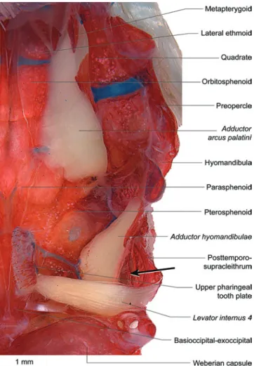
Fig. 6. Ventral view of left half of neurocranial floor and suspensorium of Copionodon pecten (Copionodontinae),
LIRP 1012 (49.5 mm SL). Internal suspensorial muscles and upper pharyngeal tooth plate with attached levator internus 4 retained; remaining elements of branchial arches removed.
Levator operculi originating from lateroventral region of
pterotic and inserting onto the dorsomedial region of the opercle (Fig. 7) (see also comments about this muscle under the “Discussion: Nematogenyidae”).
Adductor operculi posteromedial to levator operculi
(Fig. 7). Sites of origin and insertion of the muscle comparable to in the Copionodontinae.
Trichomycterinae
Adductor mandibulae with two main subdivisions - A2
and A3 - partially continuous with each other (Figs. 9-12). The degree of fusion between A2 and A3 is similar or, in many cases, greater than that occurring in Nematogenys.The A2
originates from the ventrolateral region of the suspensorium on the hyomandibula, preopercle and quadrate. In some cases the A2 lacks any obvious subdivision (Fig. 9), whereas in others it is barely subdivided into A2' and A2" portions near to the area of insertion (e.g. Scleronema angustirostris, Trichomycterus areolatus, T. cf. iheringi, some T. brasiliensis;
some T. davisi, T. maracaya, T. stawiarski, and T. zonatus)
(Figs. 10, 11). The A2'inserts, often tendinously (Figs. 9-11), onto the posterolateral region of the coronoid process of the angulo-articular. In Trichomycterus cf. iheringi the fibers of
the lateral surface of the A2' are arranged into a tripennate configuration (Figs. 10, 11), thus resembling the condition reported by Adriaens et al. (2010) for T. guianensis (see
“Discussion: Trichomycterinae”). As in the Nematogenyidae, the A2" is also tenuously discernible from the A2' by its deflection ventrad (Fig. 11) near to its insertion onto the posteromedial rim of the coronoid process of the dentary (Fig. 12). The A3 originates from the anterodorsal region of the suspensorium, always from the hyomandibula and usually also from the quadrate and metapterygoid (Fig. 10). Insertion is onto the medial face of the angulo-articular (Fig. 12). Although the A3 section never exhibit complete subdivisions, a ventral (A3') and a dorsal (A3") portion may be distinguished especially at their origins (Fig. 12). As in the Nematogenyidae, the origin of the A3" of most trichomycterines extends posteriorly on the hyomandibula so as to lying medial to the levator arcus palatini
(Figs. 10, 12). In other trichomycterines (Bullockia, Hatcheria,
Trichomycterus areolatus, T. chiltoni, T. immaculatus, and T. zonatus) that origin is not posteriorly expanded, so that the
A3" originates only anterior to the levator arcus palatini (a
condition similar to that of trichogenines and some sarcoglanidines). The A3' always originates anteroventrally to the levator arcus palatini and is more associated with the A2
(Fig. 10). Fibers of A3', however, are never exposed on the lateral surface of the adductor mandibulae as occurs in the
Nematogenyidae. Section Aω is absent (Fig. 12).
The major interspecific variations in the adductor mandibulae encountered among the members of the
Trichomycterinae involve the degree of hypertrophy of the A2 section and the overall differentiation of the sections that compose that muscle. Some trichomycterines (Ituglanis cf. amazonicus, Trichomycterus brasiliensis, T. davisi, and, in a
lesser degree, T. cf. iheringi) have a distinctly enlarged A2,
with the result that in many cases their eyes cannot be seen from a lateral view (eyes facing upwards). This feature Fig. 8. Medial view of left adductor mandibulae and attached
lower jaw of Trichogenes longipinnis (Trichogeninae), LIRP 1059 (60.8 mm SL).
Fig. 9. Left lateral view of head of Trichomycterus brasiliensis (Trichomycterinae), LIRP 1968 (76.4 mm SL). Core of nasal
apparently shows a positive allometry during ontogeny in these species. Other trichomycterines, including some that also exhibit a hypertrophied A2, possess the subdivisions of the A2 (into A2" and A2') more obviously differentiated from each other than in other species. Among the examined trichomycterines, there is a plenty of intermediate conditions between the less and the most extreme states of development of the A2, as well as variable degrees of differentiation of its subdivisions. This variation renders difficult to delimit discreet states to be incorporated into a phylogenetic analysis.
Levator arcus palatini with the same general
configuration as in the Nematogenyidae, Copionodontinae and Trichogeninae. In most species this muscle is positioned between the dorsal portions of the A2 and A3 sections of the
adductor mandibulae (Fig. 10) (like in Nematogenyidae and
Copionodontinae) while in others, it lies posterior to the A3 section (like in Trichogeninae).
The adductor arcus palatini originates from the
ventrolateral region of the neurocranium on the
orbitosphenoid, parasphenoid and sphenotic-prootic-pterosphenoid (Figs. 9, 13). Some fibers of this muscle may also arise from the connective membrane that joins the bones of the suspensorium (Fig. 13). Insertion is onto the medial surfaces of the metapterygoid, quadrate, and hyomandibula.
Extensor tentaculi usually originating only from the
ventrolateral regions of the lateral ethmoid and orbitosphenoid (Figs. 9, 10). In Trichomycterus davisi and T. stawiarski this
muscle is considerably enlarged compared to that in other trichomycterids, with the presence of a ventral bundle which also originates from the anterodorsal region of the suspensorium on the metapterygoid and, rarely, the hyomandibula. Insertion is onto the posterolateral region of the autopalatine.
Adductor hyomandibulae is absent (Fig. 13).
Protractor operculi with origin on the midlateral region
of the preopercle (just dorsal to its articulation with the interopercle) and on a tiny tendon shared with the A2 section of the adductor mandibulae (Figs. 9, 10, 14). Insertion is onto
the midventral region of the lateral face of the opercle just anterior to its opercular patch of odontodes.
Dilatator operculi with its primary and secondary
sections separated at their origin but partially continuous at their insertions (Figs. 9, 10, 14). The primary section passes medial to the levator arcus palatini and lateral to the adductor mandibulae. In some species it originates solely from the
ventral surface of the postorbital process of skull whereas, in others, it extends anteriorly to involve also the midlateral region of the frontal (the portion just anterior to the postorbital process). Insertion is as in the Trichogeninae. The secondary section originates from the otic region of the cranial roof,
mainly on the sphenotic-prootic-pterosphenoid, along with the adjacent regions of the frontal, hyomandibula, and, sometimes, the parieto-supraoccipital. Insertion is onto the dorsal process of the opercle. Despite being undifferentiated in some species (Fig. 10), in most cases, a division of the secondary section into dorsal and ventral portions is evident, at least, in the region of its insertion (Figs. 9, 14). In such cases, the secondary-dorsal portion attaches more obviously to the tip of the dorsal process of the opercle, while the secondary-ventral part extends ventrally over the lateral face of the same process. In Bullockia maldonadoi, Hatcheria macraei, Trichomycterus areolatus, T. chiltoni, T. immaculatus, and T. zonatus the origin of the
secondary-ventral section of the dilatator operculi is much
anteroventrally expanded, with its anterior limit approximately at the vertical through the middle of the protractor operculi.
As a consequence the secondary-ventral section of the
dilatator operculi assumes a triangular shape (Fig. 14).
The levator operculi (Figs. 9, 10, 14) originates from the
lateroventral region of the pterotic and inserts onto the
Fig. 11. Left lateral view of region of adductor mandibulae
region of Trichomycterus cf. iheringi (Trichomycterinae), LIRP 3182 (72.2 mm SL). Arrow indicates posteroventral bundle of A2' which apparently corresponds to the “A1-OST” of Adriaens et al.(2010) (see “Discussion: Trichomycterinae”).
Fig. 12. Medial view of left adductor mandibulae and
attached lower jaw of Trichomycterus brasiliensis
(Trichomycterinae), LIRP 1968 (95.1 mm SL). Arrows indicates space occupied by levator arcus palatini.
Fig. 13. Ventral view of left half of neurocranial floor of
Ituglanis cf. gracilior (Trichomycterinae), MZUSP 86821 (53.2
mm SL). Internal suspensorial muscles and upper pharyngeal tooth plate with attached levator internus 4 retained;
dorsomedial region of the opercle (see also some pertinent comments about this muscle under “Discussion: Nematogenyidae”).
Adductor operculi situated medial to the levator operculi
(Figs. 9, 10, 14). The adductor operculi originates from the
ventral face of the pterotic (Fig. 13) passing medial to the hyomandibula (Fig. 10) to insert onto the dorsal face of the medial crest of opercle. Separation from the levator operculi
seems to be incomplete in Ituglanis and some Trichomycterus.
Sarcoglanidinae
The adductor mandibulae of Sarcoglanis is similar to
that of nematogenyids and most trichomycterines, notably for the levator arcus palatini which lies between the
dorsoposterior parts of the A2 and A3. The A2 of Sarcoglanis
originates from the ventral part of suspensorium on the hyomandibula, preopercle, and quadrate (Fig. 15). Insertion is onto the posterior portion of the coronoid process. That section exhibits a much tenuous anterior differentiation into A2' and A2", with the latter deflecting ventrad towards its insertion. At insertion, the A2' is more associated with the angulo-articular and the A2" with the dentary. The A3 of
Sarcoglanis has two recognizable portions, both originating
from the hyomandibula. The ventral A3' lies anteroventral to the levator arcus palatini and inserts onto the anteromedial
region of the angulo-articular. The A3" originates medial to the levator arcus palatini and attaches to a small tendon that
embeds into the buccopalatal membrane.
In Microcambeva and Stauroglanis the adductor mandibulae lacks clear subdivisions (Fig. 16). However,
comparisons with other taxa, especially Sarcoglanis and
glanapterygines, lead us to infer that both A2 and A3 sections are present in these genera. These sections in Microcambeva
and Stauroglanis are fully continuous with each other along their entire length except near their insertion. The group of fibers apparently corresponding to the A2 of these genera lacks any evident subdivision into A2' and A2", but retains the same primary sites of origin and insertions of the A2 of
Sarcoglanis. The A3 of Microcambeva and Stauroglanis
also exhibits an anterior distinction into A3' and A3". As in
Sarcoglanis, the A3" inserts onto the buccopalatal membrane whereas the A3' inserts onto the angulo-articular (Fig. 17). In contrast to the condition of Sarcoglanis, the A3 of
Microcambeva and Stauroglanis is not divided posteriorly, having a common origin on the region of the hyomandibula just anterior to the levator arcus palatini (Fig. 15). The section Aω is absent (Fig. 17).
The levator arcus palatini in Microcambeva and
Stauroglanis is roughly conical, although anteriorly more elongated than in the Nematogenyidae, Copionodontinae, Trichogeninae, and Trichomycterinae (Fig. 16). That muscle in Microcambeva and Stauroglanis is located completely posterior to the adductor mandibulae with its origin and insertion comparable to that in the Nematogenyidae and the basal members of the Trichomycteridae. In Sarcoglanis this muscle is strap-like, having an origin and insertion of about the same width (Fig. 15). The origin of that muscle is from the dorsolateral region of the frontal, in the region just anterior to the postorbital process of skull, with the insertion comparable to that in the other nematogenyids and trichomycterids described.
Fig. 14. Left lateral view of opercular region of Trichomycterus immaculatus (Trichomycterinae), LIRP 285 (83.7 mm SL).
Fig. 15. Left lateral view of head of Sarcoglanis simplex
Adductor arcus palatini (Fig. 16) originating from the lateral region of the parasphenoid and adjacent portions of the orbitosphenoid and sphenotic-prootic-pterosphenoid. The insertion is solely onto the dorsal portion of the medial face of the hyomandibula (mainly on its conspicuous anterior process) in Microcambeva. In Stauroglanis and Sarcoglanis, in addition to an insertion onto the dorsal portion of hyomandibula, this muscle also inserts onto the suspensorial membrane with some fibers reaching the dorsal portion of the quadrate.
Extensor tentaculi with an origin from the ventrolateral regions of the lateral ethmoid and orbitosphenoid and insertion onto the posterior region of the autopalatine (Fig. 16).
The adductor hyomandibulae is absent.
In Microcambeva the protractor operculi is posterodorsally fused to the highly modified anteroventral part of the dilatator operculi, forming a partially continuous complex muscle mass (Fig. 16). Despite that, comparisons with other trichomycterids permit us to infer the homologies of the different parts that form this compound muscle. As is
other trichomycterids, the protractor operculi originates from posteroventral region of the suspensorium, on the preopercle and hyomandibula, and from a tendon shared with the A2 section of the adductor mandibulae. Posteriorly the
the region of the frontal just anterior to the postorbital process of skull, then passes anterolaterally to the levator arcus palatini. The posterior most group of fibers originates from the ventral surface of the same process, lateral to the levator arcus palatini. The homology of these bundles with the primary section of the dilatator operculi is inferred based on their sites of origin and insertion which are basically the same as in the Nematogenyidae, Copionodontinae, Trichogeninae, and Trichomycterinae, in spite of passing dorsolateral to the
levator arcus palatini (see below). A dorsoposterior bundle of the dilatator operculi is much better differentiated from the remaining muscle mass. That bundle certainly corresponds to the dorsal portion of the secondary division of the dilatator operculi inasmuch it retains the same basic form, position, and sites of attachment than in most other trichomycterids. The origin is from the otic region of the neurocranial roof, involving the sphenotic-prootic-pterosphenoid, frontal, and parieto-supraoccipital. Insertion is both tendinous and musculous onto the posterodorsal part of the dorsal process of the opercle. Note that, if our hypotheses of homology among the portions of the dilatator operculi are correct, differently of the basal trichomycterids, the primary section in Microcambeva passes lateral to the levator arcus palatini
(as in other members of the TSVSG clade; see below). Apparently coupled with that feature, the secondary section of the dilatator operculi becomes split into dorsal and ventral parts by the interposing primary section (see Character 26).
In Stauroglanis and Sarcoglanis (Fig. 15),the protractor operculi and dilatator operculi are completely separated from each other. The protractor operculi in Stauroglanis is quite similar to that of trichomycterines, having the same overall shape (fusiform) and attachment sites on the skeleton as in the members of that subfamily. In Sarcoglanis it is a much flattened muscle with fibers arranged in parallel. Origin is from the posterior region of the preopercle and insertion onto the ventrolateral region of the opercle (Fig. 15).
In both Stauroglanis and Sarcoglanis, the dilatator operculi apparently lacks a primary section (Fig. 15). The secondary section is present and exhibits dorsal and ventral portions readily recognizable by the distinct orientation of their superficial fibers. The secondary-ventral portion
originates from the anterolateral region of the sphenotic-prootic-pterosphenoid and inserts onto the ventral region of the dorsal process of the opercle, contacting ventrally the
protractor operculi. The secondary-dorsal section is basically
comparable to that in Microcambeva.
Levator operculi (Fig. 16) is basically like that in the
Trichomycterinae. In Sarcoglanis this muscle seems to be
attached more laterally onto the opercle (Fig. 15). This condition, however, is more related to modifications of the opercle in that genus than it is to changes in the insertion of the muscle (see “Discussion: Nematogenyidae”).
The adductor operculi (Fig. 16) is similar to that of the
Trichomycterinae. The separation from the levator operculi
is incomplete in Sarcoglanis.
Glanapteryginae
The adductor mandibulae hastwo principal sections, A2
and A3, both located lateral to the levator arcus palatini
(Fig. 18). These sections are partially continuous with each other posteriorly, but more obviously separated anteriorly (Fig. 19). The A2 has its origin on the posteroventral region of the suspensorium, involving the hyomandibula, preopercle, and quadrate (Fig. 18). As in the Nematogenyidae, most Trichomycterinae, and the sarcoglanidine Sarcoglanis, this
section of the muscle is partially differentiated into an A2' and A2" anteriorly near to its area of insertion. The A2' inserts mainly onto the posterolateral region of the coronoid process of the angulo-articular, while the A2" deflects ventrally and inserts primarily onto the medial face of the coronoid process of the dentary (Fig. 19). The A3 originates from the anterodorsal region of the hyomandibula. Despite being somewhat continuous near its origin, the A3 is well differentiated anteriorly into A3' and A3" (especially in Listrura tetraradiata). As in sarcoglanidines, the A3' inserts onto the
medial aspect of the angulo-articular, while the fibers of A3" converge onto a tendon that embeds into the buccopalatal membrane (Figs. 18, 19). Section Aω is absent (Fig. 19).
The Levator arcus palatini originates from the
ventrolateral regions of the sphenotic-prootic-pterosphenoid and frontal, extending anteriorly beyond the postorbital process of skull (more obviously in Listrura picinguabae)
(Fig. 18). Its insertion, always wider than its origin, is onto the anterodorsal region of the hyomandibula, primarily onto the conspicuous anterior process of that bone.
Adductor arcus palatini (Fig. 18) with an origin like that
in the Sarcoglanidinae. The insertion is onto the suspensorial membrane and the dorsomedial region of the hyomandibula, especially onto its conspicuous anterior process.
Extensor tentaculi originating from the ventrolateral parts
of the lateral ethmoid and orbitosphenoid (Fig. 18). The insertion is onto the posterolateral region of the autopalatine.
The adductor hyomandibulae is absent.
Protractor operculi comparativelyslender with an origin
from the lateral region of the preopercle and an insertion onto the midventral region of the opercle just anterior to its patch of odontodes (Fig. 18).
Fig. 17. Medial view of left adductor mandibulae and attached lower jaw of Microcambeva ribeirae
The dilatator operculi lacks any evident subdivision
(Fig. 18). Comparisons with other taxa, however, lead us to infer that the groups of fibers that correspond to both the primary and secondary sections of the dilatator operculi of other trichomycterids are present in the
Glanapteryginae. Existence of a secondary-dorsal portion is evidenced by the presence of a posterodorsal group of fibers that originates from dorsolateral region of sphenotic-prootic-pterosphenoid and inserts onto the tip of the dorsal process of opercle. An anterior most group of fibers originating from the lateral region of the frontal just anterior to the postorbital process of skull and inserting onto the anterodorsal region of the dorsal process of opercle indicates the existence of a primary section. Ventral to the primary portion, there is a group of fibers that inserts more ventrolaterally over the dorsal process of the opercle, a feature typical of the secondary-ventral portion of other trichomycterids. Note that, as in the sarcoglanidine Microcambeva, tridentines, and many
stegophilines and vandelliines, the primary section of the
dilatator operculi of Listrura would pass dorsolateral to
the levator arcus palatini and is located between the
dorsal and ventral portions of the secondary section (see Character 26).
Levator operculi and adductor operculi (Fig. 18) as in
the Trichomycterinae and Sarcoglanidinae, being always fully separated from each other.
Fig. 19. Medial view of right adductor mandibulae and
attached lower jaw of Listrura tetraradiata (Glanapteryginae),
MNRJ 31534 (paratype; 39.0 mm SL). Image flipped horizontally to facilitate comparisons.
Tridentinae
Members of this subfamily possess comparatively enormous eyes placed ventrolaterally on their heads. This arrangement is coupled with profound modifications of several cephalic muscles in tridentines. The adductor mandibulae,
for example, is “squeezed” by the large eyeballs, being comparatively the smallest of these muscles among all taxa examined herein (Fig. 20). Section A2 is fully separated from A3 (Fig. 21). The A2 also has almost fully separated A2' and A2" portions. The former originates from the anteroventral region of suspensorium on the quadrate and hyomandibula and inserts onto the posteromedial region of the angulo-articular. The A2" lies almost completely dorsal to the remaining sections of the adductor mandibulae, originating
from both lateral and medial faces of the conspicuous posteriorly oriented anterodorsal process of the hyomandibula
(Fig. 20). Near to its insertion, the fibers of the A2" undergo the typical ventral deflection to allow its insertion primarily along the posteromedial rim of the dentary. The A3 is much smaller than the A2, having a nearly triangular shape (Fig. 21). It has a small partially tendinous origin on the hyomandibula and a broad insertion onto the ventromedial face of the angulo-articular. One might cast doubts on the identity of that section in tridentines because of its reduced size and narrow origin. However, in spite of these modifications, that section exhibits the same medial position and the same insertion site of the A3 of most other trichomycterids. Moreover, the two other lateral sections of the adductor mandibulae of tridentines clearly correspond
The levator arcus palatini is broader at its origin than at the insertion, acquiring an inverted trapezoid shape from a lateral view. The origin is from the ventrolateral region of the frontal and the sphenotic-prootic-pterosphenoid. Insertion is onto the middorsal region of the hyomandibula (Fig. 20).
Adductor arcus palatini with an origin as in the Sarcoglanidinae and Glanapteryginae. The insertion is onto the dorsomedial part of the hyomandibula, mainly onto the conspicuous dorsoposterior process of the anterior part of this bone.
The extensor tentaculi (Fig. 20) originates from the lateral regions of the lateral ethmoid and orbitosphenoid. Insertion occurs onto the posterodorsal region of the autopalatine.
The adductor hyomandibulae is absent.
The protractor operculi originates primarily from the fascia of the levator arcus palatini, posteromedial to the eyeball (Fig. 20). Some of the deepest fibers attach to the hyomandibula and preopercle. The insertion is onto the lateral face of the opercle.
Dilatator operculi with subdivisions poorly differentiated from each other (Fig. 20), especially near its origin. A larger anterior most group of fibers originates far anteriorly on neurocranium from lateral portions of the mesethmoid and frontal. Some fibers of this part of the muscle insert onto the anterodorsal region of the dorsal process of the opercle, while other fibers attach to an elongate tendon that inserts onto the posterolateral region of the opercle. Although this section of the muscle cannot be readily subdivided into two distinct parts, its sites of origin and especially of insertion are typical of the primary and secondary-ventral sections of the dilatator operculi. These facts, together with an overall topological correspondence of this muscle with those observed in other trichomycterids, especially the closely related stegophilines and vandelliines, lead us to infer that this part of the muscle probably is a compound section formed by the fusion of the primary and secondary-ventral sections of the dilatator
operculi. The fact of the origin of that compound section extends
much more anteriorly compared to the basal trichomycterids is presumably attributed to an anterior expansion of the primary section of the dilatator operculi since that is the condition
encountered in all stegophilines and vandelliines (see descriptions of those taxa below and Character 27). A closest association of the primary section with the secondary-ventral section is also usual in most members of the Sarcoglanidinae, Glanapteryginae, Stegophilinae, and Vandelliinae. The secondary-dorsal section is more distinctly separated from the remaining muscle mass, originating mainly from the lateral regions of the sphenotic-prootic-pterosphenoid and frontal, with a small posterior bundle originating from the hyomandibula. The insertion is onto the tip of the dorsal process of opercle.
The levator operculi is relatively slender (Fig. 20)
compared with that of other trichomycterids. Sites of origin and insertion of this muscle are, however, basically the same as those in the Trichomycterinae, Sarcoglanidinae, and Glanapteryginae.
The adductor operculi is also comparatively slender and
apparently partially continuous with the levator operculi
along its anterior border. Sites of origin and insertion are basically the same as in the Trichomycterinae, Sarcoglanidinae, and Glanapteryginae.
Stegophilinae
Adductor mandibulae with the A2 and A3 sections fully
separated from each other across their entire lengths (Figs. 22-24). The A2 is almost fully separated into ventral (A2') and dorsal (A2") parts. The A2' originates from the preopercle and quadrate and inserts mainly onto the posterior region of the laterally oriented coronoid process of the angulo-articular (Figs. 22, 25). In Ochmacanthus, Parastegophilus, Pseudostegophilus, and Pareiodon the anterodorsal most
fibers of this section tenuously attach to a short tendon (Fig. 25) that embeds into the buccopalatal membrane (Fig. 26). The A2" is, at least, partially bipennate in most examined taxa (Fig. 25). It originates primarily from the posterodorsal region of the hyomandibula, with adjacent areas of the preopercle and sphenotic also involved in some cases. Fibers of this section converge anteriorly onto a long tendon that inserts onto the posterolateral rim of the dentary. As in tridentines, the A3 is much smaller than the other sections of the adductor mandibulae, being approximately triangular and notably
anteriorly displaced on the suspensorium (Figs. 22-25). Its narrow origin is both musculous and tendinous in
Haemomaster but only tendinous in all other stegophilines
examined. The A3 originates from a small area on the anterodorsal region of the hyomandibula and sometimes also the quadrate. The insertion is broad onto the medial region of the lower jaw, reaching the borders of the dentary symphysis in most cases. The Aω section is absent (Fig. 24).
The levator arcus palatini (Fig. 22) is comparable to that
in the Tridentinae. In Parastegophilus, Pseudostegophilus,
and especially Pareiodon, however, this muscle is further
expanded anteriorly. Fig. 21. Medial view of right adductor mandibulae and
The adductor arcus palatini originates from the
parasphenoid, sphenotic-prootic-pterosphenoid, and sometimes also the orbitosphenoid and lateral ethmoid (Fig. 22). Insertion is primarily onto the medial face of the hyomandibula and quadrate with the metapterygoid sometimes also involved.
The extensor tentaculi originates from the ventrolateral
regions of the orbitosphenoid and lateral ethmoid (Fig. 22) with the parasphenoid sometimes also involved. In
Haemomaster this muscle inserts onto the posterior margin
and dorsal face of the autopalatine. In other stegophilines it attaches solely onto the posterior border of this bone.
The adductor hyomandibulae is absent.
The protractor operculi is shorter and deeper compared
to in other trichomycterids, often acquiring a parallelogram shape from a lateral view (Fig. 22). It originates from the posterolateral region of the preopercle and usually also from a shared tendon with the A2 part of the adductor mandibulae
(as in Trichomycterinae). In Haemomaster the dorsal portion
of the protractor operculi is covered by the superficial fibers
of the secondary-ventral section of the dilatator operculi.
Some fibers of the protractor operculi in this genus also
intermingle with both the secondary-ventral section of the
dilatator operculi and the adductor mandibulae (A2"
section). In Pareiodon most of the fibers of the muscle
originate from the lateral fascia of the adductor mandibulae
(Fig. 27). The insertion is onto the central region of the lateral face of the opercle, ventrally to its dorsal process and anteriorly to its patch of odontodes.
The dilatator operculi exhibits many modifications in
several groups of the subfamily. In Haemomaster all three
sections (primary, dorsal, and secondary-ventral) are well differentiated although continuity along their insertions persists to some extent (Figs. 22, 23). The primary section is distinctly anteriorly elongate, being situated dorsolateral to the levator arcus palatini and
prootic-pterosphenoid. Insertion is onto the dorsomedial region of the dorsal process of the opercle. The secondary-ventral section is deep and flat, attaching anteriorly to the eyeball capsule (a translucent membranous tissue that enwraps the eyeball; Fig. 23). Posteriorly, that section covers the dorsal part of the protractor operculi, being somewhat continuous with that muscle in this area. The insertion is onto the central region of the opercle just ventral to its dorsal process.
In Homodiaetus, Ochmacanthus, Parastegophilus, and
Pseudostegophilus, the secondary-ventral section is much smaller than in Haemomaster, fusing anterodorsally with the primary section (Fig. 26) and, sometimes, also tenuously attaching to the eyeball capsule. In Pareiodon the secondary-ventral section is continuous with the secondary-dorsal one, forming a single secondary section as in the basal most trichomycterids (Fig. 27). The insertion of the secondary-ventral section in all these cases is onto the ventral region of the dorsal process of the opercle. The primary section in these taxa is distinctly anteromedially expanded, contacting its counterpart along the mid-sagittal plane and attaching to the frontal and mesethmoid or lateral ethmoid (Fig. 27). As in Haemomaster, fibers of the primary section pass dorsolateral to the levator arcus palatini and merge into a tendon that inserts on the tip of the dorsal process of opercle. In Homodiaetus and
Ochmacanthus the secondary-dorsal section has basically the same configuration than that of Haemomaster (Fig. 26). In Parastegophilus and, more significantly in
Pseudostegophilus and Pareiodon, this section is distinctly anteromedially expanded towards the mid-sagittal plane, attaching to the sphenotic-prootic-pterosphenoid, frontal, and sometimes the hyomandibula (Fig. 27).
Fig. 23. Dorsal view of head of Haemomaster venezuelae (Stegophilinae), LIRP 7438 (36.9 mm SL). Left eye removed. Arrow
indicates anterior projection of eyeball capsule anchoring to tip of mesethmoid cornu.
Fig. 24. Posterior view of left adductor mandibulae and attached lower jaw of Haemomaster venezuelae