The caffeine-binding adenosine
A
2A
receptor induces age-like
HPA-axis dysfunction by targeting
glucocorticoid receptor function
Vânia L. Batalha
1,2, Diana G. Ferreira
1,3,4, Joana E. Coelho
1, Jorge S. Valadas
1,†, Rui Gomes
1,5,
Mariana
te
mido-Ferreira
1, Tatiana Shmidt
6, Younis Baqi
7,8, Luc Buée
9, Christa E. Müller
7,
Malika Hamdane
9, Tiago F. Outeiro
3,10,11, Michael Bader
6,12,13, Sebastiaan H. Meijsing
2,
Ghazaleh Sadri-Vakili
14, David Blum
9& Luísa V. Lopes
1Caffeine is associated with procognitive effects in humans by counteracting overactivation of the adenosine A2A receptor (A2AR), which is upregulated in the human forebrain of aged and Alzheimer’s disease (AD) patients. We have previously shown that an anti-A2AR therapy reverts age-like memory deficits, by reestablishment of the hypothalamic-pituitary-adrenal (HPA) axis feedback and
corticosterone circadian levels. These observations suggest that A2AR over-activation and glucocorticoid dysfunction are key events in age-related hippocampal deficits; but their direct connection has never been explored. We now show that inducing A2AR overexpression in an aging-like profile is sufficient to trigger HPA-axis dysfunction, namely loss of plasmatic corticosterone circadian oscillation, and promotes reduction of GR hippocampal levels. The synaptic plasticity and memory deficits triggered by GR in the hippocampus are amplified by A2AR over-activation and were rescued by anti-A2AR therapy; finally, we demonstrate that A2AR act on GR nuclear translocation and GR-dependent transcriptional regulation. We provide the first demonstration that A2AR is a major regulator of GR function and that this functional interconnection may be a trigger to age-related memory deficits. This supports the idea that the procognitive effects of A2AR antagonists, namely caffeine, on Alzheimer’s and age-related cognitive impairments may rely on its ability to modulate GR actions.
Excessive glucocorticoid production associated with chronic or severe stress impairs hippocampal neuronal func-tion and predisposes the organism to neurodegenerafunc-tion1. Release of cortisol from the adrenal cortex is under
tight regulation of this hypothalamic–pituitary–adrenal (HPA) axis. The hippocampus plays a crucial role in regulating HPA axis2 and excessive glucocorticoid production disrupts the regulatory circuit that connects the
hippocampus and the hypothalamus.
1instituto de Medicina Molecular, faculdade de Medicina de Lisboa, Universidade de Lisboa, Portugal. 2Max Planck
institute for Molecular Genetics, Berlin,Germany. 3Department of neuroDegeneration and Restorative Research, University Medical Center Goettingen, Waldweg 33, 37073 Göttingen, Germany. 4instituto de farmacologia e terapêutica, faculdade de Medicina do Porto, Universidade do Porto, Portugal. 5faculdade de ciências de Lisboa, Universidade de Lisboa, Portugal. 6Max-Delbrück-center for Molecular Medicine (MDc), Berlin, Germany. 7Pharmacenter Bonn, Pharmazeutische chemie i, Pharmazeutisches institut, University of Bonn, Bonn, Germany. 8Department of chemistry, faculty of Science, Sultan Qaboos University, Muscat, Oman. 9Univ. Lille, inserm, CHU Lille, UMR-S 1172, Alzheimer & Tauopathies, Lille, France. 10Max Planck institute for experimental Medicine, Goettingen, Germany. 11ceDOc, centro de estudos de Doenças crónicas, Lisbon, Portugal. 12charité-University Medicine Berlin, Germany. 13institute of Biology, University of Lübeck, Germany. 14MassGeneral institute for Neurodegenerative Disease, Massachusetts General Hospital, Boston, MA, United States of America. †Present
address: ViB, center for the Biology of Disease; and KU Leuven, center for Human Genetics and Leuven Research institute for neuroscience and Disease (LinD), Leuven, Belgium. correspondence and requests for materials should be addressed to D.B. (email: [email protected]) or L.V.L. (email: [email protected])
Received: 07 April 2016 Accepted: 01 July 2016
Published: 11 August 2016
Age-related disorders are associated with downregulation of glucocorticoid receptors (GR) in the hippocam-pus, and subsequent desensitization of the regulatory feedback to the hypothalamus3. Accordingly, in a large
study of elder humans aged 50–70 years, elevated salivary levels of cortisol were found to be correlated with poor cognitive function4. Increased glucocorticoid activity has also been associated with greater hippocampal atrophy
and memory impairment in the elderly3. This is probably a consequence of dendritic retraction and hippocampal
dysfunction that we have shown to occur upon chronic stress1. Moreover, higher cortisol levels have been also
associated with more rapid Alzheimer’s disease (AD) progression5 and systemic administration of glucocorticoids
or stress were shown to potentiate memory impairments, hippocampal damage, β -amyloid formation and Tau accumulation in transgenic AD mice6–8.
In the recent years, multiple lines of evidence have suggested an association between adenosine modulation and stress response. In particular, activation of the adenosine A2A receptor (A2AR) was shown to contribute to the
stress response by inducing corticosterone secretion9 and by mimicking GR effects10. Moreover, we have recently
shown that oral administration of an A2AR antagonist restores morphological, behavioral, and synaptic deficits
induced by HPA-axis dysfunction in rodents1. As observed for HPA axis, we and others have demonstrated that
A2A receptors are dysregulated in the rat or human brain upon aging and AD11–13.
There is a striking parallel between A2AR over-activation/over-expression and impaired GR receptor function,
as evidenced by the similar ability of A2AR and GR antagonists to improve cognitive deficits as well as to mitigate
amyloid and Tau pathologies reminiscent of AD14–17. Altogether, such observations strongly suggest that A 2AR
over-activation and GR dysfuntion are key events in age-related hippocampal deficits and raise the possibility that both pathways might be interconnected.
In the present study, we provide the first demonstration of the instrumental impact of A2AR modulation of
GR function, a mechanism never hypothesized before. We specifically report that A2AR overexpression in
fore-brain neurons is sufficient to promote HPA-axis dysfunction, namely loss of plasmatic corticosterone circadian oscillation, and reduced GR hippocampal levels, both being age-related phenotypes18. Further, we show that
A2AR activation modulates GR-induced deficits in hippocampal synaptic plasticity, increasing susceptibility to GR
activation. Finally, we demonstrate that A2AR modulation impacts GR nuclear translocation and transcriptional
activity.
Materials and Methods
Animals.
All experimental procedures were carried strictly within the rules of the Portuguese official vet-erinary department, which complies with European Directive 2010/63/EC and the Portuguese law transposing this Directive (DL 113/2013); and approved by the Instituto de Medicina Molecular Internal Committee and the Portuguese Animal Ethics Committee (Direcção Geral de Veterinária). Environmental conditions were kept con-stant: food and water ad lib, 21 ± 0.5 °C, 60 ± 10% relative humidity, 12 h light/dark cycles, 2 to 3 animals per cage. The animals were killed by decapitation after anesthesia under halothane atmosphere. Male WT Sprague-Dawley and Tg(CaMKII-hA2AR) rats were used in the 8–14 week-old age range for all the experiments described.Generation and maintenance of transgenic animals.
Transgenic rats overexpressing the human aden-osine A2A receptor (A2AR) under the control of the CaMKIIα promotor, tg(CaMKII-hA2AR), were generated bymicroinjection of a linearized DNA construct into the male pronucleus of Sprague–Dawley rat zygotes with estab-lished methods19. The construct contained a full-length human A
2AR cDNA cloned into an expression vector with
the 8.5 kb mouse CaMKIIα promoter20 and a polyadenylation cassette of bovine growth hormone (see Fig. 1a).
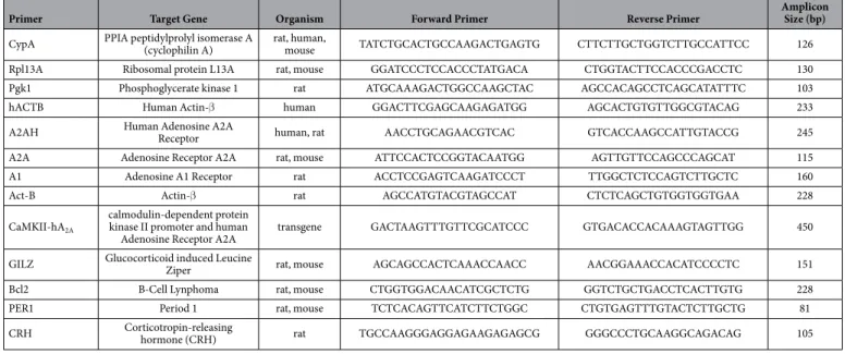
Sprague Dawley wild type (WT) male rats were used as controls. Genotyping: Transgenic rats were identified by PCR (30 cycles, 58 °C annealing temperature) of their genomic DNA isolated from ear biopsies by the use of the transgene-specific primers CaMKII-hA2A and rat β -actin primers as an internal control (Invitrogen, see Table 1). Breeding efficiency and litter size was not affected in tg(CaMKII-hA2AR) animals. The average weight of
the animals was also similar between WT 282.9 ± 37.7 g and tg(CaMKII-hA2AR) 286.7 ± 22.8 g at the age tested.
RNA extraction and quantitative real-time PCR analysis (RT-qPCR).
Total RNA was extracted and purified using the RNeasy Lipid Tissue Mini Kit (Qiagen) for tissue samples and with NucleoSpin RNA kit (Macherey-Nagel) for neuronal cultures. RNA quality was assessed by NanoDrop 2000 (Thermo Scientific) analysis (A260/A280 ≈ 2; 260/235 > 1.8). Total RNA (2 µ g) was reverse-transcribed using random primers and SuperScript™
First-Strand Synthesis System for RT-PCR (Invitrogen). RT-qPCR analysis was performed on a ABI 7900 HT (Applied Biosystems) using a home mix consisting of 100 mM Tris pH 8.3, 6 mM MgCl2, 1 mg/mlBSA, 4 mM dNTPs, 0.66x SYBR Green and 1x ROX reference dye. PPIA (cyclophilin A) and β -actin were used as reference genes for human tissues whereas PPIA, Rpl13A (ribosomal protein L13A) and Pgk1 (phosphoglycerate kinase 1) were used for rat tissues and PPIA (cyclophilin A) for primary cultures. The relative expression of target genes was determined by the comparative CT method21.
Corticosterone quantification.
Blood was collected from the tail in animals previously handled to min-imize stress and without anesthesia at two different time points, 8 AM, and 8 PM as in1. The plasma was isolatedby centrifugation at 2000 g, 4 °C for 15 min and corticosterone quantified by radioimmunoassay using the rat corticosterone 3H kit (MP Biomedicals), according to the manufacturer’s protocol.
Dissection and tissue collection.
After decapitation the brain was rapidly removed and the hippocampi were dissected free in ice-cold Krebs solution composed of (mM): NaCl 124; KCl 3; NaH2PO4 1.25; NaHCO3 26;MgSO4 1; CaCl2 2; and glucose 10, previously gassed with 95% O2 and 5% CO2, pH 7.4). One hippocampus was
Behavioural assessments.
10–14 weeks old WT and tg(CaMKII-hA2AR) rats treated either with vehicleor KW 6002, were first handled for 5 days before testing in the behavior assays. The Y-maze was performed in a two-trial recognition test in a Y-shaped maze with 3 arms (each with 35 cm length x 10 cm width x 20 cm height), angled at 120°; on the first trial (learning trial), the animal explored the maze for 10 min with only two arms opened (start and other arm); after 1 h, the animal is re-exposed to the maze for 5 min (test trial) with the novel arm available, the preference for the novel arm is considered a measure of short-term reference memory. The number of transitions was used to evaluate motor performance. The maze was cleaned with a 70% ethanol solu-tion between each animal. Rat tracings during the learning task were continuously monitored by an automated tracking system (Smart 2.5, PanLab, Barcelona) and the time spent exploring each arm was quantified.
Electrophysiological recordings.
Slices (400 µ m thick) were obtained from the same animals used for CORT analysis and behavior testing, with a McIlwain tissue chopper, left to recover for at least 1 h in Krebs solution and field excitatory postsynaptic potentials (fEPSPs) were recorded as previously described1 in the CA1Figure 1. Tg(CaMKII-hA2AR) rats overexpress hA2AR in forebrain areas. (a) Construct used to generate
Tg(CaMKII-hA2AR) rats. (b,c) Animals present an overexpression of total A2AR in the forebrain confirmed
by qPCR and Western blotting. The endogenous (right axis) rA2AR mRNA levels were not modified in the
hippocampus as assessed with specific rat A2Aprimers. (d) A2AR protein levels increase from 2 weeks old
onwards in the hippocampus and (e) no changes were detected in adenosine A1R mRNA levels for 12–14 weeks
stratum radiatum. Long term potentiation (LTP, 100 Hz, 1s) was recorded as previously described at 32 °C with a constant flux of 3 mL/min1. Whenever indicated, drugs were preincubated at 32 °C. The intensity of the stimulus
was maintained during the induction protocol. LTP was quantified as the % of change in the average slope of the fEPSP measured during the 5 data points immediately preceeding the induction of LTP, comparing to the fEPSP measured from 46 to 60 min (5 data points of 8 averages each) after LTP induction. In each individual experiment, the same LTP-inducing paradigm was delivered to each pathway. Hippocampal slices were incubated with dexa-methasone 100 nM (at 32 °C), for the time periods indicated (20′ and 60′ ), always followed by 60 min resting prior to recording, time necessary to ensure nuclear translocation (as controled in cell cultures) and gene-dependent effects. Antagonists (50 nM SCH58261; 100 nM RU486) were applied 15–20 min before treatment and agonists (CGS21680, 30 nM) at the same time.
Sample preparation.
Nuclear/cytoplasmic fraction enrichment was performed by differential centrifuga-tion. Samples were homogenized with a 29G syringe and centrifuged at 1000 g for 10 min. The supernatant is the cytoplasmic fraction; the pellet was resuspended in 100 µ L of sucrose buffer (0.32 M sucrose, 50 mM Tris, pH 7.6), homogenized and centrifuged again to ensure a minimum contamination with cytoplasm. 150 µ L of 1.5x sample buffer (350 mM Tris, 30% glycerol, 10% SDS, 600 mM dithiothreitol and 0.012% bromophenol blue, pH 6.8) were added to the nuclear fraction and 15 µ L were used for immunoblot detection. The cytoplasmic fraction was pre-pared with 20 µ L of sample and 5 µ L of 5x sample buffer. Tissue homogenates of WT and tg(CaMKII-hA2AR) wereprepared from frozen samples. Briefly samples were homogenized by sonication in immunoprecipitation-assay (RIPA) buffer (50 mM Tris, 1 mM EDTA, 150 mM NaCl 0.1% SDS, 1% NP 40, pH 8.0)22. Protein was quantified
using the BioRad Protein DC assay based on Lowry23. The appropriate volume of sample was completed with
sample buffer.
Western Blotting.
Samples were denatured by heating to 95 °C for 5 min or at 70 °C for 30 min for A2ARdetection. Samples and molecular weight markers were resolved by SDS-PAGE (8% or 10% for resolving and a 5% for stacking gels) in denaturing conditions and electro-transferred to PVDF membranes (Millipore). Membranes were blocked with 5% non-fat dry milk in TBS-T (Tris buffer saline with 0.1% Tween-20, 200 nM Tris, 1.5 M NaCl). After washing with TBS-T, membranes were incubated with primary antibody in TBS-T with 3% BSA. Secondary antibody incubation was in 5% non-fat dry milk in TBS-T. Primary antibodies were rabbit GR specific M20 (1:750/1:1000 sc-1004, Santa Cruz Biotechnology), rabbit lamin A/C specific (1:2000, #2032, Cell Signaling), rabbit pan-cadherin specific (1:20000, abcam ab6529) rabbit α Tubulin specific (1:2000, abcam, ab4074), mouse GAPDH specific (1:1000, ambion, AM4300) and mouse A2AR specific (1:2000, Upstate/Millipore - 05-717),
sec-ondary antibodies conjugated with horseradish peroxidase were goat, rabbit, or mouse specific antibodies (Santa Cruz Biotechnology, Heidelberg, Germany). Chemiluminescence detection was performed with ECL-PLUS west-ern blotting detection reagent (GE Healthcare) using X-Ray films (Fujifilm). Optical density was determined with Image-J software.
Cell culture.
N1E-115 mouse neuroblastoma cells (CRL-2263) were cultured in Dulbecco’s modified Eagle’s medium (DMEM) without pyruvate supplemented with 10% (v/v) fetal bovine serum (FBS), 100 U/ml penicillin-streptomycin, and 2 mM L-glutamine (Gibco). Cells were plated into 6-well plates for 24 h to reach 60% confluence before transfection with Exgene 500 (Euromedex). Briefly 4 µ g of pGL3(GRE)3_TK_Luc (GRE_Luc) plasmid (kindly given by Dr. Philippe Lefevbre, Inserm U1011) were mixed in 400 µ L of non-supplemented DMEM with 20 µ L of Exgene 500 (the mix volume/well) and incubated for 15 min at RT. Cells were incubatedPrimer Target Gene Organism Forward Primer Reverse Primer
Amplicon Size (bp)
CypA PPIA peptidylprolyl isomerase A (cyclophilin A)
rat, human,
mouse TATCTGCACTGCCAAGACTGAGTG CTTCTTGCTGGTCTTGCCATTCC 126 Rpl13A Ribosomal protein L13A rat, mouse GGATCCCTCCACCCTATGACA CTGGTACTTCCACCCGACCTC 130
Pgk1 Phosphoglycerate kinase 1 rat ATGCAAAGACTGGCCAAGCTAC AGCCACAGCCTCAGCATATTTC 103
hACTB Human Actin-β human GGACTTCGAGCAAGAGATGG AGCACTGTGTTGGCGTACAG 233
A2AH Human Adenosine A2A
Receptor human, rat AACCTGCAGAACGTCAC GTCACCAAGCCATTGTACCG 245
A2A Adenosine Receptor A2A rat, mouse ATTCCACTCCGGTACAATGG AGTTGTTCCAGCCCAGCAT 115
A1 Adenosine A1 Receptor rat ACCTCCGAGTCAAGATCCCT TTGGCTCTCCAGTCTTGCTC 160
Act-B Actin-β rat AGCCATGTACGTAGCCAT CTCTCAGCTGTGGTGGTGAA 228
CaMKII-hA2A
calmodulin-dependent protein kinase II promoter and human
Adenosine Receptor A2A
transgene GACTAAGTTTGTTCGCATCCC GTGACACCACAAAGTAGTTGG 450
GILZ Glucocorticoid induced Leucine
Ziper rat, mouse AGCAGCCACTCAAACCAACC AACGGAAACCACATCCCCTC 151
Bcl2 B-Cell Lynphoma rat, mouse CTGGTGGACAACATCGCTCTG GGTCTGCTGACCTCACTTGTG 228
PER1 Period 1 rat, mouse TCTCACAGTTCATCTTCTGGC CTGTGAGTTTGTACTCTTGCTG 81
CRH Corticotropin-releasing hormone (CRH) rat TGCCAAGGGAGGAGAAGAGAGCG GGGCCCTGCAAGGCAGACAG 105
for 3 h with the transfection mix before completing the volume to 3 mL. Drug treatments were performed 24 h after transfection in two technical replicates. Primary neuronal cultures. Cortical neurons from 18 days Sprague Dawley rat embryos (Harlan, Barcelona) were cultured according to Valadas24. Briefly, the embryos were
col-lected in Hanks’ balanced salt solution (HBSS) and rapidly decapitated. Meninges and white mater were removed and whole cortices were fragmented and cells were isolated by trypsinization in HBSS Ca2+/Mg2+ (1 mM/1 mM,
0.025% trypsin) and centrifuged at 200 rpm. Cells were washed with HBSS Ca2+/Mg2+ supplemented with 10%
FBS and resuspended in Neurobasal medium. Cells were plated on poly-L-lysine-coated coverslips in 6-well plates at density of 1 × 10⁶ cells/well. Neurons were grown in Neurobasal medium with 2% B-27 supplemented
with 25 µ M glutamate, 0.5 mM glutamine and 2 U/mL penicillin/streptomycin, in the absence of any positive selection for neurons. The medium was replaced at day 4 (without glutamate). Drug treatments were performed at day 8, 1 h after replacing the medium by Neurobasal without B27. All cells were kept in a 5% CO2 humidified
incubator at 37 °C.
Drug treatments.
Cell treatments were performed as in Valadas24. Drug treatments were vehicle matchedto drugs, with a max DMSO of 0.001%. Briefly, mouse neuroblastoma N1E115 cells were treated with dexameth-asone 100 nM for 24 h; antagonists (SCH58261 10–100 nM, KW6002 30 nM and RU486 100 nM) were applied 15–20 min before treatment and agonists (CGS21680, 10–50 nM) were co-applied with dexamethasone. After treatment cells were washed in ice-cold PBS and processed for luciferase assay. Primary neuronal cultures were treated with dexamethasone 100 nM for different periods of time 0, 5, 10, 15, 30, 60, 90 min; the A2AR
antago-nist SCH58261 (50 nM) was applied 15–20 min before dexamethasone. After treatment, cells were washed in ice-cold PBS and resuspended in 200 µ L of sucrose solution (0.32 M sucrose, 50 mM Tris, pH 7.6) supplemented with protease inhibitors (Roche). Hippocampal slices incubated with dexamethasone 100 nM (at 32 °C), for the time periods indicated (20′ and 60′ ), always followed by 60 min resting prior to recording, time necessary for nuclear translocation (as controled in cell cultures). Antagonists (50 nM SCH58261; 100 nM RU486) were applied 15–20 min before treatment and agonists (CGS21680, 30 nM) at the same time. In vivo therapy: KW6002 (istra-defylline, a selective A2AR antagonist) or vehicle were orally administered in the drinking water (5 mg/kg/day) to
WT male rats as described1.
Luciferase assay.
Luciferase activity was evaluated with the luciferase assay system (Promega) according to the manufacture’s procedure. Briefly, N1E115 cells were lysed in 150 µ L luciferase cell culture lysis reagent for 15 min at 4 °C. The supernatant was collected after 2 min, centrifuged at 12,000 g at 4 °C and 5 µ L were used for the assay, each technical replicate was assayed in duplicate. Luciferase activity was measured on a Mithras Microplate Reader LB 940 (Berthold Technologies).Drugs.
The A2AR selective antagonist 2-(2-furanyl)-7-(2-phenylethyl)-7H-pyrazolo[4,3-e][1,2,4]triazolo[1,5-c]pyrimidin-5-amine (SCH58261), the A2AR selective agonist 4-[2-[[6-amino-9-(N-ethyl-β
-D-ribofuranuronamidosyl)-9H-purin-2-yl]amino]ethyl]benzene propanoic acid (CGS21680) and the GR antagonist (11β ,17β )-11-[4-(dimeth-ylamino)phenyl]-17-hydroxy-17-(1-propynyl)-estra-4,9-dien-3-one (RU486) were purchased from Tocris Cookson, UK. These drugs were diluted in the assay solution from 5 mM or 10 mM (for RU486) stock ali-quots made in DMSO stored at − 20 °C. The GR agonist (11β ,16α )-9-fluoro-11,17,21-trihydroxy-16-methylpregna-1,4-diene-3,20-dione,9α -fluoro-16α -methyl-11β ,17α ,21-trihydroxy-1,4-pregnadiene-3,2 0-dione,9α -fluoro-16α -methylprednisolone (dexamethasone) was purchased from Sigma (Spain), diluted from 10 mM stock in DMSO and stored at − 20 °C. The A2AR selective antagonist, (E)-8-[2-(3,4-dimethoxyphenyl)vin
yl]-1,3-diethyl-7-methyl-3,7-dihydropurine-2,6-dione (KW6002, istradefylline) was synthesized according to a published procedure25. The purity of the product was determined by HPLC analysis coupled to electrospray
ion-ization mass spectrometry and was greater than 98%. For in vitro assays a fresh 10 mM stock solution in DMSO was prepared, used only for 1 week and stored at − 20 °C. All other reagents used were of the highest purity avail-able either from Merck, Germany or Sigma Aldrich, Spain.
Statistics.
Values presented are mean ± SEM of n experiments. To test the significance of the differences between groups in Western blotting experiments, unpaired Student’s t-test was used. To compare within slice drug treatments in LTP, paired Student’s t-test was used. In all other experiments, when comparing 3 or more groups a one-way ANOVA was used, followed by a Bonferroni’s Multiple Comparison post hoc test. For the anal-ysis of the primary neuronal cultures and corticosterone levels, a two-way ANOVA repeated measures test was used. Values of P < 0.05 were considered to be statistically significant.Results
Overexpression of A
2AR in forebrain neurons impairs HPA-axis and plasticity, rescued by an
in vivo
anti-A
2AR therapy.
We have previously demonstrated that HPA-axis function and GR hippocampal levelscould be restored through blockade of A2AR activation1. We now tested if the forebrain A2AR overexpression –
similar to what is found in aged and AD human brain - could be sufficient to drive HPA-axis dysfunction and hippocampal synaptic impairments. We generated transgenic rats that selectively overexpress human A2AR in
neurons under the control of the CaMKIIα promoter [Tg(CaMKII-hA2AR); Fig. 1a] that display cognitive
impair-ments26. As expected, expression of A
2AR was achieved in forebrain areas (Fig. 1b), mainly in the hippocampus
and cortex, though we also detected a slight increase A2AR mRNA levels in other areas of the nervous system
(Fig. 1b). Peripheral expression assessed in the spleen and adrenal glands did not reveal significant changes in A2AR protein levels (Fig. 1c). We have observed a temporal profile of protein overexpression in the hippocampus
from 2 week-old onwards reaching a plateau at 12 week-old with a 4.9 fold increase in immunoreactivity (Fig. 1d), which is comparable to levels found in physiological aging27. Importantly, there was no change in adenosine A
1
We found that A2AR overexpression triggered a decrease in GR protein and mRNA levels in the
hippocam-pus (Fig. 2a,b), as well as an increase in CRH mRNA in the hypothalamus, compared to WT animals (Fig. 2c). Additionally, transgenic animals present higher basal levels of corticosterone in the morning (AM) and a loss of the corticosterone circadian oscillation characteristic of WT rats (Fig. 2d). These features are typical of aged animals18. In order to understand the impact of A
2AR overexpression in hippocampal function, fEPSP recordings
were performed in the CA1 pyramidal neurons, while stimulating the Schaffer collaterals projections.
Hippocampal slices from [tg(CaMKII-hA2AR)] have a decreased post-tetanic potentiation (PTP), whereas
long-term potentiation (LTP) was not significantly changed (Fig. 2e,g) compared to WT animals (at 32 °C). To unmask any effect hidden by endogenous adenosine release - which inhibits LTP- we have decreased the adenosine endogenous levels28, by lowering the perfusion temperature to 30.5 °C. In these conditions, we were
able to reveal an overexcitability of LTP (Fig. 2f,g) in [tg(CaMKII-hA2AR)] animals, similar to what we
pre-viously described in the aged hippocampus29. Accordingly, the input-output curve from Tg(CaMKII-hA 2AR)
rats is shifted to the left compared to WT animals (Fig. 2h). We then investigated if in vivo long-term therapy with an A2AR blocker would revert synaptic impairments induced by A2AR overexpression. The treatment of
Figure 2. Neuronal overexpression of adenosine A2A receptor (A2AR) disrupts HPA-axis function. (a) GR
protein and (b) mRNA levels are decreased in the hippocampus (n = 5) of Tg(CaMKII-hA2AR) compared to
WT animals, and (c) CRH mRNA levels are increased in the hypothalamus (n = 5) of Tg(CaMKII-hA2AR)
compared to WT animals, calculated using a unpaired Student’s t-test. (d) Corticosterone levels evaluated at 8 AM and 8 PM are elevated in Tg(CaMKII-hA2AR) and do not oscillate in a circadian manner (n = 6–9); Results
were analysed with a two-way ANOVA followed by a Bonferroni post hoc. * P < 0.05 compared to WT, #P < 0.05
compared with WT at AM. High frequency stimulation (HFS: 100 Hz, 1s) was used to evaluate synaptic plasticity in hippocampal rat slices according to scheme depicted in lower panel (f). (e) At 32 °C, Tg(CaMKII-hA2AR) animals present a lower post-tetanic potentiation (PTP) and no effect on long term potentiation (LTP).
(f) At 30.5 °C, Tg(CaMKII-hA2AR) animals present higher LTP magnitude which was rescued by one-month
oral administration of an A2AR selective antagonist, KW 6002 (5mg/Kg/day). LTP and PTP magnitudes were
quantified in (g). * P < 0.05 unpaired Student’s t test, compared to WT; and are presented as mean ± SEM of (n = 6–11) experiments; #P < 0.05 using one-way ANOVA, followed by a Bonferroni post hoc and (h) Input/
Output (I/O) curve corresponding to fEPSP slope evoked by different stimulation intensities (0.6 – 3 mA). There is a shift to the left in the curve from Tg(CaMKII-hA2AR) animals. (i) Short-term reference memory,
using the modified Y-maze test. Impairments in Tg(CaMKII-hA2AR) animals were rescued by oral KW6002
administration (n = 10–16). Results were analysed with a one-way ANOVA followed by a Bonferroni post hoc
[tg(CaMKII-hA2AR)] rats for 1 month with the selective A2AR antagonist, KW6002 [5 mg/kg/day, orally, in the
same dose we have shown to rescue GR expression upon stress1], rescued LTP amplitude in [tg(CaMKII-hA 2AR)]
to values close to those of WT animals (Fig. 2e,g). Accordingly, when tested for short-term reference memory, using the modified Y-maze test, Tg(CaMKII-hA2AR) animals performed worse than wild type (WT), revealing
no preference for the novel arm. This effect was rescued by one-month oral tratment with KW6002 (5 mg/kg/day) (Fig. 2i). We found no changes on the total number of transitions, which could compromise the tests and indicate striatum-associated secondary effects.
Overexpression of A
2AR increases hippocampal susceptibility to dexamethasone.
We tested ifA2ARs were involved in the known GR gene-dependent effects on synaptic plasticity by applying dexamethasone
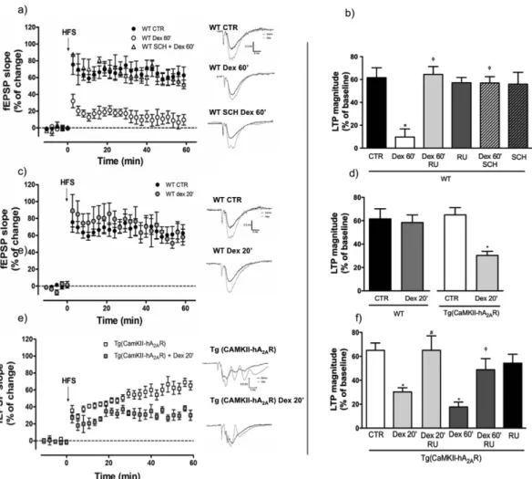
for the time periods indicated, followed by one-hour resting prior to recording, to allow gene-dependent effects. Prolonged exposure (for 60 min) of WT hippocampal slices to a synthetic GR agonist, dexamethasone (100 nM), abolished long-term potentiation (LTP) (Fig. 3a,b) an effect prevented by the GR antagonist, RU486 (Fig. 3b). Interestingly, the blockade of A2AR with SCH58261 (50 nM) prevented these GR-induced effects as efficiently as
RU486 (Fig. 3a,b), whereas neither GR nor A2AR antagonists alone had an effect on control (vehicle-treated) slices
(Fig. 3b). We then tested whether, conversely, A2AR overexpression increased susceptibility to dexamethasone.
Figure 3. Dexamethasone induced deficits in synaptic plasticity are prevented by adenosine A2A receptor
(A2AR) blockade and exacerbated by A2AR overexpression. High frequency stimulation (HFS: 100 Hz, 1s)
was used to evaluate synaptic plasticity in hippocampal slices. (a) Incubation of slices from WT rats with dexamethasone (100 nM) for 1 h decreases LTP magnitude an effect prevented by the A2AR antagonist
SCH58261 (50 nM). (b) Magnitude of the effects of dexamethasone, RU486 (100 nM) and SCH58261 (n = 3–8) in hippocampal slices from WT rats. Results are presented as mean ± SEM of n experiments analyzed using a one-way ANOVA followed by a Bonferroni post hoc test. * P < 0.05 compared to CTR; φP < 0.05 compared with
dexamethasone 60 min. (c) Incubation of slices with dexamethasone (100 nM) for 20 minutes has no effect on LTP magnitude in WT animals (n = 3–8), whereas (e) in Tg(CaMKII-hA2AR) animals is sufficient to induce
a significant decrease in LTP magnitude (n = 6–9). (d) Bar plots of the effects of dexamethasone and (f) the prevention of these effects by the GR antagonist RU486 (n = 3–9). Results are presented as mean ± SEM of n experiments analyzed using an unpaired t-test for comparisons between WT and Tg(CaMKII-hA2AR) and
one-way ANOVA followed by a Bonferroni post hoc test for drug effects.* P < 0.05 compared to control, #P < 0.05
Using a shorter exposure - 20 min - to dexamethasone (100 nM), we found that dexamethasone treatment had no impact on LTP magnitude in WT animals (Fig. 3c,d). In contrast, such shorter exposure time was sufficient to decrease LTP magnitude in tg(CaMKII-hA2AR) animals (Fig. 3d,e), an effect completely prevented by the GR
antagonist RU486 (Fig. 3f).
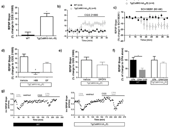
We further explored whether the A2AR signaling in these animals followed an aging-like pattern. In
Tg(CaMKII-hA2AR) animals, A2AR tonically increase excitatory transmission, an effect revealed by the inhibitory
effect of the A2AR selective antagonist SCH58261 (50 nM) on basal synaptic transmission (Fig. 4c), that was not
observed in WT animals. The effect of CGS 21680 (30 nM), a selective A2AR agonist on basal synaptic
transmis-sion was much higher in tg(CaMKII-hA2AR) than in WT animals (Fig. 4a,b). This effect was abolished by H89, a
protein kinase A (PKA) inhibitor, but not by GF 109203x, a protein kinase C (PKC) inhibitor (Fig. 4d). To eval-uate if the tonic adenosine inhibitory tonus was altered in tg(CaMKII-hA2AR) animals, we activated A2AR while
blocking A1R with a selective antagonist, DPCPX (50 nM). A1R blockade did not prevent A2AR effects on basal
synaptic transmission (Fig. 4e). Finally we explored if the A2AR- A1R crosstalk, shown to disappear in normal
aging30, is lost in the rats that overexpress A
2AR. While in WT animals, A1R activation by CPA (30 nM) causes a
strong inhibition of synaptic transmission that is attenuated when A2AR are simultaneously activated with CGS
21680; in tg(CaMKII-hA2AR) animals, the A2AR activation did not modify A1R mediated effects (Fig. 4f,g).
Figure 4. Overexpression of adenosine A2A receptor (A2AR) induces age-like modifications in adenosine
neuromodulation. (a,b) The A2AR selective agonist CGS21680, 30 nM, has an effect on basal fEPSP slope in
Tg(CaMKII-hA2AR) but not in WT animals ; Results were analyzed using a unpaired Student’s t-test (* P < 0.05
comparing to WT); (c) A2AR tonically increase excitatory transmission in Tg(CaMKII-hA2AR) animals, an
effect revealed by the inhibitory effect of the A2AR selective antagonist SCH58261 (50 nM) on basal synaptic
transmission, that was not observed in WT animals. (d) The effect of CGS21680, 30 nM is blocked by H89 (1 µ M), a PKA antagonist, but not GF (1 µ M) a PKC antagonist. Results were analyzed using One-way ANOVA followed by a Bonferroni’s multiple comparison post hoc test (#P < 0.05 comparing with Tg(CaMKII-hA
2AR with
CGS21680 alone). (e) The effect of A2AR activation does not change in the presence of the selective adenosine
A1R antagonist DPCPX (100 nM;n = 3); P > 0.05 using a paired Student’s t-test analysis). (f) The effect of
CGS58261 upon CPA, 30 nM, on fEPSP is lost in Tg(CaMKII-hA2AR) animals. Results were analyzed using a
Adenosine A
2AR regulates GR transcriptional activity and translocation.
We then evaluated if thisincreased sensitivity to dexamethasone in tg(CaMKII-hA2AR) animals is linked to an ability of A2AR to alter
tran-scriptional regulation by GR. Therefore, mouse neuroblastoma N1E115 cells were transiently transfected with the plasmid pGL3(GRE)3_TK_Luc (GRE_Luc) that contains the glucocorticoid response elements (GRE, at which GR binds to regulate gene transcription) coupled to the luciferase gene. Exposure to dexamethasone (100 nM for 24 h) triggered the expected increase in luciferase expression, an effect blocked by the GR selective antagonist RU486 (100 nM; Fig. 5a). Notably, A2AR blockade with SCH58261 (10–100 nM) or KW6002, (30 nM) reduced the
dexamethasone-induced increase of luciferase expression (Fig. 5b). Conversely, A2AR activation with the selective
agonist CGS21680 (10–50 nM), increased the dexamethasone-induced luciferase expression (Fig. 5c). Finally, even in the absence of an exogenous GR activation, A2AR blockade decreased luciferase expression (Fig. 5d) while
A2AR activation increased it (Fig. 5e); this effect of A2AR on the endogenous GR activity was prevented by RU486
(Fig. 5f).
In addition, we explored the ability of A2AR to control the nuclear translocation of GR in primary cortical
neuronal cultures. As expected, dexamethasone (100 nM) induced a significant enrichment of GR in the nuclear
Figure 5. Adenosine A2A receptors (A2AR) modulate glucocorticoid response element (GRE) regulated
luciferase expression in N1E115 cells and promote dexamethasone induced Glucocorticoid Receptor (GR) translocation to the nucleus. (a) Dexamethasone induced an increase in luciferase activity (n = 8–17) (b) which is decreased upon A2AR blockade by two antagonists, SCH 58261 (10–100 nM) and KW 6002 (50 nM)
(n = 5–11) and (c) increased upon direct A2AR activation with CGS 21680 (10–50 nM) (n = 3–9), as depicted in
the upper schemes. Activation of A2AR alone is sufficient to modulate endogenous GR transcriptional activity
(d) A2AR antagonist decreases luciferase activity (n = 6–14) while (e) A2AR agonist increases it (n = 3–11).
(f) A2AR effects are prevented by the glucocorticoid receptor (GR) antagonist, RU 486 (100 nM, n = 5–10).
Results are presented as mean ± SEM of n experiments. In (d–f) results were normalized to CTR, i.e., to the condition without dexamethasone, while in b) and c) results are normalized to DEX-induced activity. * P < 0.05 compared to control, ΦP < 0.05 compared with dexamethasone induced luciferase activity calculated using a
one-way ANOVA followed by a Bonferroni post hoc test. (g) Left panel illustrates the gradual enrichment of GR in the nuclear fraction of neuronal cultures over time of exposure to dexamethasone (n = 2–4). This increase is completely prevented by blocking A2AR with SCH 58261- 50 nM (in right panel). Results are presented
as mean ± SEM of n experiments. * P < 0.05 compared to control, #P < 0.05 compared with dexamethasone
calculated using two-way ANOVA followed by a Bonferroni post hoc. (h) Expression levels of GR and A2AR in
versus cytoplasmic fraction in a time-dependent manner, which was maximal after 90 minutes; this effect was completely inhibited upon A2AR blockade (Fig. 5g), in line with data obtained in N1E115 cells.
Overexpression of A
2AR receptors downregulates the expression of GR target genes.
In orderto test whether A2ARs are impacting on GR-dependent regulation of endogenous target genes, we evaluated the
effect of selective A2AR inhibition on GR-dependent gene regulation in primary neuronal cultures. Quantitative
PCR experiments targeting GR-activated genes (GILZ; PER1 and Bcl2)31–33, containing GRE motifs34–36 were run
following 1h incubation with dexamethasone (100 nM), in the presence or absence of the A2AR selective
antago-nist SCH51280 (50 nM). Arguing for a physiological role for A2AR in modulating GR activity, we found that the
increased GILZ, PER1 and Bcl2 mRNA expression induced by dexamethasone was significantly reduced when-ever A2AR are blocked with SCH 58261 (Fig. 6a–c).
Finally, we assessed the impact of the age-like GR downregulation and A2AR overactivation seen in the
hip-pocampus of [tg(CaMKII-hA2AR)] (see Fig. 1), on the expression of the same GR-target genes (Fig. 6d). As
pre-dicted, we found that expression levels for both GILZ and Bcl2 were decreased in [tg(CaMKII-hA2AR)] compared
to WT animals, whereas for PER1 we could not detect significant changes.
Discussion
Our findings demonstrate for the first time that GR transcriptional activity and nuclear localization are modu-lated by adenosine A2A receptors, thereby affecting GR function. Importantly, we here show that neuronal A2AR
overexpression is sufficient to impair the HPA-axis function and decrease GR hippocampal levels. Furthermore, the combined evidence that A2AR over-expression increases the susceptibility to GR agonists on one hand, and
that A2AR blockade prevents the detrimental synaptic effects of GR activation on the other hand suggests that
A2AR play a critical role in the control of memory dysfunction by modulating GR expression and activation.
Figure 6. Adenosine A2A receptors (A2AR) regulates transcription of GR target genes. mRNA levels of GILZ
(a), PER1 (b) and Bcl2 (c) increase in primary neurons after incubation with dexamethasone (100 nM) for 1 h an effect prevented by the presence of the selective A2AR antagonist SCH 51280 (50 nM), n = 3–5. * P < 0.05
compared to control, calculated using one-way ANOVA followed by a Bonferroni post hoc test (d) mRNA levels of GILZ, PER1 and Bcl2 in the hippocampus of Tg(CaMKII-hA2AR) compared to WT animals. Results are
This is the first time that the link between the well-documented hippocampal increase in A2AR expression
and the decrease in GR density and associated HPA-axis dysfuntion - features of aging and Alzheimer’s disease (AD) - is established. This also sustains the novel hypothesis that A2AR up-regulation through modulation of GR
function is sufficient to trigger synaptic dysfunction and subsequent memory impairments. This novel A2AR-GR
interaction has far-reaching implications in multiple pathologies in which corticosteroids play a pivotal role and that are alleviated by A2AR antagonists, notably caffeine.
Stress hormones and HPA-axis dysfunction have long been recognized as a critical feature underlying brain aging and pathology37. Indeed, altered cortisol levels are observed in post-traumatic stress syndrome or major
depression38 and elevated salivary levels of cortisol were found to be correlated with poor cognitive function in
a large study of humans aged 50–70 years4. Increased glucocorticoid activity has a predominant impact on the
hippocampus, which plays an inhibitory role in regulating the HPA axis2 and controls mood and memory39. Thus,
chronic exposure to glucocorticoids leads to cell death and hippocampal atrophy40,41 and is associated with
mem-ory impairment in the elderly3. Accordingly, recent evidence supports a pivotal role of stress hormones in
neu-rodegenerative diseases, namely in AD42. This is re-enforced by the following observations: 1) administration of
the GR antagonist, RU486, reverts multiple features of AD pathology15,16; 2) repeated stress worsens AD-induced
deficits43; 3) elevated cortisol levels are associated with a faster disease progression in AD5; 4) systemic
adminis-tration of glucocorticoids or stress potentiate memory impairments, hippocampal damage, β -amyloid formation and Tau pathology in transgenic AD mice6,8,44.
Interestingly, in aging or in other brain pathologies where a dysfunction of the HPA-axis is present, there is also an upsurge of A2AR in the hippocampus1,11 and their blockade has proven to be beneficial, but the
mech-anisms remained unknown1,14. We now provide the first demonstration that A
2AR modulation of GR may be
essential for the efficacy of A2AR blockade, a mechanism never hypothesized before.
The data we present clearly show that a specific increase in A2AR in forebrain neurons is able to impair the stress
response system. A2AR neuronal overexpression disturbed HPA-axis and increased plasma corticosterone levels,
providing a tentative connection between the adenosine neuromodulation system and the control of GR signaling. Our observations are in agreement with previous reports that A2AR activation in a model of spinal cord
injury mimicked the effects of GR activation in attenuating neuronal damage10. In particular, we showed that
A2ARs modulate GR transcriptional activity, an effect reversed by a GR antagonist (Fig. 5a–c). This decrease
in GR-dependent transcriptional regulation could be a consequence of the observed effect of A2AR on the
sub-cellular localization of GR (Fig. 5g). The reduced nuclear localization upon A2AR inhibition provides a
straightforward explanation for the reduced GR-dependent regulation of genes involved in different functions such as apoptosis (Bcl2) and the circadian clock (PER1)32,33. Our results indeed show that the mRNA levels for
both GILZ and Bcl-2 are decreased in the hippocampus of the transgenic rats as compared to WT animals, while PER1 levels remain unchanged (Fig. 6d).
PER1, being the only clock gene in the group, exhibits a robust amplitude of rhythmic expression, initiated in the SCN and PVN45. In all other brain regions, such as the hippocampus while still rhythmic, driven by CORT
and GRE-dependent effects, have attenuated amplitudes45. Accordingly, other authors failed to see significant
rhythmic expression of Per1 mRNA in whole hippocampus of rodents46. One can especulate that although we
observe GR downregulation in Tg(CaMKII-hA2AR), the levels are not sufficient to disturb Per1 expression. The recently described ability of caffeine to impact on the circadian clock47 strengthens our hypothesis since
corticosterone circadian oscillation is tightly controlled by clock gene expression. Furthermore, we not only show that A2AR overexpression impairs circadian corticosterone, we also show that A2AR blockade was sufficient to
prevent the deleterious impact of a synthetic GR agonist (dexamethasone) on hippocampal synaptic plasticity (Fig. 3a,b). This tight A2AR-GR interaction discloses a new view on how HPA-axis dysfunction emerges, and also
supports the therapeutic utility of A2AR antagonists as an important alternative to GR antagonists in
reestablish-ing HPA-axis dysfunction, which occurs in multiple clinical conditions1. In fact, the therapeutic interest of using
selective A2AR antagonists against multiple pathologies is increasing and A2AR antagonists have been recently
approved as co-adjuvant therapy for Parkinson’s disease48. Various studies also support the ability of caffeine
and A2AR blockade to prevent memory impairment in various conditions49, and recent work revealed that
caf-feine can even have pro-cognitive effects50. A
2AR antagonism was also proposed for the treatment of depression
and anxiety-like disorders51 in agreement with the decreased incidence of depression in individuals consuming
caffeine52. However, the lack of knowledge with regarding the mechanism of action of A
2AR antagonists
compro-mised their acceptance for clinical use. The present report shows that A2AR not only regulate HPA-axis function,
but also directly modulate GR, which represent key findings for understanding the mechanisms by which A2AR
antagonism is effective. Our data complete and strengthen our previous demonstration that A2AR blockade
over-came stress effects by reestablishing the HPA-axis and GR levels in the hippocampus1. These findings are critical,
not only for possible treatment strategies of the memory dysfunction associated with psychopathologies, but also in the context of aging and other circumstances in which the glucocorticoid response is impaired.
We have also observed that the A2AR synaptic responses in tg(CaMKII-hA2AR) are dependent on PKA activation
and that in vivo administration of an A2A blocker rescues the higher LTP recorded in these animals back to WT
lev-els (panel f), Fig. 2). This supports the crucial contribution of the A2A-PKA interplay to LTP induction mechanisms,
as proposed recently53. Furthermore, the features of adenosine neuromodulation evaluated in Tg(CaMKII-hA 2AR)
follow an aging-like profile. Namely, A2AR activation in Tg(CaMKII-hA2AR) animals has a direct effect in basal
synaptic transmission, mediated by PKA and not PKC, and independent of A1R inhibition. This reproduces what is
observed in aged animals30. Since A
1 receptor levels (Fig. 1e) are not changed in transgenic animals, and we obtain
the same A2AR effect on synaptic transmission regardless of A1R blockade with DPCPX (Fig. 4e), this does not seem
to depend on endogenous adenosine levels. Taken together, the results now presented suggest that an increase in neuronal A2AR is sufficient to drive age-like changes also in adenosine modulation and consequent LTP impairment.
similar to the LTP observed previously in aged animals29,54,55 which typically present memory deficits in a variety of
tasks. The fact that this ‘aged LTP’ was normalized upon A2AR blockade29,54, in a similar way we have shown to occur
now in Tg(CaMKII-hA2AR) animals, further reinforces this relationship.
Previous studies already hinted on a direct A2AR-HPA axis link, but data were contradictory. Global knockout
of the A2AR was shown to impact on both plasma corticosterone levels and melanocyte stimulating hormone levels
of pro-opiomelacortin expression56. This was mainly mediated by a profound effect on adenosine tonic modulation
by the deletion of the gene during embryonic and postnatal development, which occurs both centrally and system-ically. Exposure to higher levels of corticosterone in early-life, as occurs in these KO animals, induces an independ-ent disruption of the inhibitory hippocampal-hypothalamic feedback control of corticosterone release resulting in chronic higher levels of this hormone in plasma, as we have demonstrated previously1. Due to the intrinsic
limitations of that constitutive model, the authors could not prove whether the central effects would simply derive from knocking out A2AR KO in the adrenal glands. Our current model overcomes this confounding factor, since
CAMKIIa activity which drives A2AR expression occurs only from postnatal day 4 onwards, mainly in
glutama-tergic neurons of the forebrain57, thus inducing an age-like physiological pattern of A
2AR overexpression (Fig. 1d).
The transducing pathways by which A2AR trigger GR/GRE transcriptional activity remain to be elucidated.
Given the complexicity of A2AR signaling58 and its engagement in numerous signalosome protein complexes59,
this has been difficult to dissect. A2AR can recruit multiple signaling pathways, being the most common in the
hippocampus the cAMP/PKA/CREB, PKC and MAPK pathways60. Our data indicate that A
2AR effects are
prob-ably associated to the activation of PKA rather than PKC. The effects observed in GILZ and Bcl-2 expression levels in Tg(CaMKII-hA2AR) (Fig. 6d) may thus result either from the modulation of GR actions on the
glu-cocorticoid response element (GRE), or reflect an independent action following A2AR-driven CREB activation
and binding to cyclic AMP response elements (CREs). It is known that CREB is a positive activator of Bcl-2 expression in rat61. On the other hand, Per1 gene contains CRE responsive elements and its levels are elevated by
activation of CREB62. If A
2AR would act directly through CRE in our model, one would expect an up-regulation
of both Bcl-2 and Per1 in Tg(CaMKII-hA2AR) animals. However, we observed a clear down-regulation of Bcl-2
and no differences in Per1 expression, which is consistent with a response to the GR downregulation and less GRE-mediated gene expression, rather than a CRE- activation for these two genes. Furthermore, there is no evi-dence of putative binding sites for CRE in the rat GILZ, neither in the literature nor in databases such as TRED (Transcriptional Regulatory Element Database). Alternatively, a direct protein kinase A (PKA) modulation of GR binding to GRE is also possible, as previously described63. PKA exerts a regulatory role in the activation of
multi-ple nuclear hormone receptors. For exammulti-ple, it has been shown that PKA activates GR-dependent DNA binding in cotransfection studies. In addition, PKA can directly phosphorylate GR in vitro, enhances transcription by the retinoic acid receptor, and regulates dimerization of human estrogen receptor-α (revised in64) It has been
shown that PKA associates with GR and potentiates GR-dependent transcription, namely that PKA attenuates GR cross-repression64. Our observation that tg(CaMKII-hA
2AR) have an enhanced A2AR-PKA activation associated
to an increased sensitivity to dexamethasone favours the latter hypothesis.
Finally, there is an apparent paradox that arises from the present study: the fact that both stress and A2AR
upregulation decrease GR levels in the hippocampus, while simultaneously potentiating GR activation. The for-mer is however reconciled by the fact that GR activation is an important pathway to decrease GR expression and activation effects65–67, particularly upon chronic exposure to glucocorticoids68 as in chronic stress or aging.
Indeed, this combination of GR downregulation and increased levels of cortisol is observed upon aging: while GR mRNA is significantly reduced in several hippocampal subfields (i.e. stratum granulosum and temporal hip-pocampus proper) of aged cognitively impaired rats compared to young animals, their cortisol levels take signif-icantly longer to return to baseline following an acute stressor, and this was signifsignif-icantly correlated with poorer spatial learning ability18. Therefore, by increasing GR nuclear location and GR-mediated transcription, A
2AR not
only increase the susceptibility to stress, but also, through the same pathway, contribute to the downregulation of GR. Whether GR are more prone to activation in these conditions due to increased nuclear localization, a higher affinity for DNA, or to faster kinetics, remains to be clarified. Additionally, a redistribution of GR receptors in the hippocampus was also observed after exposure to corticosteroids68 which may also account for a modified
susceptibility upon higher circulating corticosterone levels.
In summary, our results provide the first evidence for a functional interaction between GR and A2ARs,
reveal-ing that A2AR modulate GR transcriptional activity and nuclear localization directly affecting GR target genes.
Moreover, we demonstrate that this interaction impacts GR-mediated effects on synaptic plasticity, and that A2AR
blockade prevents the deleterious effects associated with GR activation/function. Our data also reveal that this interaction may be particularly important in situations where A2AR are over-activated, such as in chronic stress or
aging, possibly due to an activation of the PKA pathway.
The beneficial effects of A2AR antagonists, namely caffeine, against cognitive impairments may be, at least
partially, due to the now reported effects on GR. The expansion of this interaction to the immune response, cell proliferation, tumor response and other cellular functions that imply GR or corticosteroids use in therapeutics, could have an enormous clinical impact.
References
1. Batalha, V. L. et al. Adenosine A(2A) receptor blockade reverts hippocampal stress-induced deficits and restores corticosterone circadian oscillation. Mol Psychiatry18, 320–331, doi: 10.1038/mp.2012.8 (2013).
2. Jacobson, L. & Sapolsky, R. The role of the hippocampus in feedback regulation of the hypothalamic-pituitary-adrenocortical axis. Endocr Rev12, 118–134, doi: 10.1210/edrv-12-2-118 (1991).
3. Lupien, S. J. et al. Cortisol levels during human aging predict hippocampal atrophy and memory deficits. Nat Neurosci1, 69–73, doi: 10.1038/271 (1998).
4. Lee, B. K. et al. Associations of salivary cortisol with cognitive function in the Baltimore memory study. Arch Gen Psychiatry64,
5. Csernansky, J. G. et al. Plasma cortisol and progression of dementia in subjects with Alzheimer-type dementia. Am J Psychiatry163,
2164–2169, doi: 163/12/2164 (2006).
6. Green, K. N., Billings, L. M., Roozendaal, B., McGaugh, J. L. & LaFerla, F. M. Glucocorticoids increase amyloid-beta and tau pathology in a mouse model of Alzheimer’s disease. J Neurosci26, 9047–9056, doi: 26/35/9047 (2006).
7. Caroll, D. & Zhang, B. Primer and interviews: advances in targeted gene modification. Interview by Julie C. Kiefer. Dev Dyn240,
2688–2696, doi: 10.1002/dvdy.22780 (2011).
8. Yao, Y. Y., Wu, Q. S., Li, W. Z. & Li, W. P. Dexamethasone potentiated Abeta-induced learning and memory impairment in rats. Neurol Res33, 371–380, doi: 10.1179/016164110× 12816242542698 (2011).
9. Chen, Y. C., Huang, S. H. & Wang, S. M. Adenosine-stimulated adrenal steroidogenesis involves the adenosine A2A and A2B receptors and the Janus kinase 2-mitogen-activated protein kinase kinase-extracellular signal-regulated kinase signaling pathway. Int J Biochem Cell Biol40, 2815–2825, doi: 10.1016/j.biocel.2008.05.016 (2008).
10. Okonkwo, D. O. et al. A comparison of adenosine A2A agonism and methylprednisolone in attenuating neuronal damage and improving functional outcome after experimental traumatic spinal cord injury in rabbits. J Neurosurg Spine4, 64–70, doi: 10.3171/ spi.2006.4.1.64 (2006).
11. Lopes, L. V., Cunha, R. A. & Ribeiro, J. A. Increase in the number, G protein coupling, and efficiency of facilitatory adenosine A2A receptors in the limbic cortex, but not striatum, of aged rats. J Neurochem73, 1733–1738 (1999).
12. Albasanz, J. L., Perez, S., Barrachina, M., Ferrer, I. & Martin, M. Up-regulation of adenosine receptors in the frontal cortex in Alzheimer’s disease. Brain Pathol18, 211–219, doi: 10.1111/j.1750-3639.2007.00112.x (2008).
13. Batalha, V. L., Valadas, J. S., Baqi, Y., Radjainia, H. & Lopes, L. V. Adenosine A2A receptor activation-trigger to aging-like modifications on adenosine modulation in the hippocampus. Journal of Neurochemistry125, 153 (2013).
14. Arendash, G. W. et al. Caffeine protects Alzheimer’s mice against cognitive impairment and reduces brain beta-amyloid production. Neuroscience142, 941–952, doi: S0306-4522(06)00937-7 (2006).
15. Baglietto-Vargas, D., Medeiros, R., Martinez-Coria, H., LaFerla, F. M. & Green, K. N. Mifepristone alters amyloid precursor protein processing to preclude amyloid beta and also reduces tau pathology. Biol Psychiatry74, 357–366, doi: 10.1016/j.biopsych.2012.12.003 (2013).
16. Lante, F. et al. Subchronic glucocorticoid receptor inhibition rescues early episodic memory and synaptic plasticity deficits in a mouse model of Alzheimer’s disease. Neuropsychopharmacology40, 1772–1781, doi: 10.1038/npp.2015.25 (2015).
17. Laurent, C. et al. A2A adenosine receptor deletion is protective in a mouse model of Tauopathy. Mol Psychiatry21, 149, doi: 10.1038/ mp.2015.115 (2016).
18. Bizon, J. L. et al. Hypothalamic-pituitary-adrenal axis function and corticosterone receptor expression in behaviourally characterized young and aged Long-Evans rats. Eur J Neurosci14, 1739–1751 (2001).
19. Popova, E. A., Krivokharchenko, A. S. & Vil’ianovich, L. I. [In vitro development of murine embryos using different types of microinjections]. Ontogenez33, 107–110 (2002).
20. Mayford, M., Bach, M. E. & Kandel, E. CaMKII function in the nervous system explored from a genetic perspective. Cold Spring Harb Symp Quant Biol61, 219–224 (1996).
21. Schmittgen, T. D. & Livak, K. J. Analyzing real-time PCR data by the comparative C(T) method. Nat Protoc3, 1101–1108 (2008). 22. Palacios, S. D. et al. Role of p38 mitogen-activated protein kinase in middle ear mucosa hyperplasia during bacterial otitis media.
Infect Immun72, 4662–4667, doi: 10.1128/IAI.72.8.4662-4667.2004 (2004).
23. Lowry, O. H., Rosebrough, N. J., Farr, A. L. & Randall, R. J. Protein measurement with the Folin phenol reagent. J Biol Chem193,
265–275 (1951).
24. Valadas, J. S. et al. Neuroprotection afforded by adenosine A2A receptor blockade is modulated by corticotrophin-releasing factor (CRF) in glutamate injured cortical neurons. J Neurochem123, 1030–1040, doi: 10.1111/jnc.12050 (2012).
25. Hockemeyer, J., Burbiel, J. C. & Muller, C. E. Multigram-scale syntheses, stability, and photoreactions of A2A adenosine receptor antagonists with 8-styrylxanthine structure: potential drugs for Parkinson’s disease. J Org Chem69, 3308–3318, doi: 10.1021/ jo0358574 (2004).
26. Batalha, V. F., Coelho, D. G. , Valadas, J. E, Bader, J. S., Gomes, M., R. A. Blum, D. & Lopes, L. V. Adenosine neuromodulation of hippocampal synaptic transmission in chronic stress: aging-like effect. Paper presented at FENS Forum 2014; Barcelona. Publisher: http://fens2014.meetingxpert.net/FENS_427/poster_100713/program.aspx/anchor100713 (2014).
27. Diogenes, M. J., Assaife-Lopes, N., Pinto-Duarte, A., Ribeiro, J. A. & Sebastiao, A. M. Influence of age on BDNF modulation of hippocampal synaptic transmission: interplay with adenosine A2A receptors. Hippocampus17, 577–585, doi: 10.1002/hipo.20294 (2007).
28. Masino, S. A., Latini, S., Bordoni, F., Pedata, F. & Dunwiddie, T. V. Changes in hippocampal adenosine efflux, ATP levels, and synaptic transmission induced by increased temperature. Synapse41, 58–64, doi: 10.1002/syn.1060 (2001).
29. Diogenes, M. J. et al. Enhancement of LTP in aged rats is dependent on endogenous BDNF. Neuropsychopharmacology36,
1823–1836, doi: 10.1038/npp.2011.64 (2011).
30. Lopes, L. V., Cunha, R. A. & Ribeiro, J. A. Cross talk between A(1) and A(2A) adenosine receptors in the hippocampus and cortex of young adult and old rats. J Neurophysiol82, 3196–3203 (1999).
31. D’Adamio, F. et al. A new dexamethasone-induced gene of the leucine zipper family protects T lymphocytes from TCR/CD3-activated cell death. Immunity7, 803–812, doi: S1074-7613(00)80398-2 (1997).
32. Feng, Y. et al. Dexamethasone-induced neuroprotection in hypoxic-ischemic brain injury in newborn rats is partly mediated via Akt activation. Brain Res1589C, 68–77, doi: S0006-8993(14)01347-X (2014).
33. Reddy, T. E., Gertz, J., Crawford, G. E., Garabedian, M. J. & Myers, R. M. The hypersensitive glucocorticoid response specifically regulates period 1 and expression of circadian genes. Mol Cell Biol32, 3756–3767, doi: 10.1128/MCB.00062-12 (2012).
34. van der Laan, S. et al. Chromatin immunoprecipitation scanning identifies glucocorticoid receptor binding regions in the proximal promoter of a ubiquitously expressed glucocorticoid target gene in brain. J Neurochem106, 2515–2523, doi: 10.1111/j.1471- 4159.2008.05575.x (2008).
35. Polman, J. A. et al. A genome-wide signature of glucocorticoid receptor binding in neuronal PC12 cells. BMC neuroscience13, 118, doi: 10.1186/1471-2202-13-118 (2012).
36. Li, Q., Dashwood, W. M., Zhong, X., Nakagama, H. & Dashwood, R. H. Bcl-2 overexpression in PhIP-induced colon tumors: cloning of the rat Bcl-2 promoter and characterization of a pathway involving beta-catenin, c-Myc and E2F1. Oncogene26, 6194–6202, doi: 10.1038/sj.onc.1210438 (2007).
37. Porter, N. M. & Landfield, P. W. Stress hormones and brain aging: adding injury to insult? Nat Neurosci1, 3–4, doi: 10.1038/196 (1998).
38. Gerritsen, L., Comijs, H. C., Deeg, D. J., Penninx, B. W. & Geerlings, M. I. Salivary cortisol, APOE-epsilon4 allele and cognitive decline in a prospective study of older persons. Neurobiol Aging32, 1615–1625, doi: 10.1016/j.neurobiolaging.2009.09.007 (2011). 39. Fanselow, M. S. & Dong, H. W. Are the dorsal and ventral hippocampus functionally distinct structures? Neuron65, 7–19, doi:
10.1016/j.neuron.2009.11.031 (2010).
40. Sapolsky, R. M. & Meaney, M. J. Maturation of the adrenocortical stress response: neuroendocrine control mechanisms and the stress hyporesponsive period. Brain Res396, 64–76 (1986).
42. Rothman, S. M. & Mattson, M. P. Adverse stress, hippocampal networks, and Alzheimer’s disease. Neuromolecular Med12, 56–70, doi: 10.1007/s12017-009-8107-9 (2010).
43. Joshi, Y. B., Chu, J. & Pratico, D. Stress hormone leads to memory deficits and altered tau phosphorylation in a model of Alzheimer’s disease. J Alzheimers Dis31, 167–176, doi: 10.3233/JAD-2012-120328 (2012).
44. Chadwick, W. et al. Amitriptyline-mediated cognitive enhancement in aged 3xTg Alzheimer’s disease mice is associated with neurogenesis and neurotrophic activity. PLoS One6, e21660, doi: 10.1371/journal.pone.0021660 (2011).
45. Chun, L. E., Woodruff, E. R., Morton, S., Hinds, L. R. & Spencer, R. L. Variations in Phase and Amplitude of Rhythmic Clock Gene Expression across Prefrontal Cortex, Hippocampus, Amygdala, and Hypothalamic Paraventricular and Suprachiasmatic Nuclei of Male and Female Rats. Journal of biological rhythms30, 417–436, doi: 10.1177/0748730415598608 (2015).
46. Shieh, K. R., Yang, S. C., Lu, X. Y., Akil, H. & Watson, S. J. Diurnal rhythmic expression of the rhythm-related genes, rPeriod1, rPeriod2, and rClock, in the rat brain. Journal of biomedical science12, 209–217, doi: 10.1007/s11373-004-8176-6 (2005). 47. Burke, T. M. et al. Effects of caffeine on the human circadian clock in vivo and in vitro. Sci Transl Med7, 305ra146, doi: 10.1126/
scitranslmed.aac5125 (2015).
48. Chen, J. F., Eltzschig, H. K. & Fredholm, B. B. Adenosine receptors as drug targets–what are the challenges? Nat Rev Drug Discov12,
265–286, doi: 10.1038/nrd3955 (2013).
49. Cunha, R. A. & Agostinho, P. M. Chronic caffeine consumption prevents memory disturbance in different animal models of memory decline. J Alzheimers Dis20 Suppl 1, S95–116, doi: 10.3233/JAD-2010-1408 (2010).
50. Borota, D. et al. Post-study caffeine administration enhances memory consolidation in humans. Nat Neurosci17, 201–203, doi: 10.1038/nn.3623 (2014).
51. Cunha, R. A., Ferre, S., Vaugeois, J. M. & Chen, J. F. Potential therapeutic interest of adenosine A2A receptors in psychiatric disorders. Curr Pharm Des14, 1512–1524 (2008).
52. Lucas, M. et al. Coffee, caffeine, and risk of depression among women. Arch Intern Med171, 1571–1578, doi: 10.1001/archinternmed. 2011.393 (2011).
53. Baudry, M. et al. Multiple cellular cascades participate in long-term potentiation and in hippocampus-dependent learning. Brain Res
1621, 73–81, doi: 10.1016/j.brainres.2014.11.033 (2015).
54. Costenla, A. R., de Mendonca, A. & Ribeiro, J. A. Adenosine modulates synaptic plasticity in hippocampal slices from aged rats. Brain Res851, 228–234, doi: S0006-8993(99)02194-0 (1999).
55. Sousa, V. C. et al. Maternal separation impairs long term-potentiation in CA1-CA3 synapses and hippocampal-dependent memory in old rats. Neurobiol Aging35, 1680–1685, doi: 10.1016/j.neurobiolaging.2014.01.024 (2014).
56. Jegou, S. et al. Adenosine A2A receptor gene disruption provokes marked changes in melanocortin content and pro-opiomelan- ocortin gene expression. Journal of neuroendocrinology15, 1171–1177 (2003).
57. Burgin, K. E. et al.In situ hybridization histochemistry of Ca2+ /calmodulin-dependent protein kinase in developing rat brain. J Neurosci10, 1788–1798 (1990).
58. Fredholm, B. B., Chern, Y., Franco, R. & Sitkovsky, M. Aspects of the general biology of adenosine A2A signaling. Progress in neurobiology83, 263–276, doi: 10.1016/j.pneurobio.2007.07.005 (2007).
59. Keuerleber, S., Gsandtner, I. & Freissmuth, M. From cradle to twilight: the carboxyl terminus directs the fate of the A(2A)-adenosine receptor. Biochimica et biophysica acta1808, 1350–1357, doi: 10.1016/j.bbamem.2010.05.009 (2011).
60. Ribeiro, J. A. & Sebastiao, A. M. Modulation and metamodulation of synapses by adenosine. Acta physiologica199, 161–169, doi: 10.1111/j.1748-1716.2010.02115.x (2010).
61. Kim, S. J., Nian, C., Widenmaier, S. & McIntosh, C. H. Glucose-dependent insulinotropic polypeptide-mediated up-regulation of beta-cell antiapoptotic Bcl-2 gene expression is coordinated by cyclic AMP (cAMP) response element binding protein (CREB) and cAMP-responsive CREB coactivator 2. Mol Cell Biol28, 1644–1656, doi: 10.1128/MCB.00325-07 (2008).
62. Tischkau, S. A., Mitchell, J. W., Tyan, S. H., Buchanan, G. F. & Gillette, M. U. Ca2+ /cAMP response element-binding protein (CREB)-dependent activation of Per1 is required for light-induced signaling in the suprachiasmatic nucleus circadian clock. J Biol Chem278, 718–723, doi: 10.1074/jbc.M209241200 (2003).
63. Rangarajan, P. N., Umesono, K. & Evans, R. M. Modulation of glucocorticoid receptor function by protein kinase A. Molecular endocrinology6, 1451–1457, doi: 10.1210/mend.6.9.1435789 (1992).
64. Doucas, V. et al. Cytoplasmic catalytic subunit of protein kinase A mediates cross-repression by NF-kappa B and the glucocorticoid receptor. Proceedings of the National Academy of Sciences of the United States of America97, 11893–11898, doi: 10.1073/ pnas.220413297 (2000).
65. Surjit, M. et al. Widespread negative response elements mediate direct repression by agonist-liganded glucocorticoid receptor. Cell
145, 224–241, doi: 10.1016/j.cell.2011.03.027 (2011).
66. Ramamoorthy, S. & Cidlowski, J. A. Ligand-induced repression of the glucocorticoid receptor gene is mediated by an NCoR1 repression complex formed by long-range chromatin interactions with intragenic glucocorticoid response elements. Mol Cell Biol
33, 1711–1722, doi: 10.1128/MCB.01151-12 (2013).
67. Oakley, R. H. & Cidlowski, J. A. Homologous down regulation of the glucocorticoid receptor: the molecular machinery. Crit Rev Eukaryot Gene Expr3, 63–88 (1993).
68. Herman, J. P. & Spencer, R. Regulation of hippocampal glucocorticoid receptor gene transcription and protein expression in vivo. J Neurosci18, 7462–7473 (1998).
Acknowledgements
The authors acknowledge J. Baião for technical assistance in the animal house and C. Batalha for the figures layout. VLB and DGF were supported by a grant from Fundação para a Ciência e Tecnologia and an EMBO fellowship (Harvard Medical School-GSV lab- exchange period); LVL is an Investigator FCT, funded by Fundação para a Ciência e Tecnologia (PTDC-099853/2009). TFO is supported by the DFG Center for Nanoscale Microscopy and Molecular Physiology of the Brain. MH, LB and DB thank the following supports: LabEx (excellence laboratory) DISTALZ (Development of Innovative Strategies for a Transdisciplinary approach to Alzheimer’s disease), Inserm, CNRS, Université Lille 2, Région Nord/Pas-de-Calais, DN2M, ANR (ADORATAU) and FUI MEDIALZ. DB, CEM and YB are grateful for support by the LECMA/Alzheimer Forschung Initiative (AFI). DB and LVL were recipients of PHC FCT/Pessoa program and are currently recipients of an international exchange program (AAP Internationalisation / LIA) funded by the University of Lille.
Author Contributions
V.L.B. has written the draft, designed and performed most of the experimental work. L.V.L coordinated and designed the study. V.L.B. and R.G. performed the qPCR supervised by S.H.M., V.L.B. and M.H. performed the N1E115 experiments under supervision of D.B. and L.B. Tg(CaMKII-hA2AR) line was developed by T.S. and M.B.
electrophysiology experiments. V.L.B., S.H.M., G.S.V., T.F.O., D.B. and L.V.L. designed experiments and wrote the manuscript. The manuscript has been read and approved by all named authors.
Additional Information
Competing financial interests: The authors declare no competing financial interests.
How to cite this article: Batalha, V. L. et al. The caffeine-binding adenosine A2A receptor induces age-like
HPA-axis dysfunction by targeting glucocorticoid receptor function. Sci. Rep.6, 31493; doi: 10.1038/srep31493 (2016).
This work is licensed under a Creative Commons Attribution 4.0 International License. The images or other third party material in this article are included in the article’s Creative Commons license, unless indicated otherwise in the credit line; if the material is not included under the Creative Commons license, users will need to obtain permission from the license holder to reproduce the material. To view a copy of this license, visit http://creativecommons.org/licenses/by/4.0/