111 111 111 111 111 Mem Inst Oswaldo Cruz, Rio de Janeiro, Vol. 102(1): 111-115, February 2007
Impairment of cell division of
Trypanosoma cruzi
epimastigotes
Michele A Zacks
Department of Pathology, University of Texas Medical Branch, 301 University Boulevard, Galveston, TX 77555-0609, US
The mechanisms that facilitate the adaptation of Trypanosoma cruzi to two distinct hosts, insect and verte-brate, are poorly understood, in part due to the limited ability to perform gene disruption studies by homolo-gous recombination. This report describes a developmentally-defective phenotype that resulted from integra-tion of a drug marker adjacent to the GAPDH gene in T. cruzi.
Key words: Trypanosoma cruzi - life cycle - differentiation
troporation with a neomycin-resistance (neor)-based
construct targeting TcGPI8 and subsequent drug selec-tion of transfectants resulted in genomic integraselec-tion of the neor cassette, as confirmed by Southern blot
analy-sis (Fig. 1) and PCR amplification of the 1.3 kilobase GAPDH-IR-neor fragment from gDNA of the
trans-fectants (data not shown). However, GAPDHIR-neor did
not integrate into the TcGPI8 gene and the 3' and 5' ends of TcGPI8 were absent (data not shown). Genome walk-ing and PCR-clonwalk-ing identified the site of insertion of the GAPDHIR/neor cassette (Fig. 2A-B) as adjacent to
Financial support:American Heart Association, John Sealy Me-morial Endowment Fund for Biomedical Research, American Health Assistance Foundation, the National Institutes of Health (AI053098-01, AI054578-01), the James W. McLaughlin Predoctoral Fellowship Fund, the Department of Pathology and the Graduate School of Biomedical Sciences at the University of Texas Medical Branch
E-mail: mazacks@utmb.edu. Received 18 May 2006 Accepted 29 November 2006
Trypanosoma cruzi is a protozoan parasite of the ancient branch of eukaryotes (Kingdom Eukaryota, Or-der Kinetoplastida) (Stevens et al. 1999) and is endemic in South and Central America, and Mexico. T. cruzi is transmitted to vertebrates, including humans, predomi-nantly by insect vector (subfamily Triatoma, family Reduviidae) and also occurs via blood transfusion, or-gan transplantation, and congenital routes. In humans, 30% of chronically infected individuals are estimated to develop Chagas disease, a distinct form of cardiomy-opathy (Miles 2003). Despite the description of the as-sociation between T. cruzi transmission via the triatomine insect vector and heart disease in 1909 and decades of research illuminating the extraordinary mechanism of host cell infection (Hall 1993, Burleigh & Andrews 1998), no vaccine is available and only two anti-parasitic drugs have been licensed for treatment. However, these drugs are effective mainly at the acute stage of infection and are highly toxic (Barrett et al. 2003). The mechanisms that facilitate the adaptation of T. cruzi to two distinct hosts, insect and vertebrate, are poorly understood, in part due to the limited ability to perform gene disruption studies by homologous recom-bination. This report describes a developmentally-defec-tive phenotype that resulted from integration of a drug marker adjacent to the GAPDH gene in T. cruzi. This study was initiated to evaluate the role of surface ex-pressed glycosylphophatidylinositol (GPI)-anchored proteins in the complex life cycle of T. cruzi and uti-lized a homologous recombination-mediated approach to targeted disruption of the TcGPI8 gene in the para-site, described in Zacks and Garg (2006) (Fig. 1).
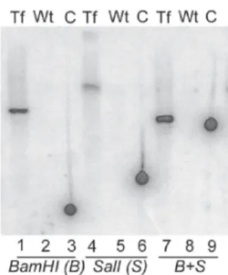
Elec-Fig. 1: genetic analysis of stable transfectants.Constructs for disruption of TcGPI8 were designed such that drug resistance genes, neoror bler,
were flanked by 400-600 base pairs of the 5' and 3' ends of the TcGPI8
gene. The GAPDH-intergenic region (GAPDH-IR) sequence, present upstream of the neor or bler genes, provides the necessary splice accep-tor site for mRNA processing (Nozaki & Cross 1995). Trypanosoma cruzi
SylvioX10/4 strain (American Type Culture Collection, Manassas, VA) epimastigotes were electroporated with the linearized disruption cas-sette (10 µg) and incubated in selective drug (G418, 60 µg/ml) (Kelly et al. 1992). Following positive selection, as evidenced by the death of mock-transfected parasites cultured at the same drug concentration, the drug pressure for selection of transfectants was increased to 200-400 µg/ml; after greater than one month, chromosomal DNA (gDNA) was extracted from these stable transfectants (Medina-Acosta & Cross 1993). For South-ern blot analysis, gDNA isolated from stable transfectants or from wild type, untransfected parasites was digested with restriction enzymes (BamHI and/or SalI) and resolved via agarose gel electrophoresis. Fol-lowing DNA transfer to nylon membrane, hybridization was performed using a 32P-labeled neor probe (random primer labeling) at 68oC. After 6 days exposure, the image was scanned by phosphorimager. gDNA from
stable transfectants hybridized with the neor probe (lanes 1, 4, and 7) indicating that neor is present in the transfectant genome. As expected,
112 112 112 112
112 T. cruzi developmental mutants • Michele A Zacks
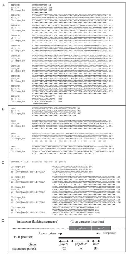
Fig. 2: identification of the site of integration of GAPDHIR-neor cassette in Trypanosoma cruzi genome. Genome walking via an arbitrarily primed PCR approach was used to determine the site of integration (Parker et al. 1991, Liu & Whittier 1995). Briefly, random, degenerate primers (Liu & Whittier 1995) were used in pairs with neor-specific primers (nested primers R1, TTTCGCTTGGTGGTCGAATGGGCAGGTA-3'; R2, 5'-GCACAGCTGCGCAAGGAACGCCC-3': R3, 5'-GCCGCGCTGCCTCG-3') to amplify fragments from gDNA of stable neor-TcGPI8 transfectants
contain-ing the unknown sequence flankcontain-ing GAPDHIR-neorusing modified cycling parameters (Liu & Whittier 1995). PCR fragments were cloned and sequenced (UTMB Protein Core Facility). Three informative clones were obtained, e.g., clones in which the DNA sequence matched to the expected
GAPDHIR, and neor portions and in which additional flanking sequence was present. T. cruzi blast search was performed using these flanking sequences. The sequence of the third clone was identical to c1-8_-4. Sequence alignments of TAIL-PCR clones with A) GAPDHIR, B) neor, and C)
113 113 113 113 113 Mem Inst Oswaldo Cruz, Rio de Janeiro, Vol. 102(1), February 2007
the 3' end of GAPDH (Fig. 2C) (Kendall et al. 1990). The orientation of the insertion is shown in Fig. 2D.
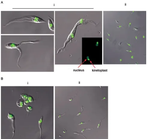
The morphology of epimastigote-stage transfectants was documented via confocal microscopy (Fig. 3). Stable neor-TcGPI8 transfectants (Fig. 3A) showed unusual
morphologies, first with the appearance of thin extended forms that appeared to be reduced in motility. Highly motile parasites with two or three flagella were present with frequent observation (> 2 per microscopy field) of two parasites with fused membranes, either in apical or longitudinal style. The nucleic acid staining pattern ob-served in doublet parasites incubated with the fluores-cent Syto11 dye indicated that duplication of kinetoplast and nuclei was not impaired. However, in many cases, cellular membranes appeared to be fused. The possibil-ity that these represent intermediate morphological forms in normal T. cruzi division cannot be excluded. However, such parasites were not found in either rou-tine (e.g. daily) light microscope examination of non-transformed cultures during consideration over an ex-tensive period of cultivation with selective drug (e.g., G418 for minimum of 2-3 months) or without selective drug while maintained in parallel cultures using liver
in-fusion tryptose medium. Nor, as presented here, were such forms documented during confocal microscopy. This observation is consistent for this study as well as for cultures of pTEX transformed cultures that were continuously cultured for > 2 years in the presence of selective drug (e.g., G418, as reported here), under daily to weekly monitoring. At the time this phenotype be-came pronounced, growth of the transformant popula-tion declined and could not be further maintained, most likely as a result of their inability to complete their rep-lication cycle. Thus, at the epimastigote stage, a striking defect in cell division occurred in transfectants.
The effect of this transformation on the life cycle development of transfectants, e.g., differentiation of epimastigote to the infective metacyclic forms and in-fection of mammalian cells, was further evaluated. Transfectant epimastigotes and, as control, wild type epimastigotes were grown in liver infusion tryptose medium for ≥ 10 days without addition of media to achieve stationary-growth phase for the conversion of parasites to metacyclic trypomastigotes and were used to infect fibroblast monolayers at a parasite to cell ratio of 50:1. At 24 h after infection, medium containing free
Fig. 3: morphological characteristics of stable transfectants. Stable neor-TcGPI8 transfectants were generated and genetically characterized, as
described in Figs 1 and 2.Parasites were harvested by centrifugation and washed in ice-cold PBS. Epimastigote morphology was documented via confocal microscopy of live parasites. To visualize kinetoplast and nuclear DNA, epimastigotes were incubated on ice for 10 min with Syto11 (Molecular Probes, 1:200 dilution), a cell-permeant nucleic acid binding fluorescent dye (excitation λMax, 515 nm; emission λMax, 543 nm). Confocal images were captured on a Zeiss LSM 510 UV Meta Laser Scanning Confocal Microscope (UTMB Optical Imaging Core Facility) at a magnification of 63X. Signals were overlaid with Nomarski differential interference using Zeiss AxioVision Viewer software. A: stable neor-TcGPI8 transfectants. i: transfectants; ii: wild type untransfected T. cruzi. Kinetoplast and nuclear DNA, which replicated in doublet parasites, are indicated by arrow; B: neor
114 114 114 114
114 T. cruzi developmental mutants • Michele A Zacks
parasites was replaced and cells monitored for the ap-pearance of trypomastigotes. Three independent in vitro infection experiments were performed in two different fibroblast cell lines (C2C12 and BHK21) using station-ary-phase cultures of stable transfectants or of wild type untransfected T. cruzi. Transfectants did not convert to the typical morphology of the infective metacyclic form following ≥ 10 days of cultivation without addition of new growth medium. No trypomastigotes were seen when monitoring fibroblasts infected with the station-ary phase cultures of these neor-TcGPI8 transfectants
during a 10-12 day incubation period. However, wild type, untransfected parasites were capable of infecting either fibroblast line, with appearance of trypomastigotes and amastigotes within 6 and 11 days, respectively. In sum-mary, morphological transformation of the neor-TcGPI8
transfectant epimastigotes into metacyclic trypo-mastigotes in culture conditions of “starvation” did not occur, unlike for wild type parasites. In conclusion, these stable neor-TcGPI8 transfectants were not infective for
mammalian cells in vitro.
In effort to obtain homozygous transfectants, the stable neor-TcGPI8 transfectants were subsequently
electroporated with a similar disruption construct con-taining the phleomycin-resistance (bler) gene in place
of the neor gene. During drug selection (500 µg/ml ble),
drastic changes from normal epimastigote morphology in axenic cultures were observed in these transfectants (Fig. 3B, panel i) but not in wild type parasites, which were cultured in parallel without selective drug (Fig. 3B, panel ii). Amastigote-like forms with short, retracted flagella were abundant (> 50% of the culture) among the transfected epimastigote population (Fig. 3B, panel i), whereas the morphology of the parallel wild-type untransfected culture was normal (Fig. 3B, panel ii). Uptake of propidium iodide in these forms indicated that parasite membranes were disrupted (data not shown). These parasites could not be maintained in culture, pre-cluding isolation of gDNA for further genetic analysis. In summary, this report documents an interesting and selective defect in which organelle duplication (kineto-plast and nuclei) does not appear to be blocked, in which the flagella replicate but in some cases, in which the plasma membranes do not appear to separate effectively. From these studies, it can be surmised that cell division of epimastigotes was impaired selectively. To date, few studies have been published on the mechanism of cell division of T. cruzi (Gomez et al. 1998, 2001, Bogitsh et al. 1999, Grellier et al. 1999a, b, Orr et al. 2000, Santori et al. 2002). The pattern of development, e.g., the time course of DNA replication, organelle duplica-tion and cell division of T. cruzi cannot be assumed to occur as has been demonstrated in elegant studies of the related trypanosomatid, T. brucei (Hendriks et al. 2000, McKean 2003). In T. cruzi, targeted disruption of genes for functional analysis remains a challenge and induc-ible expression systems have not been possinduc-ible (DaRocha et al. 2004). In addition to the deficit of tools for genetic manipulation of T. cruzi, we lack an under-standing of the mechanisms regulating T. cruzi gene
ex-pression and governing the cellular processes that en-able the parasite to transform morphologically. However, recently, the genome sequence of T. cruzi CL-Brener strain was completed (El-Sayed et al. 2005). It is antici-pated that future developments in transformation, gene disruption and/or inducible expression systems, com-bined with a comparative genomics approach (Parsons et al. 2005) will enhance our understanding of the com-plex developmental cycle and morphological transfor-mation required for T. cruzi survival and may identify specific drug targets. Further molecular-genetic studies will be required to evaluate the potential role of the flanking sequences of GAPDH, such as promoter func-tions, in the regulation of cell division and differentia-tion in T. cruzi.
REFERENCES
Barrett MP, Burchmore RJ, Stich A, Lazzari JO, Frasch AC, Cazzulo JJ, Krishna S 2003. The trypanosomiases. Lancet 362: 1469-1480.
Bogitsh BJ, Middleton OL, Ribeiro-Rodrigues R 1999. Effects of the antitubulin drug trifluralin on the proliferation and metacyclogenesis of Trypanosoma cruzi epimastigotes.
Parasitol Res 85: 475-480.
Burleigh BA, Andrews NW 1998. Signaling and host cell inva-sion by Trypanosoma cruzi. Curr Opin Microbiol1: 461-465.
DaRocha WD, Otsu K, Teixeira SM, Donelson JE 2004. Tests of cytoplasmic RNA interference (RNAi) and construction of a tetracycline-inducible T7 promoter system in Trypano-soma cruzi. Mol Biochem Parasitol133: 175-186. El-Sayed NM, Myler PJ, Bartholomeu DC, Nilsson D, Aggarwal
G, Tran AN, Ghedin E, Worthey EA, Delcher AL, Blandin G, Westenberger SJ, Caler E, Cerqueira GC, Branche C, Haas B, Anupama A, Arner E, Aslund L, Attipoe P, Bontempi E, Bringaud F, Burton P, Cadag E, Campbell DA, Carrington M, Crabtree J, Darban H, da Silveira JF, de Jong P, Edwards K, Englund PT, Fazelina G, Feldblyum T, Ferella M, Frasch AC, Gull K, Horn D, Hou L, Huang Y, Kindlund E, Klingbeil M, Kluge S, Koo H, Lacerda D, Levin MJ, Lorenzi H, Louie T, Machado CR, McCulloch R, McKenna A, Mizuno Y, Mottram JC, Nelson S, Ochaya S, Osoegawa K, Pai G, Par-sons M, Pentony M, Pettersson U, Pop M, Ramirez JL, Rinta J, Robertson L, Salzberg SL, Sanchez DO, Seyler A, Sharma R, Shetty J, Simpson AJ, Sisk E, Tammi MT, Tarleton R, Teixeira S, Van Aken S, Vogt C, Ward PN, Wickstead B, Wortman J, White O, Fraser CM, Stuart KD, Andersson B 2005. The genome sequence of Trypanosoma cruzi, etio-logic agent of Chagas disease. Science 309: 409-415. Gomez EB, Kornblihtt AR, Tellez-Inon MT 1998. Cloning of a
cdc2-related protein kinase from Trypanosoma cruzi that interacts with mammalian cyclins. Mol Biochem Parasitol 91: 337-351.
Gomez EB, Santori MI, Laria S, Engel JC, Swindle J, Eisen H, Szankasi P, Tellez-Inon MT 2001. Characterization of the
Trypanosoma cruzi Cdc2p-related protein kinase 1 and iden-tification of three novel associating cyclins. Mol Biochem Parasitol113: 97-108.
115 115 115 115 115 Mem Inst Oswaldo Cruz, Rio de Janeiro, Vol. 102(1), February 2007
trypomastigotes to amastigotes. Mol Biochem Parasitol98: 239-252.
Grellier P, Sinou V, Garreau-de Loubresse N, Bylen E, Boulard Y, Schrevel J 1999b. Selective and reversible effects of vinca alkaloids on Trypanosoma cruzi epimastigote forms: block-age of cytokinesis without inhibition of the organelle duplica-tion. Cell Motil Cytoskeleton42: 36-47.
Hall BF 1993. Trypanosoma cruzi: mechanisms for entry into host cells. Semin Cell Biol4: 323-333.
Hendriks E, van Deursen FJ, Wilson J, Sarkar M, Timms M, Matthews KR 2000. Life-cycle differentiation in Trypano-soma brucei: molecules and mutants. Biochem Soc Trans 28: 531-536.
Kelly JM, Ward HM, Miles MA, Kendall G 1992. A shuttle vec-tor which facilitates the expression of transfected genes in
Trypanosoma cruzi and Leishmania. Nucleic Acids Res 20: 3963-3969.
Kendall G, Wilderspin AF, Ashall F, Miles MA, Kelly JM 1990.
Trypanosoma cruzi glycosomal glyceraldehyde-3-phosphate dehydrogenase does not conform to the ‘hotspot’ topogenic signal model. Embo J9: 2751-2758.
Liu YG, Whittier RF 1995. Thermal asymmetric interlaced PCR: automatable amplification and sequencing of insert end frag-ments from P1 and YAC clones for chromosome walking.
Genomics25: 674-681.
McKean PG 2003. Coordination of cell cycle and cytokinesis in
Trypanosoma brucei. Curr Opin Microbiol 6: 600-607. Medina-Acosta E, Cross GA 1993. Rapid isolation of DNA from
trypanosomatid protozoa using a simple ‘mini-prep’ proce-dure. Mol Biochem Parasitol59: 327-329.
Miles M 2003. American trypanosomiasis (Chagas disease). In G Cook, A Zumla (eds), Manson’s Tropical Disease, El-sevier Science, London, p. 1325-1337.
Nozaki T, Cross GA 1995. Effects of 3' untranslated and intergenic regions on gene expression in Trypanosoma cruzi. Mol Biochem Parasitol75: 55-67.
Orr GA, Werner C, Xu J, Bennett M, Weiss LM, Takvorkan P, Tanowitz HB, Wittner M 2000. Identification of novel serine/ threonine protein phosphatases in Trypanosoma cruzi: a potential role in control of cytokinesis and morphology. In-fect Immun 68: 1350-1358.
Parker JD, Rabinovitch PS, Burmer GC 1991. Targeted gene walking polymerase chain reaction. Nucleic Acids Res19: 3055-3060.
Parsons M, Worthey EA, Ward PN, Mottram JC 2005. Compara-tive analysis of the kinomes of three pathogenic trypanosomatids: Leishmania major, Trypanosoma brucei
and Trypanosoma cruzi. BMC Genomics 6: 127.
Santori MI, Laria S, Gomez EB, Espinosa I, Galanti N, Tellez-Inon MT 2002. Evidence for CRK3 participation in the cell division cycle of Trypanosoma cruzi. Mol Biochem Parasitol121: 225-232.
Stevens JR, Noyes HA, Dover GA, Gibson WC 1999. The an-cient and divergent origins of the human pathogenic trypano-somes, Trypanosoma brucei and T. cruzi. Parasitology118: 107-116.