Iheringia, Série Zoologia, Porto Alegre, 105(1):76-83, 31 de março de 2015
Shape and size variations of Aegla uruguayana (Anomura, Aeglidae)
under laboratory conditions: A geometric morphometric approach to
the growth
Valeria P. Diawol1, Federico Giri1,2 & Pablo A. Collins1,3
1. Instituto Nacional de Limnología (CONICET - Universidad Nacional del Litoral), Santa Fe, Argentina. (valeriadiawol@hotmail.com) 2. Facultad de Humanidades y Ciencias (Universidad Nacional del Litoral), Santa Fe, Argentina. (fgiri@inali.unl.edu.ar) 3. Facultad de Bioquímica y Ciencias Biológicas (Universidad Nacional del Litoral), Santa Fe, Argentina. (pagcollins@gmail.com)
ABSTRACT. Crustacean growth studies typically use modal analysis rather than focusing on the growth of individuals. In the present work, we use geometric morphometrics to determine how organism shape and size varies during the life of the freshwater crab, Aegla uruguayana Schmitt, 1942. A total of 66 individuals from diverse life cycle stages were examined daily and each exuvia was recorded. Digital images of the dorsal region of the cephalothorax were obtained for each exuvia and were subsequently used to record landmark configurations. Moult increment and intermoult period were estimated for each crab. Differences in shape between crabs of different sizes (allometry) and sexes (sexual dimorphism; SD) were observed. Allometry was registered among specimens; however, SD was not statistically significant between crabs of a given size. The intermoult period increased as size increased, but the moult frequency was similar between the sexes. Regarding ontogeny, juveniles had short and blunt rostrum, robust forehead region, and narrow cephalothorax. Unlike juveniles crabs, adults presented a well-defined anterior and posterior cephalothorax region. The rostrum was long and stylised and the forehead narrow. Geometric morphometric methods were highly effective for the analysis of aeglid-individual- growth and avoided excessive handling of individuals through exuvia analysis.
KEYWORDS. Crustacea, cephalothorax, ontogeny, intermoult, morphometrics.
RESUMEN. Variaciones de forma y tamaño deAegla uruguayana (Anomura, Aeglidae): Una aproximación desde la morfometría geométrica al crecimiento.Los estudios de crecimiento en crustáceos típicamente utilizan análisis modal en lugar de focalizarse en el crecimiento individual de los organismos. En el presente trabajo, utilizamos morfometría geométrica para determinar cómo varia la forma y el tamaño a lo largo de la vida del cangrejo de agua dulce, Aegla uruguayana Schmitt, 1942. Un total de 66 individuos, en diferentes etapas del ciclo de vida, se examinaron diariamente, registrándose la presencia de exuvias. Imágenes digitales de cada muda fueron obtenidas de la región dorsal del cefalotórax y se utilizaron para registrar las configuraciones de landmarks. El incremento por muda y el período de intermuda se estimaron para cada cangrejo. Diferencias de forma entre tamaños (alometría) y sexos (dimorfismo sexual; SD) se observaron. Se registró la presencia de alometría entre los especímenes; sin embargo, el SD no fue estadísticamente significativo respecto del tamaño. El período intermuda aumentó de manera directamente proporcional al tamaño, pero la frecuencia de muda fue similar entre los sexos. Durante la ontogenia, los juveniles presentaron rostro corto y romo, frente robusta, y ancho del cefalotórax estrecho. Los adultos presentaron la región anterior y posterior del cefalotórax bien definido en relación con los juveniles. El rostro fue largo y estilizado y la frente estrecha. Los métodos de morfometría geométrica fueron muy efectivos para el análisis del crecimiento individual en aéglidos y permitieron evitar la manipulación excesiva de los individuos a través del análisis de las mudas.
PALABRAS-CLAVE. Crustáceos, cefalotórax, ontogenia, intermuda, morfometría.
Generally, species shape varies from the birth to death of each individual according to the development of the growth that characterises the species. These ontogenetic variations represent different physiological, morphological, ethological, and/or population events or conditions of species (e.g., puberty, adults, hierarchy, kairomone, and reproduction) (Klingenberg, 1998). Some impermanent
variations are initiated by outside factors and revert over time, while others represent a definitive shift to a new life stage (AdAmset al., 2004).
Like many other freshwater animals, the growth of crustaceans is a discontinuous process that occurs in cycles due to the shedding of the exoskeleton in each ecdysis event (KurAtA, 1962; PetriellA & boschi, 1997;
luPPiet al., 2004). There are two basic components to
this phenomenon, both of which are regulated by both exogenous and endogenous factors; the increase in size during each moult and the intermoult period. Each of these periods of transformation that occur between the two moults
marks a full course of morphological, physiological and biochemical factors that are responsible for the growth and the shape of the individual (drAch, 1939; hArtnoll,
1982; Wenner, 1985). Identification of how these factors
interact with moult increment in individuals is relevant to understanding growth.
Direct or indirect methods must be adapted to gain insight into growth in the absence of permanent structures. Typically, crustacean growth is evaluated by one morphological dimension, such as length or width (hArtnoll, 1978; PetriellA & boschi, 1997).
However, such an analysis does not consider variation in shape throughout the life of these species. Geometric morphometrics capture the geometry of structures and maintains this information across analyses, combining geometry, statistics and biology. Furthermore, it promotes a more integral understanding of growth that includes changes in shape through the life of a species (rohlf &
mArcus, 1993; AdAmset al., 2004).
The family Aeglidae is one of the six decapod families observed in the continental aquatic environments of South America (Perez-losAdAet al., 2004, 2009; bond
-bucKuPet al., 2010; sAntoset al., 2010). Is the only one
Anomura life cycle in freshwater and exhibits an endemic distribution in the tropical, temperate and cold areas of southern South America (mArtin & Abele, 1986). Aeglidae
are represented by a single living genus, Aegla Leach, 1820, which includes 75 species, each with an endemic distribution (sAntoset al., 2013; 2014) in a variety of
environments, such as lakes, swamps, caves, rivers, streams and lagoons. Alternatively: a small number of these species, including Aegla uruguayana Schmitt, 1942, have a wider distribution (schmitt, 1942; loPretto, 1978; hobbs, 1979;
bond-bucKuP & bucKuP, 1994; bond-bucKuP, 2003; giri
& collins, 2004; Almerãoet al., 2009).
Although many Aeglidae species have been described, studies on the growth of these organisms are scarce. The majority of these studies used modal analysis rather than focusing on the individual growth of each organism (VAz-ferreirAet al., 1945; bAhAmonde& lóPez,
1961; lóPez, 1965; buenoet al., 2000; sWiech-Ayoub
& mAsunAri, 2001; noro & bucKuP, 2003; bosset al.,
2006; silVA-cAstiglioniet al., 2006; gonçAlVeset al.,
2009; treVisAn & sAntos, 2011). While these studies can
provide relative growth rates for portions of the population (by sex or maturity), they cannot provide size increments or the intermoult period for individuals (steVens, 2012).
There are no studies documenting Aeglidae growth under laboratory conditions that focus on moults.
The aim of our work was to identify and characterize the changes in shape and size of the cephalothorax of the freshwater Anomura A. uruguayana during its ontogeny. Therefore, this study was the first to analyse the variation in the size and shape of A. uruguayana individuals at different developmental stages by tracking individual moult cycles and considering exuvia as evidence of changes in growth.
MATERIALS AND METHODS
Field collection and laboratory maintenance. Sixty six A. uruguayana individuals of both sexes were analysed, including specimen from each size range recorded in their habitat. Specimen were separated into three categories, juveniles (N = 45; LC range: 2.99-10.71 mm), males (N = 13; LC range: 12.21-28.66 mm) and females (N = 8 non-ovigerous; LC range: 12.05-19.02 mm) according to cephalothorax length (LC) following the sexual maturity criteria outlined by ViAuet al. (2006).
Specimens were collected at Las Pencas Stream, in Entre Ríos province, Argentina (32°17’23.8”S, 60°26’30.53”W). Individuals were transported to the Instituto Nacional de Limnología (INALI-CONICET-UNL) in plastic containers filled with stream water. Specimens were placed in an aquarium containing small shelters (rocks, vegetation, etc.) that were brought from the sampling site in accordance with the ecological requirements of
these animals (teodósio & mAsunAri, 2009). Specimens
were acclimatised to laboratory conditions for five days under controlled conditions: temperature (25 ± 1°C), light (photoperiod: 12h/12h light-darkness) and constant aeration. After this period, individuals were separated, and the sex of each crab was identified following the morphological criteria (MArtin & Abele, 1988). The crabs
were maintained in individual aquaria and fed daily with pellet food designed for crustaceans (collins & PetriellA,
1996). Each aquarium was cleaned prior to feeding. The isolated individuals were observed daily and the presence of exuvia was recorded: subsequently, exuvia were carefully removed and maintained in alcohol (96%). This preservation method had no effect on shape (rufino
et al., 2004).
Image acquisition and landmark definition. A total of 159 photographs of cephalothorax exuvia were obtained using a SONY Cyber-shot® digital camera and a stereoscopic magnifying glass with a built-in MOTIC® camera. Subsequently, 21 landmarks were recorded on the dorsal cephalothorax. Landmarks (LM), defined as “points of correspondence on each object that match between and within populations,” (dryden & mArdiA, 1998) were
identified and digitalised (TpsDig program, rohlf, 2004)
on the exuvia. Cephalothorax size was represented by a calculation using the centroid size (CS): the square root of the sum of the squares of the distances between the centroid and each point of the homologue object (booKstein, 1991).
This was used as a measure of the crab’s size.
Measurement error (photograph and landmark location) and side-individual variation were tested by Procrustes ANOVA, photographed twice and digitized fourfold for 13 specimens. The cephalothorax is a structure with object symmetry; because of this spatial arrangement, the trough symmetric sides are patterned and partially redundant (Klingenberget al., 2002). This
allowed us preform the analysis using only one-half of the cephalothorax, as defined by the axis of symmetry (landmarks 1 LM, 10 LM, and 11 LM). This reduced the number of variables required to increase the statistical power (i.e., a greater number of landmarks correspond to a greater number of shape variables, and therefore, more specimens would be needed to equilibrate the matrix for the multivariate analysis) and to avoid algebraic problems (rufinoet al., 2006).
Finally, 12 landmarks representing the half of the cephalothorax were included in the analysis. The following step consisted of removing unwanted parameters, such as position and size by General Procrustes Analysis (GPA) (MorphoJ Klingenberg, 2011). The allometry among
individuals was analysed using a regression of shape on CS values or each individual (MorphoJ Klingenberg, 2011).
Iheringia, Série Zoologia, Porto Alegre, 105(1):76-83, 31 de março de 2015
The size increase was expressed as:
Rate of increase: (Cs2 - Cs1) / Cs1
where: Cs1 is the value of the centroid from an individual first moult and Cs2 corresponds to the centroid of the second moult. This formula was applied to all moults. The intermoult period was assessed during a daily follow-up of the individuals.
Data analyses. Statistical analysis of the data was performed using R software version 2.6.2 (r deVeloPment
core teAm, 2008). With data from the animals isolation,
a Wilcoxon test (W) was used to compare cephalothorax size, increase rates, and intermoult time between juveniles
vs. adults and males vs. females because the data were not normally distributed and/or the variances were not homogeneous. A MANCOVA was conducted comparing the shapes of the individual moults and comparing the male and female shapes.
RESULTS
Error measurement: the relationship between photograph and landmark location. The error of measurement was acceptable; the mean squares for individual variation were greater than the mean squares of other effects (side, individual-side and error). Side variation by specimen was not statistically significant (Appendix I).
Shape and size variations during ontogeny. Different size crabs had distinct cephalothorax shapes and displayed ontogenetic, allometric changes. This variation was explained in 4.38% of individuals (P < 0.0001) (Fig. 2). Furthermore, individual changes at each moult (growth) were similar for juveniles and adults, with certain changes in cephalothorax shape that characterised each ontogenetic phase (MANCOVA: Wilks’ λ = 0.56, FGL1 = 5.34, P =
9.03e-10). Cephalothorax size, identified by Cs, was also statistically significant in these groups (W= 20.0, P < 2.2e-16), establishing a relationship between shape and centroid size (Fig. 2).
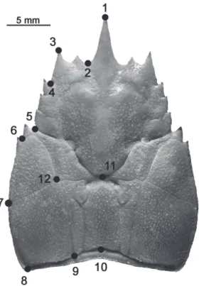
Compared to adults, the anterior and posterior (divided by the landmarks L6-L12) regions of the cephalothorax of smaller individuals were less defined. Juveniles had a particularly short and blunt rostrum (L1), and the forehead region (L1-L3) was more robust than in adults. Additionally, cephalothorax width (L7) was narrower in juveniles. Larger individuals presented well-defined anterior and posterior cephalothorax regions. The Fig. 1. Location of the 12 landmarks (LM) left dorsal half cephalothorax
of Aegla uruguayana Schmitt, 1942.
Tab. I. Description and location of landmarks (LM) in Aegla uruguayana
Schmitt, 1942.
Number Location
1LM Tip of the rostrum 2LM Orbital sine
3LM Tip of the anterolateral spine
4LM Union between the first and posterior end of the anterolateral lobe 5LM Union between the third hepatic lobe and the epibranchial 6LM Union between the epibranchial and the lateralis linea aeglica
7LM Union between the branchial line and the posterior of the linea aeglica lateralis 8LM Posterior vertices of the cephalothorax
9LM Posterior extreme of the longitudinal dorsal line 10LM Centre-posterior extremes of the cephalothorax 11LM Centre-anterior extremes of the areola 12LM Anterior extremes of the bar line
rostrum was longer and stylised (L1) and the forehead (L1-L3) was narrower in adults than in juveniles (Fig. 3). Regarding sexual dimorphism (SD), differences in shape were observed between the cephalothorax of males and females (MANCOVA: Wilks’ λ = 0.49, FGL1 = 2.14, P
= 0.02). The first Relative Warp (RW1) explained 21.45 % of the variation in shape and the second Relative Warp (RW2) explained 14.08 %. However, the allometry was not statistically significant between the sexes, explaining 2.78 % of the variation (P = 0.0562) (Fig.4). Furthermore, the variation in cephalothorax size between the males and females was not statistically significant (W = 549.0, P = 0.51). Males possessed a greater maximum width (L7), the longest rostrum (L1) and a narrower front (L3). In general,
the rostrum front (L1-L3) was more robust in males (Fig. 5). Growth rate in relationship to size and sex. Individual growth rates decreased as size increased, shifting with an increase in CS. Furthermore, smaller individuals exhibited greater variability in growth (Fig. 6). While the mean growth rate in adults was significantly lower than that of juveniles (W = 770.0, P = 0.04), the difference in growth rate of males and females was not statistically significant (W = 131.0, P = 0.48).
Intermoult period. For all groups, intermoult time increased with size (Fig. 7). Beginning in the postmoult stage, juveniles of 1.11 ± 0.39 CS passed an average of 32.00 ± 14.45 days between ecdyses at 25 ± 1°C, whereas adults, with a centroid size of 2.45 ± 0.56, had an average intermoult time of 52 ± 14.05 days. The mean intermoult
Fig. 3. Deformation grids adult (left black), juvenile (right black) and consensus configuration (grey) of Aegla uruguayana Schmitt, 1942 (scale factor 35).
Fig. 4. Spatial variations in the shape of the cephalothorax explained by relative warp (RW) 1 and 2 for males (up gray triangle) and female (down black triangle) of Aegla uruguayana Schmitt, 1942.
Fig. 5. Deformation grids males (left black), and females (right black) and consensus configuration (grey) of Aegla uruguayana Schmitt, 1942 (scale factor 0.05).
Iheringia, Série Zoologia, Porto Alegre, 105(1):76-83, 31 de março de 2015
values were 46 ± 3.56 days for males and 56 ± 17.44 days for females (average size (CS) 2.49 ± 0.62 and 2.38 ± 0.49, respectively). The difference in intermoult time for juveniles and adults was statistically significant (W = 38.5, P = 0.0004). However, the variation between the sexes was not statistically significant (W = 13, P = 0.52).
DISCUSSION
We observed that during ontogeny the cephalothorax undergoes changes in size and shape. The changes cephalothorax shape are related to the different stages of development (juveniles and adults) and to sexual dimorphism in adults.Sexual dimorphism manifests though variations cephalothorax shape but not size.
The present study observed variations in the size and shape of individuals through multiple moults; this design permitted an original perspective and the recognition of different aspects of growth than are commonly reported. Our growth study during the molt and intermolt periods differ from traditional methods both in controlled conditions and the natural environment (renzulli & collins, 2000;
steVens, 2012; gonçAlVeset al., 2009; treVisAn& sAntos,
2011), which have allowed an integral identification of growth.
The relationship between the one-dimensional measurement, as size represented by the centroid size and the entire shape of the cephalothorax, represented by landmarks, allows to study growth as an integral approximation. In this context, we could identify the degree of the shape change during growth in different regions of the cephalothorax. These differences could reflect internal growth (e.g., gonad and muscle) or hierarchy and agonistic behaviour in the population (e.g different dimensions in cephalothorax, armament and chelae) (Williner & collins,
2000; colPoet al., 2005; giri & collins, 2004; ViAuet al.,
2006; Ayres-Pereset al., 2011). Furthermore, differences
in the growth of shape across several species could provide information regarding group evolution or interaction with the environment (hArtnoll, 1982; collinset al., 2007).
Differences in the size and shape of the rostrum and the posterior area of the aeglid cephalothorax were observed between juveniles and adults. These observations are consistent with the location in which the puberty moult occurs. teodósio & mAsunAri (2009) observed changes in
the size and shape of the rostrum of A. schmitti Hobbs III, 1979. Working with juveniles, these authors found that larger individuals had proportionally longer rostrums. Therefore, according to the authors, variation in body proportion is related to the ontogenetic development of the species, which is consistent with variations observed in this analysis. bond-bucKuP & bucKuP (1994) describe variations in the
anterior region of the cephalothorax (pre-cervical width/ forehead width). In our analysis, allometric differences were observed throughout the cephalothorax when juvenile and adult data were analysed through geometric morphometric methods. Regarding sexual dimorphism, differences in shape were observed in the rostrum and more clearly in the posterior region of the cephalothorax. mArtin &
Abele (1988) characterised the aeglid anterior region as
narrow, and posterior region as wide. These features are associated with reproduction because these decapods have large eggs with direct development and the females keep early juveniles in the abdomen (bond-bucKuPet al., 1996;
bueno & bond-bucKuP, 1996). giri & collins (2004)
observed differences in cephalothorax shape between the sexes in some populations of A. uruguayana. Similar to this study, the authors reported that this distinction is most obvious at the posterior vertex of the cephalothorax. Sexual dimorphism was observed throughout the entire cephalothorax in other species, specifically that the posterior lateral region is wider in females than in males (lóPez,
1965; loPretto, 1978; bond-bucKuP & bucKuP, 1994;
JArA, 1994; bond-bucKuPet al., 2008; giri & loy, 2008;
treVisAnet al., 2012; treVisAn & sAntos, 2012). These
findings will allow us to identify the moment of transition between juveniles and reproductive adults in future studies.
Regarding the relative size of males and females, similar values were recorded for A. leptodactyla (noro &
bucKuP, 2003) and A. marginata Bond-Buckup & Buckup,
1994 (treVisAnet al., 2012). However, in the biometric
analysis of A. uruguayana individuals, VAz-ferreirAet
al. (1945) observed that males were wider and longer than females in the area of the junction between third hepatic lobe and the epibranchial area (landmark 5 in this study). However, variations in this region were not evident in this study. Other authors (schmitt, 1942; ringuelet,
1948; bAhemonde & lóPez, 1961; rodrigues & hebling,
1978; JArA, 1980; bond-bucKuP& BucKuP, 1994; sWiech
-Ayoub & mAsunAri, 2001; giri & collins, 2004; boss
et al., 2006; silVA-cAstiglioniet al., 2006; giri & loy,
2008; gonçAlVeset al., 2009; bArríAet al., 2011) have
Fig. 7. Relationship between intermoult time (days) and size (CS) of
agreed that males are larger than females. According to silVA-cAstiglioniet al. (2006), the larger size of males is
most likely because they invest their energy primarily in somatic growth. Females are smaller than males because they invest most of their energy in reproduction (gonad maturation and egg production) at the expense of body growth. Corroborating the findings of the present study, F. Giri (unpublished data) did not find differences in the sizes of males and females of A. uruguayana but observed larger sizes in female A. platensis and A.scamosa Ringuelet, 1948. buenoet al. (2000) recorded larger A. platensis
females than males. The authors attributed this difference to the fact that the largest males of the population were rare during sampling. The results obtained here may indicate that male and female of A. uruguayana present differences in cephalothorax shape but not size.
Regarding the growth rates found for males and females, there was similarity and consistency with observations of other crustaceans (e.g., crab A. leptodactyla; prawn Macrobrachium borellii Nobili, 1896 and crayfish Parastacus pugnax Poepping, 1835) (collins, 1996; noro
& bucKuP, 2003; ibArrA & ArAnA, 2011). However, the
growth rate for females was slightly higher in other species, such as A. paulensis (cohenet al., 2011). Conversely, in
other species, including A. platensis, A. jarai, A. longirostri, and A. itacolomiensis, growth was more intense in males than in females (buenoet al., 2000; bosset al., 2006;
silVA-cAstiglioniet al., 2006; gonçAlVeset al., 2009).
All of these observations were obtained using a classical methodology, and it is possible that morphometric geometry may cause us to consider growth as a more integral process and not as a one-dimensional event. Furthermore, this information may allow new interpretations of the groups’ phylogeny or the effects of environmental forces upon each species.
Individual tracing allowed us to determine the intermoult period and its variability as a component of growth. The similarity in the intermoult periods observed in males and females was consistent with the previous reports in the prawn M. borellii (collins, 1996). By
contrast, Palaemonetes argentines Nobili, 1901 females had longer intermoult periods than males (schuldt &
dAmboreneA, 1989), which is similar to other decapod
species (hArtnoll, 1982). The differences in intermoult
period between sexes were not observed for A. uruguayana in this study, but this may be related to the study seasonality, or to environmental conditions (e.g., temperature, salinity, and food availability) and the need for gonad maturation for the production of a new generation, as occurs in other species (sArdá, 1983; collins, 1996; renzulli & collins,
2000; VegA-VillAsAnteet al., 2006). According to KurAtA
(1962) and hArtnoll (1982), feeding is one of the most
influential factors of growth. Similarly, VegA-VillAsAnteet
al. (2006) describe the relationship between the duration of the moulting cycle and environmental factors in the habitat.
Furthermore, in A. uruguayana, as in other crustaceans (hArtnoll, 1982), growth was slowed as animal
size increased. Therefore, as hArtnoll (1985) explains,
growth in Anomura can be considered undetermined, i.e., the animal undergoes continuous ecdysis after puberty, but it does not have unlimited growth. In other decapod’s taxa, ibArrA & ArAnA (2011) observed that the growth
rate for burrowing crayfish Parastacus pugnax decreased linearly as the individuals grew and reached zero at their maximum length. Similar results were obtained for other crustaceans (collins, 1996; VegA-VillAsAnteet al., 2006),
in which the size increase was substantial for juveniles and decreased linearly with age.
Finally, we consider the techniques and procedures used in this study, which allowed separate analysis of morphological aspects of growth, such as the shape and size, enabling a thorough study of the growth process and broadening the scope of traditional methods. Additionally, the methodology of the analysis (with the exuvia of cephalothorax being removed during moulting) reduced handling of the individuals, which decreased the likelihood of inducing stress and allowed the individual to be released after the study was completed. This was a key factor for our analysis. Thus, this new approach, in combination with traditional methods offers an integral approach to the study of decapod growth.
Acknowledgments. This work was supported by the grants of the Project PICT “Diversidad biológica en ambientes dulceacuícolas a través del gradiente este-oeste de argentina: rotíferos, microcrustáceos y macrocrustáceos como grupos de estudio” (grant to PAC, ANPCyT Process #2007-01360).
REFERENCES
AdAms, d. c.; rohlf, f. J. & slice, d. e. 2004. Geometric Morphometrics:
Ten Years of Progress Following the ‘Revolution’. Italian Journal of Zoology 71:5-16.
Almerão, m.; bond-bucKuP, g. & mendonçA. Jr m. de s. 2009. Mating
behavior of Aegla platensis (Crustacea, Anomura, Aeglidae) under laboratory conditions. Journal of Ethology 28:87-94.
Ayres-Peres, l.; ArAuJo, P. b. & sAntos, s. 2011. Description of the agonistic behavior of Aegla longirostri (Decapoda: Aeglidae). Journal of Crustacean Biology 31(3):379-388.
bAhAmonde, n. & lóPez, m. t. 1961. Estudios biológicos en la población de Aegla laevis laevis (Latreille) de el Monte (Crustacea, Decapoda,
Anomura). Investigaciones Zoológicas Chilenas 7:19-58.
bArríA, e. m; sePúlVedA, r. d. & JArA, c. g. 2011. Morphologic variation in Aegla Leach(Decapoda: Reptantia: Aeglidae) from central-southern Chile: interespecific differences, sexual dimorphism, and spatial segregation. Journal of Crustacean Biology 31(2):231-239.
bond-bucKuP, g. 2003. Família Aeglidae. In: melo, G. A. S. ed. Manual
de identificação dos Crustacea Decapoda de água doce do Brasil. São Paulo, Edições Loyola, p. 21-116.
bond-bucKuP, g. & bucKuP, l. 1994. A familia Aeglidae (Crustacea, Decapoda, Anomura). Arquivos de Zoologia 32(4):159-346.
bond-bucKuP, g.; bueno, A. & KeunecKe, K. 1996. Primeiro estágio juvenil de Aegla prado Schmitt (Crustacea, Decapoda, Anomura, Aeglidae). Revista Brasileira de Zoologia 13:1049-1061.
bond-bucKuP, g.; JArA, c. g.; Pérez-losAdA, m.; bucKuP, l. & crAndAll,
K. A. 2008. Global diversity of crabs (Aeglidae: Anomura: Decapoda)
in freshwater. Hydrobiologia 595(1):267-273.
bond-bucKuP, g.; JArA, c. g; bucKuP, l.; Pérez-losAdA, m.; crAndAll,
Iheringia, Série Zoologia, Porto Alegre, 105(1):76-83, 31 de março de 2015 booKstein, f. l. 1991. Morphometric tools for landmark data.
Geometry and biology. New York, Cambridge University Press. 435p.
boss, h. J.; silVA-cAstiglioni, d.; schAcht, K.; bucKuP, l. & bond
-bucKuP, g. 2006. Crescimento de Aegla jarai Bond-Buckup & Buckup (Crustacea, Anomura, Aeglidae). Revista Brasileira de Zoologia 23(2):490-496.
bueno, A. A. P. & bond-bucKuP, g. 1996. Os estágios juvenis iniciais de Aegla violacea Bond-Buckup & Buckup (Crustacea, Anomura, Aeglidae). Nauplius 4:39-47.
bueno, A. A. P.; bond-bucKuP, g. & bucKuP, l. 2000. Crescimento de
Aegla platensis Schmitt em ambiente natural (Crustacea, Decapoda, Aeglidae). Revista Brasileira de Zoologia 17(1):51-60.
cohen, f. P. A.; tAKAno, b. f.; shimizu, r. m. & bueno, s. l. s. 2011. Life Cycle and Population Structure of Aegla paulensis (Decapoda: Anomura: Aeglidae). Journal of Crustacean Biology 31(3):389-395.
collins, P. A. 1996. Ablación unilateral en el camarón de agua dulce
Macrobrachium borellii. In:Acuicultura en Latinoamérica. IX Congreso latinoamericano de acuicultura. 2° Simposio Avances y Perspectivas de la Acuicultura en Chile, p. 131-135.
collins, P. A. & PetriellA, A. 1996. Crecimiento y supervivencia del camarón Macrobrachium borellii (Decapoda: Palaemonidae) alimentado con dietas artificiales. Neotropica 42(107-108):3-8.
collins, P. A.; Williner, V. & giri, f. 2007. Littoral Communities. Macrocrustaceans. In:iriondo, m. h.; PAggi, J. c. & PArmA, m. J. eds. The Middle Paraná River: Limnology of a subtropical Wetland. Heidelberg, Springer-Verlag, p. 277-301.
colPo, K. d; oliVeirA, l. r & sAntos, s. 2005. Population biology of the freshwater anomuran Aegla longirostri (Crustacea, Anomura, Aeglidae) from Ibicuí-Mirim River, Itaára, RS, Brazil. Journal of Crustacean Biology 25:495-499
drAch, P. 1939. Mueet cycle dʼintermue chez les crustacés décapodes. Annales de lʼInstitut Oceanographique 19:103-391.
dryden, i. l. & mArdiA, K. V. 1998. Statistical Shape Analysis. NewYork, John Wiley & Sons. 347p.
giri, f. & collins, P. A. 2004. A geometric morphometrics analysis of two sympatric species of the family Aeglidae (Crustacea, Decapoda, Anomura) from the La Plata basin. The Italian Journal of Zoology 71:85-88.
giri, f. & loy, A. 2008. Size and shape variation of two freshwater crabs in argentine an Patagonia: The influence of sexual dimorphism, habitat, and species interactions. Journal of Crustacean Biology 28:37-45.
gonçAlVes, r.; bond-bucKuP, g. & bucKuP, l. 2009. Crescimento de
Aegla itacolomiensis (Crustacea, Decapoda) em um arroio da Mata
Atlântica no sul do Brasil. Iheringia, Série Zoologia 99(4):397-402.
hArtnoll, r. g. 1978. The Determination of Relative Growth in Crustacea. Crustaceana 34(3):281-293.
_____. 1982. Growth,. In: Abele, l. g. ed. The biology of Crustacea. New York, Academic Press, vol. 2, p. 111-196.
_____. 1985. Growth, sexual maturity and reproductive output. In: Wenner, A. m. ed. Factors in Adult Growth 3, Crustacean Issues. Rotterdam, A. A. Balkema, p. 101-128.
hobbs, h. h iii. 1979. A new species of the endemic South American genus Aegla from Paraná, Brasil (Crustacea, Anomura, Aeglidae). Proceedings of the Biological Society of Washington91:982-988.
ibArrA, m. & ArAnA, P. m. 2011. Crecimiento del camarón excavador
Parastacus pugnax (Poeppig, 1835) determinado mediante técnica de marcaje. Latin American Journal of Aquatic Research 39 (2):378-384.
JArA, c. g. 1980. Dos nuevas especies de Aegla Leach (Crustacea, Decapoda, Anomura) del sistema hidrográfico del río Valdivia. Anales del Museo de Historia Natural de Valparaíso 13:255-266.
_____. 1994. Aegla pewenchae,a new species of central Chilean freshwater decapods (Crustacea: Anomura: Aeglidae). Proceedings of the Biological Society of Washington107:325-339.
Klingenberg, c. P. 1998. Heterochrony and allometry: the analysis of evolutionary change in ontogeny. Biological Review73:79-123.
_______. 2011. MorphoJ: an integrated software package for geometric
morphometrics. Molecular Ecology Resources 11:353-357.
Klingenberg, c. P.; bArluengA, m. & meyer, A. 2002. Shape analysis of symmetric structures: quantifying variation among individuals
and asymmetry. Evolution 56:1909-1920.
KurAtA, h. 1962. Studies on the age and growth of Crustacea. Bulletin of the Hokkaido Regional Fisheries Research Laboratory 24:1-115.
lóPez, m. t. 1965. Estudios biológicos en Aegla odebrechtii paulensis Schmitt (Crustacea, Decapoda, Anomura). Boletim de Zoologia da Facultade de Filosofia, Ciências e Letras287(25):301-314.
loPretto, e. c. 1978. Estructura exoesqueletaria y miológica del quinto par de pereiópodos del macho de la familia Aeglidae (Crustacea, Anomura). Limnobios 1:284-298.
luPPi, t. A.; sPiVAK, e. d.; bAs, c. c. & Anger, K. 2004. Molt and
growth of an estuarine crab, Chasmagnathus granulates (Brachyura: Varunidae), in Mar Chiquita coastal lagoon, Argentina. Journal of Applied Ichthyology 20:333-344.
mArtin, J. W. & Abele, l. g. 1986. Phylogenetic relationships of the genus Aegla (Decapoda: Anomura: Aeglidae), with comments on anomuran phylogeny. Journal of Crustacean Biology 6:575-616.
_____. 1988. External morphology of the genus Aegla (Crustacea: Anomura: Aeglidae). Smithsonian Contributions to Zoology 453:1-46.
noro, c. K. & bucKuP, l. 2003. O crescimento de Aegla leptodactyla
Buckup & Rossi (Crustacea, Anomura, Aeglidae). Revista Brasileira de Zoologia 20(2):191-198.
Pérez-losAdA, m.; bond-bucKuP, g.; JArA, c. g. & crAndAll, K. A.
2004. Molecular systematics and biogeography of the southern South American fresh-water “crabs” Aegla (Decapoda: Anomura: Aeglidae) using multiple heuristic tree search approaches. Systematic Biology 53:767-780.
_____. 2009. Conservation assessment of southern South American freshwater ecoregions on the basis of the distribution and genetic diversity of crabs from the Genus Aegla. Conservation Biology 23:692-702.
PetriellA, A. m. & boschi, e. e. 1997. Crecimiento en crustáceos decápodos: resultados de investigaciones realizadas en Argentina. Investigaciones Marinas 25:135-157.
r deVeloPment coreteAm 2008. R: A Language and Environment for Statistical Computing. Vienna, R Foundation for Statistical Computing. Available at <http://www.R-project.org>. Accessed on May, 2104.
renzulli, P. & collins, P. 2000. Influencia de la temperatura en el crecimiento del cangrejo Trichodactylus borellianus. Fabicib 4 :129-136.
ringuelet, r. A. 1948. Los “cangrejos” argentinos del género Aegla de Cuyo y la Patagonia. Revista del Museo de La Plata (N. S.), Zoología 5(34):297-347.
rodrigues, W. & hebling, n. J. 1978. Estudios biológicos en Aegla perobae Hebling & Rodrigues, 1977 (Decapoda, Anomura). Revista Brasileira de Biologia 38(2):383-390.
rohlf, f. J. 2004. tpsDig, Digitize Landmarks and Outlines, Version 1.40. Stony Brook, Department of Ecology and Evolution, State University of New York.
rohlf, f. J. & mArcus, l. f. 1993. A revolution in morphometrics. Trends Ecology and Evolution8:129-132.
rufino, M. M. 2006. Geographic and gender shape differences in the carapace of Liocarcinus depurator (Brachyura: Portunidae) using geometric morphometrics and the influence of a digitized method. Journal of Zoology 269:458-465.
rufino, M. M.; Abelló, P. & yule, A. B. 2004. The effect of alcohol and freezing preservation on carapace size and shape in Liocarcinus depurator (Crustacea, Brachyura). In: eleWA, A. m. t. ed. Morphometrics–applications in biology and paleontology. Heidelberg, Springer Verlag, p. 45-53.
sAntos, s; bond-bucKuP, g.; bucKuP, l.; loureiro, t. l.; gonçAlVes,
A. s.; Verdi, A.; scArAbino, f. & clAViJo, c. 2014. The aeglidae of
Uruguay (Decapoda, Anomura), with the description of a new species of Aegla. Crustaceana Monographs19:195-205.
sAntos, s; bond-bucKuP, g.; Pérez-losAdA, m.; JArA, c. g; crAndAll,
K. A. & bucKuP, l. 2010. New records and description of a new species of Aeglidae (Crustacea, Anomura) from river basins in Southern Brazil. Nauplius 18:79-86.
sAntos, s; JArA, c. g.; bArtholomei-sAntos, m. l.; Pérez-losAdA, m. &
Leach, 1820 (Crustacea, Anomura, Aeglidae) from the West-Central region of Rio Grande do Sul, Brazil. Nauplius 21(2):211-223.
sArdá, f. 1983. Determinación de los estados de intermuda en Nephrops norvegicus (L), mediante la observación de los pleópodos. Investigación Pesquera 47(1):95-112.
schmitt, W. l. 1942. The species of Aegla, endemic South American fresh-water crustaceans. Proceedings of the United States National Museum91(3132):431-520.
schuldt, m. & dAmboreneA, m. c. 1989. Infección de Palaemonetes
argentinus (Crustacea, Palaemonida) con Probopyrus cf. oviformes (Crustacea, Bopyridae) en el canal Villa Elisa (Selva Marginal de Punta Lara, provincia de Buenos Aires, Argentina). Estructura poblacional del consorcio, interacción y fluctuación. Biota 5:21-53.
silVA-cAstiglioni, c.; bArcelos, d. f. & sAntos, s. 2006. Crescimento de Aegla longirostri Bond-Buckup & Buckup (Crustacea, Anomura, Aeglidae). Revista Brasileira de Zoologia 23(2):408-413.
steVens, b. g. 2012. Growth of juvenile red king crabs, Paralithodes
camtschaticus, through sequential molts in the laboratory. Journal of Crustacean Biology 32(2):215-22.
sWiech-Ayoub, b. P. & mAsunAri, s. 2001. Flutuaçao temporal e especial de abundância e composição de comprimento da carapaça de Aegla castro Schmitt, 1942 (Crustacea, Decapoda, Aeglidae) no Buraco do Padre, Ponta Grossa, Paraná, Brasil. Revista Brasileira Zoologia 18(3):1003-1017.
teodósio, e. A. o. & mAsunAri, s. 2009. Estrutura populacional de
Aegla schmitti (Crustacea: Anomura: Aeglidae) nos reservatórios dos Mananciais da Serra, Piraquara, Paraná, Brasil. Zoologia 26(1):19-24.
treVisAn, A. & sAntos, s. 2011. Crescimento de Aegla manuinflata (Decapoda, Anomura, Aeglidae) em ambiente natural. Iheringia, Série Zoologia 101(4):336-342.
_____. 2012. Morphological sexual maturity, sexual dimorphism and
heterochely in Aegla manuinflata (Anomura). Journal of Crustacean Biology 32(4):519-527.
treVisAn, A.; mArochi, m. z.; costA, m.; sAntos, s. & mAsunAri, s. 2012. Sexual dimorphism in Aegla marginata (Decapoda: Anomura). Nauplius 20(1):75-86.
VAz-ferreirA, r.; grAy, r. & VAz-ferreirA, m. 1945. Notas biométricas sobre los crustáceos decápodos del género Aegla Leach. I. La variación de algunas magnitudes en Aegla uruguayana Schmitt. Comunicaciones Zoológicas del Museo de Historia Natural de Montevideo1(24):1-6.
VegA-VillAsAnte, f.; cortés-JAcinto, e.; gArcíA-guerrero, m. & cuPul -mAgAñA, A. 2006. Contribución al estudio de la muda y crecimiento de Callinectes arcuatusen Baja California Sur (México), como base
para la producción de Jaiba Suave. Comunicación Científica - CIVA 2006: 514-521.
ViAu, V. e.; lóPez-greco, l. s.; bond-bucKuP, g. & rodríguez, e. m.
2006. Size at the onset of sexual maturity in the anomuran crab, Aegla uruguayana (Aeglidae). Acta Zoologica 87:253-264.
Wenner, A. m. 1985. Crustacean Issues: Factors in Adult Growth 3. Rotterdam, A. A. Balkema. 362p.
Williner, V. & collins, P. 2000. ¿Existe jerarquización en las poblaciones de Palemónidos del valle aluvial del Río Paraná?. Natura Neotropicalis31(1-2):53-60.
Received 24 October 2014. Accepted 30 March 2015 ISSN 0073-4721 Article available in www.scielo.br/isz
Appendix 1. Error measurement of the photos and landmarks in Aegla uruguayana Schmitt, 1942.
Centroid size
SS MS df F P (param.)
Effect
Individual 12560824.14 1046735.34 12 4073.72 <0.0001
Error1 3340.32 256.95 13 10.05 <0.0001
Residual 664.97 25.57 26
Shape
SS MS df F P (param.)
Effect
Individual 0.02009 0.00009 228 3.77 <0.0001
Side 0.00061 0.00003 19 1.38 0.1361
Ind* Side 0.00532 0.00002 228 1.03 0.4002
Error1 0.01123 0.00002 494 3.97 <0.0001