Exploring the
Fasciola hepatica
tegument proteome
R. Alan Wilson
a,⇑, Janelle M. Wright
b, William de Castro-Borges
a,c, Sophie J. Parker-Manuel
a,
Adam A. Dowle
d, Peter D. Ashton
a, Neil D. Young
e, Robin B. Gasser
e, Terry W. Spithill
b,f aCentre for Immunology and Infection, Department of Biology, University of York, Heslington, York YO10 5DD, United KingdombSchool of Animal and Veterinary Sciences, Charles Sturt University, Wagga Wagga, NSW 2650, Australia cDepartamento de Ciências Biológicas, Universidade Federal de Ouro Preto, CEP – 35400-000 Ouro Preto, MG, Brazil d
Centre of Excellence in Mass Spectrometry, Technology Facility, Department of Biology, University of York, Heslington, York YO10 5DD, United Kingdom e
Department of Veterinary Science, The University of Melbourne, Parkville, Victoria 3052, Australia f
Department of Agricultural Sciences and Centre for AgriBioscience, La Trobe University, Bundoora, Victoria 3083, Australia
a r t i c l e
i n f o
Article history: Received 15 June 2011
Received in revised form 29 August 2011 Accepted 30 August 2011
Available online 5 October 2011
Keywords: Fasciola hepatica Liver fluke Tegument proteome Excretory–secretory proteins Tandem mass spectrometry Morphology
Vomitus
a b s t r a c t
The surface tegument of the liver flukeFasciola hepaticais a syncytial cytoplasmic layer bounded exter-nally by a plasma membrane and covered by a glycocalyx, which constitutes the interface between the parasite and its ruminant host. The tegument’s interaction with the immune system during the fluke’s protracted migration from the gut lumen through the peritoneal cavity and liver parenchyma to the lumen of the bile duct, plays a key role in the fluke’s establishment or elimination. However, little is known about proteins of the tegument surface or its secretions. We applied techniques developed for the blood fluke,Schistosoma mansoni, to enrich a tegument surface membrane preparation and analyse its composition by tandem mass spectrometry using new transcript databases for F. hepatica. We increased the membrane and secretory pathway components of the final preparation to30%, whilst eliminating contaminating proteases. We identified a series of proteins or transcripts shared with the schistosome tegument including annexins, a tetraspanin, carbonic anhydrase and an orthologue of a host protein (CD59) that inhibits complement fixation. Unique toF. hepatica, we also found proteins with lec-tin, cubulin and von Willebrand factor domains plus 10 proteins with leader sequences or transmem-brane helices. Many of these surface proteins are potential vaccine candidates. We were hampered in collecting tegument secretions by the propensity of liver flukes, unlike blood flukes, to vomit their gut contents. We analysed both the ‘vomitus’ and a second supernatant released from haematin-depleted flukes. We identified many proteases, some novel, as well as a second protein with a von Willebrand fac-tor domain. This study demonstrates that components of the tegumental surface ofF. hepaticacan be defined using proteomic approaches, but also indicates the need to prevent vomiting if tegument secre-tions are to be characterised.
Crown CopyrightÓ2011 Published by Elsevier Ltd. on behalf of Australian Society for Parasitology Inc. All rights reserved.
1. Introduction
Fasciolosis is a major zoonotic disease caused by the liver flukes
Fasciola hepaticaandFasciola gigantica, which are flatworm para-sites transmitted following ingestion of herbage contaminated with the infective stage (metacercariae). The infection is a signifi-cant constraint on ruminant productivity in Europe, Africa, Asia, the Americas and Australasia, with prevalences in some regions of 80–100%, >600 million animals at risk, and annual economic losses of >US$3 billion (Piedrafita et al., 2004, 2007). It is recogni-sed by the World Health Organization (WHO) as a major food-borne problem, with up to 17 million people infected and
180 million at risk. High but localised prevalence (72–100%) has been recorded in Bolivia (>1 million cases), Peru, Africa and the Middle East (Mas-Coma et al., 2005; McManus and Dalton, 2006) with up to 830,000 people infected in Egypt alone (Mas-Coma et al., 2005). Triclabendazole is the drug of choice for treatment of fasciolosis but resistance, first observed in Australia in 1995, is now widespread in Europe (Fairweather, 2009). In endemic areas such as Bolivia and Egypt, resistance threatens to make the disease untreatable and new methods of control are urgently needed.
The production of an effective vaccine may be a sustainable control strategy. However, although there has been a considerable effort to develop such a vaccine, current experimental approaches suffer from two limitations: efficacy is variable between animals and the level achieved to date (38–72%) falls short of the >80% protection generally agreed as necessary for a commercially viable
0020-7519/$36.00 Crown CopyrightÓ2011 Published by Elsevier Ltd. on behalf of Australian Society for Parasitology Inc. All rights reserved. doi:10.1016/j.ijpara.2011.08.003
⇑ Corresponding author. Tel.: +44 1904 328600; fax: +44 1904 328505. E-mail address:[email protected](R.A. Wilson).
Contents lists available atSciVerse ScienceDirect
International Journal for Parasitology
product for cattle (Hillyer, 2005; McManus and Dalton, 2006). Two candidate vaccines (leucine amino peptidase and SAP2) have shown an efficacy of >80% in single animal trials but require vali-dation in cattle (Piacenza et al., 1999; Espino and Hillyer, 2004; Acosta et al., 2008). The development of anti-Fasciola vaccines has been hindered by lack of insights into natural acquired im-mune mechanisms expressed by ruminant hosts against fluke infection and knowledge of immune correlates of protection. A bet-ter understanding of the targets of acquired immunity in cattle is required if we are to devise a commercial vaccine for large ruminants.
There is good evidence from several studies that livestock can acquire resistance toFasciola. Vaccination using either irradiated metacercariae, drug abbreviated infection, parasite extracts or de-fined antigens can induce 48–89% reductions in fluke burdens in ruminants (Dalton et al., 1996; Morrison et al., 1996; Piacenza et al., 1999; Hoyle et al., 2003; Piedrafita et al., 2004; Hillyer, 2005; McManus and Dalton, 2006; Golden et al., 2010). These re-sults demonstrate that Fasciolaantigens can elicit high levels of immunity in cattle and sheep, suggesting that the goal of >80% pro-tection is achievable. The key step now is to identify the parasite stage and antigens driving the acquired protective response. In cat-tle or sheep where protection has been demonstrated following vaccination or due to natural resistance, clinical serology has re-vealed that killing of parasites occurs within approximately 6 weeks of infection but only after some damage to the liver paren-chyma (Dalton et al., 1996; Roberts et al., 1997; Piacenza et al., 1999; Hoyle et al., 2003; Piedrafita et al., 2004). These observations suggest that the newly excysted juvenile (NEJ) or immature para-site migrating in the liver, not the adult fluke, is the target of the acquired immune response but the nature of this effector response acting in vivo is not clear. Moreover, a vaccine targeting the NEJ, which suppressed or eliminated invasion of the liver parenchyma, would minimise liver pathology and reduce production losses in livestock. Using in vitro studies, it has been shown thatF. hepatica
NEJs are susceptible to antibody-dependent cell cytotoxicity (ADCC) mediated by nitric oxide (NO) released by rat peritoneal macrophages (Piedrafita et al., 2001, 2004). An ADCC immune mechanism effective againstF. giganticaNEJs in vitro has also been reported in sheep and in this case killing was mediated by super-oxide radicals (not NO) produced by macrophages (Piedrafita et al., 2007). Although an ADCC mechanism effective against liver flukes has not been demonstrated in vivo, these results suggest the possibility that antigens on the surface tegument of NEJs/ immature flukes, recognised by immune sera, may represent tar-gets of the proposed ADCC mechanism and that these antigens rep-resent novel vaccine candidates. Although the immune mechanisms that kill Fasciola in cattle have not been resolved, we have proposed that they may involve an ADCC mechanism sim-ilar to that observed againstF. giganticain sheep (Piedrafita et al., 2004).
The tegument of flukes is a surface syncytial layer covering the parasite, rich in secretory inclusions and bounded externally by a plasma membrane bearing a dense glycocalyx. Important tegu-mental functions include renewal of the surface plasma membrane and the active uptake of nutrients (Dalton et al., 2004). The com-bined plasma membrane and its glycocalyx can potentially interact directly with the immune system but few studies have analysed tegument surface proteins. Surface radiolabelling was used to identify seven proteins from 10 to 78 kDa in F. hepatica NEJs, revealing that the profile changed during the first 7–14 days of infection (Lammas et al., 1985). A second labelling study demon-strated variations in surface composition of proteins from 14 to >200 kDa, between NEJ, immature and adult flukes (Dalton and Joyce, 1987) and a protein on the juvenile fluke tegument showing a repetitive sequence has been identified (Trudgett et al., 2000).
Lastly, a detergent extract ofF. hepaticategument proteins with undefined composition was shown to suppress the maturation and function of murine bone marrow-derived dendritic cells ( Ham-ilton et al., 2009). Clearly, further definition of the proteins on the tegument surface of the fluke is needed to inform vaccine development.
In contrast, the tegument proteins of the related flukes Schisto-soma mansoni(van Balkom et al., 2005; Braschi et al., 2006; Braschi and Wilson, 2006),Schistosoma japonicum(Mulvenna et al., 2010a) andOpisthorchis viverrini(Mulvenna et al., 2010b) have recently been subjected to proteomic analysis. The studies have ranged from the simple compositional analysis of material sloughed off the fluke surface by freeze/thaw/vortexing (van Balkom et al., 2005) to more sophisticated protocols that enrich for tegument membrane proteins using differential extraction (Braschi et al., 2006). Proteins accessible to impermeant probes on live schisto-somes have been tagged with biotin to facilitate their recovery and identification (Braschi and Wilson, 2006; Mulvenna et al., 2010a). Finally, live schistosomes have also been subjected to a complementary approach of enzymatic shaving that releases ex-posed proteins into the medium for recovery and identification (Castro-Borges et al., 2011). Such studies have provided a wealth of information about the surface organisation of the blood dwelling schistosomes and it is notable that two surface-exposed tegument proteins of S. mansoni have shown efficacy as vaccines in the mouse model of schistosomiasis (Tran et al., 2006; Cardoso et al., 2008). Here, as the first step towards a molecular definition of the tegument surface, the parasite-host interface, the better to understand the interaction ofF. hepaticawith the immune system, we report the analysis of tegument proteins isolated from the adult fluke.
2. Materials and methods
2.1. Biological material
Adult flukes were obtained from the bile ducts of cattle at York Abattoir (Anglo Beef Processors Ltd., United Kingdom), situated approximately 1 mile from the University of York laboratory. They were collected into ice-cold RPMI-1640 medium (Invitrogen, Pais-ley, Scotland) immediately after culling, separated from contami-nating blood, mucus and bile by washing extensively in medium, and given a pre-incubation at 37°C for 30 min prior to tegument
isolation.
2.2. RNA isolation and cDNA synthesis for 454 sequencing
Live flukes in RPMI-1640 medium were homogenised in TRIzol (3 ml per worm) (Invitrogen) and 1 ml aliquots stored at 80°C.
Total RNA was extracted by addition of 200
l
l of chloroform perml of TRIzol, mixed by inversion for 15 s, incubated at 20°C (room
temperature; RT) for 3 min, and then centrifuged at 12,000gfor 5 min at 4°C to separate the phases. The aqueous layer was
re-moved, 250
l
l each of isopropanol and high salt solution (0.8 Mso-dium citrate, 1.2 M NaCl) were added to precipitate RNA at 80°C
overnight. The sample was centrifuged at 12,000g for 30 min at 4°C and the resulting pellet washed twice with 70% ethanol, the
supernatant carefully removed and the pellet air dried at RT. The RNA was resuspended in 300
l
l of diethyl pyrocarbonate(DEPC)-treated water, quantified using a Nanodrop spectrophotometer (Invitrogen) and re-precipitated with 30
l
l of 3 M sodium acetate,pH 7.5, 990
l
l absolute ethanol. Total RNA (1l
l sample containing50–500 ng/
l
l) was quality-assessed using a Bioanalyser RNA 6000NanoChip (Agilent, Wokingham, Berks). Polyadenylated RNA was purified from a pool of total RNA derived from three flukes using
a PolyA purist kit (Ambion, Huntingdon, UK) according to the man-ufacturer’s instructions. Briefly, total RNA was incubated with poly (T) cellulose at 70°C for 5 min and then 1 h at RT. The cellulose
pel-let was washed five times and the poly(A) RNA eluted with 400
l
lof warm (70°C) 1 mM sodium citrate, before precipitation with 0.1
vol. of 5 M ammonium actetate, 1
l
l Glycoblue (15 mg/ml;Ambi-on), 2.5 vol. absolute ethanol, at 80°C, overnight.
Double-stranded cDNA was made using a SMART PCR cDNA synthesis kit, with PrimeScript reverse transcriptase and an Advan-tage 2 PCR kit (all from Clontech, Mountain View, CA, USA) to en-rich for full length transcripts. A 1
l
g sample of polyA RNA wasadded per 10
l
l reaction and the first strand synthesis allowed toproceed for 1 h at 37°C. A 2
l
l aliquot from this reaction wassub-jected to long-range PCR for 13 cycles, according to the manufac-turer’s instructions. The quality of a 5
l
l aliquot of the resultingcDNA from the 100
l
l reaction mixture was assessed on a 1.5%aga-rose gel before submission to the Genomics Laboratory within the York Technology Facility, for 454 sequencing on the Genome Se-quencer FLX platform (Roche, Branford, CT, USA).
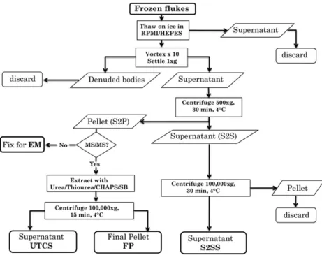
2.3. Tegument preparation by freeze/thaw/vortexing
The method for isolation of the tegument surface membranes of
F. hepatica(Fig. 1) was derived from that used for the blood flukeS. mansoni (Braschi et al., 2006), which in turn was based on the freeze/thaw/vortex (FTV) protocol developed earlier (Roberts et al., 1983). Briefly, 20 adult flukes were snap-frozen in liquid N2, then slowly thawed to 4°C in 5 ml of ice cold RPMI-1640
med-ium plus protease inhibitors (Protease inhibitor cocktail, Sigma, Poole, Dorset, UK). The tegument was detached from fluke bodies by 101 s pulses on a vortex mixer and the supernatant recovered by allowing the bodies to settle at 4°C. The membranes (S2P) were
pelleted from this supernatant by centrifugation at 500gfor 30 min at 4°C, leaving a supernatant (S2S) that was recentrifuged at
100,000g, 30 min, 4°C to produce the cytosolic fraction (S2SS).
The residual S2P pellet was washed three times in 40 mM tris(hydroxymethyl)aminomethane (Tris), pH 7.4 at 4°C, with
intervening centrifugations, to remove soluble contaminants. It was then extracted with 100
l
l 5 M urea (BDH, VWR International,Dorset, UK), 2 M thiourea (BDH) in 40 mM Tris, 4% 3-[(3-Cholami-dopropyl)dimethylammonio]-1-propanesulfonate (CHAPS; Sigma), 2% caprylyl sulfobetaine (SB 3–10; Sigma) to recover membrane-associated proteins (UTCS extract), recentrifuged at 100,000g, 30 min, 4°C and the final pellet (FP) solubilised with 50
l
l of0.1% SDS, 1% Triton X-100 in 100 mM triethylammonium bicarbon-ate (TEAB) at RT. A protein assay was performed on S2SS using Coo-massie Plus Bradford reagent (Thermo Lifescience, Basingstoke, UK). UTCS and FP samples were assayed by separation on a one dimensional (1D) SDS NuPAGE 4–12% gradient gel (Invitrogen) with 5 and 10
l
g S2SS as a comparator, stained with Sypro Rubyovernight, imaged with a Molecular FX imager and total protein content estimated by densitometry analysis using Quantity One software (all BioRad, Hemel Hempstead, UK).
2.4. Adult secreted proteins
Unlike adultS. mansoni, which are reluctant to vomit in vitro, adultF. hepaticareadily regurgitate their gut contents. A batch of newly collected flukes (washed as described in Section 2.1) was pre-incubated at 37°C for 30 min in a large culture dish to
encour-age vomiting and diminish contamination from gut contents. This provided a sample of vomitus for MS/MS analysis. The flukes were then washed three times in RPMI-1640 with a minimum of phys-ical handling before transfer to 5 cm diameter plastic culture dishes for short-term culture; only the paler flukes that had visibly emptied the pigment from their guts were used for this incubation. Groups of six flukes were incubated at 37°C for 1 h to provide total
secretions, the experiment being performed three times to provide biological replicates. The supernatant was removed and concen-trated at 4°C using a 5,000 mol. wt cut-off centrifugation device
(Vivaspin, Vivascience, Generon, Maidenhead, UK or Amicon, Milli-pore, Watford, UK).
2.5. Electron microscopy
Immediately prior to UTCS extraction, the detached tegument (S2P) sample was evaluated by transmission electron microscopy (TEM) to determine the nature of the material being subjected to
MS/MS analysis. The pellet was fixed in 2.5% glutaraldehyde/ 4% formaldehyde in 0.1 M phosphate buffer, pH 7.2, at 4°C overnight.
Flukes from the secretion experiment were similarly fixed, cut into 1–2 mm cubes after 30 min in fixative and then post-fixed in 1% aqueous osmium tetroxide at 4°C overnight. All samples were
washed three times in 0.1 M phosphate buffer, pH 7.2, at 4°C, then
post-fixed in 1% aqueous osmium tetroxide at RT for 1.5 h. After three washes in distilled water, specimens were dehydrated in a graded series of acetones and embedded in Spurr resin. Thin sec-tions (80–100 nm) were cut on an Ultracut UCT (Leica, Milton Key-nes, UK) and collected on 400-mesh hexagonal copper grids, then stained with 1% uranyl acetate in 50% ethanol and lead citrate (Reynolds, 1963). Sections were imaged on a Tecnai G2 BioTWIN operating at 120 kV (FEI, Hillsboro, Oregon).
2.6. Sample preparation and liquid chromatography
Aliquots containing 50
l
g of S2SS, 20l
g of UTCS and 20l
g S2Pprotein, respectively, were subjected to in-solution digestion. Briefly, each protein aliquot was diluted in 100
l
l of 0.4 M TEABand denatured in the presence of 0.1% SDS. Cysteine residues were reduced by adding 1 mM tris-(2-carboxyethyl) phosphine (TCEP) during incubation at 65°C for 30 min. Alkylation was then
per-formed by addition of 10 mM methyl methane-thiosulfonate (MMTS) for 1 h in the dark at RT. Trypsin (Sequencing Grade, Pro-mega, Southhampton, UK) was then added at a 1:20 (enzyme:sub-strate) ratio and the final volume of the in-solution digestion adjusted to 200
l
l with 0.4 M TEAB. Digestion was allowed topro-ceed overnight at 37°C. The digested samples were centrifuged at
10,000gfor 3 min, the supernatant concentrated using a SpeedVac (Thermo Scientific, Basingstoke, UK) and redissolved in 500
l
l ofloading solution (10 mM KH2PO4 in 25% acetonitrile (ACN), pH
3.0). Two clean-up steps were used. For the first step, to remove SDS and excess reducing and alkylating reagents, the sample was loaded onto a cation exchange cartridge-system (P/N4326747, Ap-plied Biosystems, Framingham, USA), and eluted with loading solu-tion plus 350 mM KCl (pH 3.0) according to the manufacturer’s instructions. For the second step, to remove salts, the sample was concentrated to dryness using the SpeedVac, resuspended in 1.0 ml 0.1% trifluoroacetic acid (TFA), and loaded onto a C18 col-umn (Strata C18-E, 55
l
m, 70 Å, Phenomenex, Macclesfield, UK).Bound peptides were eluted twice in 250
l
l of 50% ACN/0.1%TFA, concentrated to dryness and resuspended in 20
l
l 0.1% TFA.A 3
l
l aliquot was loaded onto an UltiMate LC system (Dionex,Camberley, UK) equipped with a polystyrene–divinylbenzene (PS-DVB) monolithic column (200
l
m internal diameter5 cm). Peptides were eluted over a linear gradient of 3–51% (v/v) ACN at a flow rate of 3l
l/min, with 0.1% heptafluorobutyric acid as thecounter ion throughout, monitoring UV absorbance at 214 nm. A Probot microfraction collector (Dionex) was used to collect 6-s fractions onto a prespotted anchor chip containing 4-hydroxy-
a
-cyano-cinnamic acid (Bruker Daltonics, Bremen, Germany). Samples of vomitus and secretion supernatants were subjected to in-solution digestion and then processed for LC–MS/MS, exactly as described above for the tegument fractions.
2.7. Tandem MS and database searching
Positive-ion MALDI-TOF-MS spectra were obtained using an Ultraflex III (Bruker Daltonics, Coventry, UK) in reflectron mode, equipped with a Nd:YAG smart beam laser. MS spectra were ac-quired over a mass range ofm/z720–4000 and calibrated exter-nally against an adjacent prespotted anchor chip containing nine mass standards (Bruker Daltonics). Monoisotopic masses were ob-tained using a SNAP averaging algorithm (C 4.9384, N 1.3577, O 1.4773, S 0.0417, H 7.7583) and a S/N threshold of 3.
For each spot, the 10 strongest peaks, with a S/N ratio >15, were selected for MS/MS fragmentation. Where similar peaks of less than 100 ppm (ppm) mass difference were observed, within six fractions, only the most intense were fragmented. Peaks present in over 70% of the fractions were considered to be background and were not selected for fragmentation. Fragmentation was per-formed in LIFT mode without the introduction of a collision gas. The default calibration was used for MS/MS spectra, which were baseline-subtracted and smoothed (Savitsky-Golay, width 0.15m/ z, cycles 4); monoisotopic peak detection used the SNAP averaging algorithm with a minimum S/N threshold of 1. Bruker Flex Analysis software was used to perform the spectral processing and peak list generation for both the MS and MS/MS spectra.
Data were searched using Mascot software (version 2.1, Matrix Science, London, UK) through the Bruker BioTools interface (ver-sion 3.2). The significance threshold of the Mascot output was ad-justed to provide an approximately 1% false discovery rate by searching of a Mascot-generated decoy database, and results fil-tered with an expect value <0.05. Databases searched were: (i) Na-tional Center for Biological Information non-redundant (NCBInr) [http://www.ncbi.nlm.nih.gov/protein]; (ii) FhA, compiled from expressed sequence tags (ESTs) deposited on the Wellcome Trust Sanger Institute (WTSI) ftp site [ftp://ftp.sanger.ac.uk/pub/patho-gens/Fasciola]; (iii) FhB, an in-house database compiled from 150,000 reads from the 454 sequencer; (iv) FhC, the new tran-script database established recently (Young et al., 2010). FhB was annotated automatically against Uniprot, NCBInr andS. mansoni
gene predictions (www.GeneDB.org), followed by manual inspec-tion to select the most credible annotainspec-tion. FhA was annotated automatically against theS. mansonigene predictions and FhC by its originators. The FhB database is available on request from R.A. Wilson ([email protected]).
The identity ofF. hepaticaproteins revealed by Mascot search-ing was first determined ussearch-ing the FhB database. The list was then augmented by adding new identities revealed by a search using FhA, host proteins and full-lengthF. hepatica cDNAs at NCBInr, and finally using the FhC database when this became available. The exponentially modified protein abundance index (emPAI; Ishi-hama et al., 2005), generated by Mascot for each protein identity, was used as an approximate guide to its abundance in all samples examined. The emPAI is based on the number of peptides actually observed, relative to the number observable per protein. All F. hepatica-specific proteins identified by MS/MS that lacked homol-ogy were subjected to a conserved domain (CDD) search for do-main structure (NCBI). They were also interrogated using SignalP (http://www.cbs.dtu.dk/services/SignalP/) to detect leader se-quences, and HMMTOP (http://www.enzim.hu/hmmtop/) for membrane spanning regions, enabling some unknown proteins to be assigned to secreted and/or membrane categories.
3. Results
3.1. Analysis of transcript data
The 353 adult F. hepatica cDNA sequences deposited in the NCBInr database at the end of 2009, with much redundancy, do not form an adequate database to undertake tegument proteomics. The additional single-pass ESTs from adult flukes available at the Wellcome Trust Sanger Institute (WTSI) ftp site improved coverage of the transcriptome, compiling into 1,064 contigs and 3,009 sing-lets (termed here the FhA database). Further to this we obtained 3,923 contigs (FhB database) from150,000 reads generated by 454 sequencing of adult fluke cDNA. We later added the 14,424 adult fluke FhC contigs and a larger number of singlets, thus achieving a total in excess of 20,000 assembled contigs for Mascot
searching of MS/MS spectra. Inevitably there is overlap in content between the different sequence sources, but collectively they should provide a good representation of the adult transcriptome. From previous work on the proteome ofS. mansoniwe compiled a list of known tegument proteins in that parasite, which com-prised seven membrane enzymes, 15 membrane-spanning trans-porters, nine transport-associated ATPases, five surface defence proteins, 14 proteins from the exocytosis pathway, eight mem-brane-associated GTPases, 13 membrane structural proteins, and five surface proteins with no homology (Braschi et al., 2006; Cas-tro-Borges et al., 2011, and our unpublished data). The three Fh databases were then interrogated using this list to find any F. hepaticaorthologues (Table 1). Remarkably, many proteins previ-ously identified in theS. mansonitegument membranes were rep-resented by orthologues encoding sequences in the F. hepatica
transcriptome. All seven of the membrane enzymes and 15 trans-porters were detected, together with all nine associated ATPases. Three of five surface defence proteins, 13 of the 13 exo/endo cyto-sis pathway proteins, all eight of theS. mansoniGTPases and 10 of the 13 membrane structural proteins were found. Additionally, two trematode-specific tegument proteins of unknown function, Sm200 and Low Molecular Weight Protein (LMWP), were repre-sented in theF. hepaticatranscriptome.
3.2. Morphology of the tegument pellet
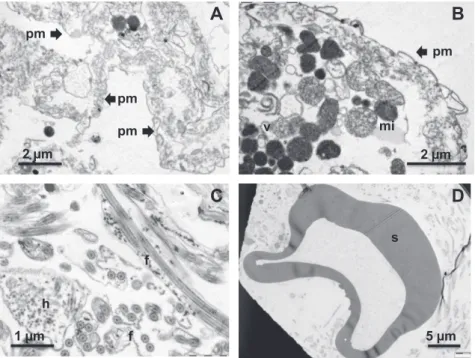
To facilitate interpretation of tegument protein composition, a tegument pellet was subjected to TEM, prior to the UTCS extraction step. It should be noted that the material had been subjected to freeze/thaw/vortexing before processing for electron microscopy so that considerable distortion of structure was inevitable. Most areas of the pellet contained sheets of membrane that convincingly approximated in appearance to detached tegument surface (Fig. 2A and B). However, small membranous vesicles (Fig. 2A) were also present, as well as more densely stained spherical inclusions (Fig. 2B). We interpret the more granular of these as mitochondrial remains while the darker inclusions probably represent secretory vesicles. The pellet was not homogeneous in composition through-out, aggregates of freeF. hepaticasperm being apparent in at least one area (Fig. 2C). These can be clearly identified by the long dou-ble flagellum of the tail in both longitudinal and cross-section, as well as the microtubule-demarcated head region. A singleF. hepat-icaegg was also located in one section, clearly identifiable by the tanned protein egg shell, but lacking internal features due to plas-molysis during processing (Fig. 2D).
3.3. Proteomic analysis of S2SS, UTCS and FP fractions
The scheme of sample processing after tegument detachment by FTV was intended to deplete the sample, first of cytosolic com-ponents and then of proteins more strongly associated with the plasma membrane, leaving a final pellet enriched in tightly-bound tegument surface proteins. A total of 229 proteins was identified from the various fractions, distributed 100, 125 and 88 between the S2SS, UTCS and FP fractions, respectively (Supplementary Ta-bles S1–S6). A measure of the effectiveness of the differential extraction protocol is provided by the number of unique identities in each fraction (58, 45 and 38, respectively), representing 57%, 35% and 44% of each sub-total. As further evidence for the success of the partitioning protocol, 19 proteins (8%) were shared between all fractions, whilst only five were present in the first S2SS and last FP fractions but not in the intermediate UTCS fraction. The smallest number of identities in the FP results from the prior removal of the bulk of proteins by the extraction process, and this is reflected in the emPAI totals for each: S2SS, 66.5; UTCS, 48.2; FP, 26.9
(repre-senting 47%, 34% and 19% of the notional total, respectively; Sup-plementary Tables S2–S6).
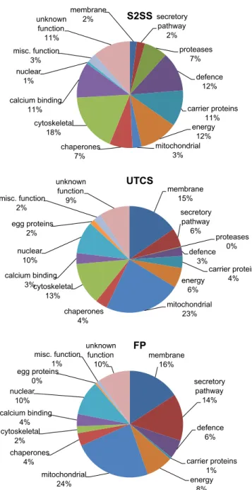
A functional annotation was provided for all but 38 constitu-ents, of which four (termed Fasciola-specific, membrane helix) could be assigned to the membrane category and six (termed Fas-ciola-specific, secreted) to the secretory pathway on the basis of the CDD/SignalP/HMMTOP searches (Table 2), leaving 28 unas-signed proteins that wereF. hepatica or trematode-specific ( Sup-plementary Tables S1–S6). The differing compositions of the three fractions was also evident when classified by function (Fig. 3;Table 2; Supplementary Table S1) and plotted by emPAI score to take account of approximate abundance. The major con-stituents of the soluble (cytosolic) S2SS fraction, were in descend-ing percentage order: cytoskeleton (18), defence (12), energy metabolism e.g. glycolytic enzymes (12), unknown function (11), carrier proteins (11), calcium-binding (11), chaperones (7), prote-ases and inhibitors (7), mitochondria (3), miscellaneous (3), mem-brane (2), secretory pathway (2) and nuclear (1). UTCS extraction of the washed (i.e. insoluble) membrane pellet increased represen-tation of membrane and secreted proteins to 15% and 6%, respec-tively, but mitochondrial proteins also increased to 23% and nuclear proteins to 10% (Fig. 3;Supplementary Table S1). Propor-tionally, other categories such as defence, energy, calcium binding, protease and carrier had all decreased, although cytoskeleton re-mained high at 13%. This presumably reflects the anchoring of many cytoskeletal proteins to membranes and organelles.
Similar trends were also seen in the composition of the FP frac-tion with mitochondrial, membrane, secretory pathway and nucle-ar proteins representing 24%, 14%, 16% and 10%, respectively (Fig. 3; Supplementary Table S1). The cytoskeletal and calcium-binding proteins as well as chaperones now each represented 2– 4% of the FP, whilst carrier proteins and proteases were virtually undetectable although one ferritin sequence was identified ( Sup-plementary Table S1). Note that histone H4 is represented by a small sequence fragment, not a full coding sequence to which the peptide hits gave almost full coverage, distorting the emPAI (6.17). As histones are in equimolar ratios in the histone core of the nucleus (Chung et al., 1978), the values for H4 were therefore replaced by the mean of the emPAIs for all other histones (0.32). Overall, the fractionation protocol succeeded in enriching for membrane and secretory pathway proteins (minus proteases and inhibitors), their proportions increasing from 2 to 15 to 16% and 2 to 6 to 14%, respectively, from S2SS to UTCS to FP. In turn the sol-uble glycolytic enzymes (energy category) were reduced from 12 to 6 to 8% and gut proteases from 7 to 0 to 0%, respectively. The mitochondrial proteins (3%, 23% and 24%) were also highly en-riched constituents in the FP.
The specific constituents of each functional category provide indicators to biological processes occurring in the tegument. Here we focus on the membrane and secreted proteins likely to be un-iquely involved in tegument function. We note only that the vast majority of proteins identified, particularly in the S2SS fraction, are a cross section of the fluke cytosol and cytoskeleton, and details are given inSupplementary Tables S1–S6. The same is true of the mitochondrial and nuclear proteins that were enriched by the FTV method and subsequent extraction steps; details of identities obtained can be found inSupplementary Tables S1–S6.
3.3.1. Membrane-associated category
Table 1
Putative orthologues ofSchistosoma mansonitegument proteins identified in theFasciola hepaticatranscriptome.
Fh Database Protein identity Smp Fh Database Protein identity Smp
A B C Orthologue A B C Orthologue
Enzymes Exo- and endo-cytosis pathway
Acetylcholinesterase Smp_136690 Plasmolipin/ lipid raft-associated protein Smp_046290
Alkaline phosphatase Smp_155890 Endophilin, BAR domain Smp_003230
ATP-diphosphohydrolase 1 Smp_042020 Endophilin, BAR domain Smp_163720
Ectonucleotide pyrophosphatase/phosphodiesterase Smp_153390 Clathrin heavy chain Smp_154240
Carbonic anhydrase Smp_168730 Sarco/endoplasmic reticulum-type Ca-2+-ATPase Smp_007260
Calpain Smp_157500 Lysosome-associated membrane glycoprotein Smp_073400
ADP ribosyl cyclase (SARC) Smp_025830 ATPase, H+ transporting, vacuolar V0 subunit a Smp_040970
Transporters Secretory carrier membrane protein Smp_127650
Aquaporin-3 Smp_005720 Phospholipid scramblase 1 Smp_008860
Aquaporin Smp_128210 Valosin, transitional endoplasmic reticulum ATPase Smp_018240
High-affinity copper uptake protein Smp_048230 RTN4-N, Reticulon Smp_020370
Multidrug resistance protein 1, 2, 3 Smp_170820 H+ Vacuolar ATP synthase subunit ac39 Smp_079950
Glucose transport protein GTP1 Smp_012440 H+ Vacuolar ATP synthase catalytic subunit A Smp_147050
Sugar transporter, not GTP1 or GTP4 Smp_171870 Membrane-associated GTPases
Cationic amino acid transporter Smp_176940 Rab 1 GTP-binding protein (GTPase) Smp_169460
Amino acid transporter SPRMhc Smp_037540 Rab-27B GTP-binding Smp_139340
Amino acid transporter Smp_147070 Rac GTPase Smp_062300
Voltage-dep anion-selective channel Smp_022990 Rho GTPase Cdc42 Smp_167030
Voltage-dep anion-selective channel Smp_091240 Rho2 GTPase Smp_072140
Voltage-gated potassium channel Smp_151810 Rho GEF Smp_126600
Sodium/chloride dependent transporter Smp_028690 Rap-1b, Ras-related protein Smp_071250
Sodium/chloride-dependent transporter Smp_131890 Rap-1b, Ras-related protein Smp_142450
Sodium/chloride-dependent transporter Smp_143800 Membrane structure
Membrane transport associated ATPases Dysferlin Smp_141010
Na+/K+ ATPase beta subunit/1 Smp_015020 Otoferlin Smp_163750
Na+/K+ ATPase beta subunit/2 Smp_143800 Tetraspanin, (Sm-TSP-2) Smp_181530
Na+/K+ ATPase alpha subunit Smp_033550 Tetraspanin Smp_140000
Phospholipid transporting ATPase Smp_192080 Tetraspanin Smp_017430
Cation/phospholipid transporting ATPase (flippase) Smp_091650 Annexin Smp_045560
Cation-transporting ATPase type 13A3 Smp_175360 Annexin Smp_077720
Plasma membrane calcium-transporting atpase Smp_176130 Annexin Smp_045500
Plasma membrane calcium-transporting atpase Smp_137170 Annexin Smp_045550
ATPase, aminophospholipid transporter Smp_104500 Annexin Smp_074140
Defence Trematode-specific
CD59 Smp_019350 Sm200 GPI-anchored surface glycoprotein Smp_017730
CD59 Smp_105220 Low molecular weight protein, cfClonorchis Smp_194860
TCIP integrin homologue Smp_194920
1352
R.A.
Wilson
et
al.
/International
Journal
for
Parasitology
41
(2011)
them anion-selective channels and the third a glucose transporter, were detected plus three membrane enzymes, a carbonic anhy-drase and two calpain proteases. A CD59 orthologue was identified, which in humans is an inhibitor of complement fixation. Although no receptors were found, a single GTPase and five other small GTPase-associated proteins all point to the occurrence of cell sig-nalling processes at the tegument surface. FourF. hepatica-specific proteins, all in the FP, were predicted by HMMTOP to encode 1–3
membrane-spanning regions so were assigned to the membrane category (Table 2).
3.3.2. Secretory protein category
A total of 21 proteins was assigned to the secreted/secretory pathway, exclusive of putative gut proteases and a Kunitz-type protease inhibitor, which were present in the S2SS fraction and are treated separately below. Nineteen of the 21 proteins were
A
D
C
B
s
f
f
h
pm
pm
pm
pm
mi
v
5 µm
1 µm
2 µm
2 µm
Fig. 2.Transmission electron micrographs of the S2P pellet prior to UTCS extraction, to determine the morphology of the material being analysed. (A) Large and continuous sheets of membrane (pm) that represent the tegument surface. (B) A membrane sheet (pm), but also mitochondria (mi) and secretory vesicles (v). (C) Section of the pellet containing sperm tail flagella (f) and heads (h). (D) A single egg, represented by its tanned protein shell (s).
Table 2
Membrane, membrane-associated and secreted proteins identified in cytosolic (S2SS), pellet extract (UTCS) and final pellet (FP) fractions of tegument.
Database Protein ID emPAI emPAI emPAI Database Protein ID emPAI emPAI emPAI
S2SS UTCS FP S2SS UTCS FP
Membrane associated FhA00116 C-type lectin domain, secreted C 0.17
FhB01398 Annexin a 0.21 FhC02107 Galectin domain 0.07 0.07
FhB02550 Annexin b 1.11 0.93 FhC02219 CUB (cubulin) domain 0.31
FhB00592 Annexin c 0.20 2.86 1.34 FhC06044 CUB (cubulin) domain 0.14
FhB00705 Tetraspanin 0.28 0.30 FhC06620 CUB (cubulin) domain 0.15
FhC14169 Otoferlin A 0.25 FhA03239 LMWP cf.Clonorchis 0.1
FhC14498 Dysferlin 0.19 0.28 FhB00114 SmKK7-like 0.77
c48411 Otoferlin B 0.37 FhC00742 von Willebrand factor A domain 0.36
FhB03799 Voltage-dep anion-selective channel 0.12 0.12 FhC05840 Vacuolar protein ATPase 0.06
FhC06452 Calcium-activated chloride channel 0.14 FhB01127 Endophilin B1 0.92 0.48 0.51
FhC00666 Glucose transporter 0.09 FhB00788 Translocon-associated protein delta 0.28
FhB00925 Carbonic anhydrase 0.29 0.14 FhB02015 Ribophorin I, oligosaccharyltransferase 0.17
FhB00712 Calpain A 0.24 FhC00286 Oligosaccharyl-transferase 0.09
FhB01987 Calpain B 0.19 FhB00004 Fasciola-specific, secreted 0.87
FhA04073 CD59 orthologue 0.22 FhC04266 Fasciola-specific, secreted 0.45
FhA03206 Rho2 GTPase, membrane signalling 0.24 FhB02167 Fasciola-specific, secreted 0.18 0.41
FhC06056 Rho GTP-binding protein 0.22 FhB01808 Fasciola-specific, secreted 0.25 0.59
FhC02604 Rab5 0.2 FhC00442 Fasciola-specific, secreted 0.37
FhC01017 Rab-2,4,14, 0.25 FhC05339 Fasciola-specific, secreted 0.23
FhC05214 Rab 11B 0.08 Proteases and inhibitors
FhC00074 Rab GDP-dissociation inhibitor 0.09 FhC02704 Kunitz-type proteinase inhibitor 2.39
FhB00010 Fasciolaspecific, membrane helix 0.12 0.12 FhB00269 Cathepsin L (Clade 1) 0.14
FhB01238 Fasciolaspecific, membrane helix 0.17 FhB01160 Cathepsin L (Clade 2) 0.17
FhB01787 Fasciolaspecific, membrane helix 0.45 FhB03790 Cathepsin L (unclassified) 1.27
FhC00749 Fasciolaspecific, membrane helix 0.11 FhB01159 Leucyl aminopeptidase 0.51
Secretory pathway FhB02190 Cathepsin A 0.07
FhB00578 C-type lectin domain, secreted A 0.16 FhB01026 DJ-1 protease (park-7) 0.26
FhB01221 C-type lectin domain, secreted B 0.09 0.09
present in the UTCS fraction or the FP. Notable members were four lectins, three with a C-type and one with a galectin domain. In addition three distinct proteins with a cubulin (CUB) domain were identified. A protein containing both anti-alpha trypsin inhibitor and von Willebrand factor type A domains was detected together with two putativeF. hepaticaorthologues of secreted proteins in other trematodes, the proposed potassium channel blocker SmKK7 and the LMWP ofClonorchis. Five proteins associated with the pro-tein export pathway were identified, comprising a vacuolar ATP-ase, an endophilin and three components of the endoplasmic reticulum/Golgi apparatus (two glycosyltransferases and a translo-con-associated protein delta). Finally, sixF. hepatica-specific pro-teins encoded a signal peptide but no transmembrane regions, so were assigned to the secretory pathway.
The presence of known gut proteases in the S2SS and UTCS but not the FP fraction indicates some contamination of the tegument
preparation (Table 2). Cathepsin L isoforms and the Kunitz-type protease inhibitor were particularly prominent, the latter being by far the most abundant constituent of the S2SS fraction. Mito-chondrial proteins were strongly represented in the UTCS fraction but sufficient diagnostic proteins remained after chaotropic/deter-gent treatment of the tegument preparation for them to constitute the major component of the FP fraction (emPAI total of 6.54; Sup-plementary Table S1). Motor proteins such as actins, tubulins and dyneins were abundant in the S2SS and UTCS fractions but virtu-ally absent from FP, so very likely extracted by the treatment; in addition to being tegument constituents, these proteins may also derive from the sperm tails visible in the pellet subjected to TEM (Fig. 2;Supplementary Table S1). The nuclear proteins, represented by histones, and ribosomal proteins involved in protein synthesis were not, in contrast, extracted by UTCS treatment and therefore remained in the FP, presumably originating in the sperm heads de-tected by electron microscopy (Fig. 2).
3.4. Morphology of flukes after in vitro incubation
We fixed flukes ex vivo before culture and at the end of the in vitro culture period, for ultrastructural examination. A clear dis-tinction was visible between the two samples. Numerous type 2 disc-shaped bodies were present in the tegument cytoplasm of the ex-vivo flukes, the surface plasma membrane was intact and the external glycocalyx was visible (Fig. 4A). An oblique section through the surface of an in vitro fluke again revealed the type 2 vesicles and an apparently intact surface, but aggregations of small ‘‘vesicles’’ could be seen in some of the surface pits (Fig. 4B). We interpret these vesicles as evidence for the leakage of cytoplasm into the pits, indicating some damage to the plasma membrane. Whilst not extensive, such leakage would have an impact on the composition of tegument secretions detected by proteomic analysis.
3.5. Proteomic analysis of secretion samples
Initial observations described above revealed that the adult flukes readily produced abundant vomitus from the gut, containing large quantities of protein (mostly cathepsins) that effectively swamped any possible contribution from the tegument secretions to the culture medium. Therefore, we attempted to ‘‘wash-out’’ the
membrane 2% secretory pathway 2% proteases 7% defence 12% carrier proteins 11% energy 12% mitochondrial 3% chaperones 7% cytoskeletal 18% calcium binding 11% nuclear 1% misc. function 3% unknown function 11%
S2SS
membrane 15% secretory pathway 6% proteases 0% defence 3% carrier proteins 4% energy 6% mitochondrial 23% chaperones 4% cytoskeletal 13% calcium binding 3% nuclear 10% egg proteins 2% misc. function 2% unknown function 9%UTCS
membrane 16% secretory pathway 14% defence 6% carrier proteins 1% energy 8% mitochondrial 24% chaperones 4% cytoskeletal 2% calcium binding 4% nuclear 10% egg proteins 0% misc. function 1% unknown function 10%FP
Fig. 3.Pie chart showing the percentage distribution of proteins identified in the cytosolic fraction (S2SS) (A), extract supernatant (UTCS) (B) and final pellet (FP) (C) preparations on the basis of the exponentially modified Protein Abundance Index (emPAI), classified by biological function.
A
B
mv t2b t2b gc p p t2b mv 200nm 200nmFig. 4.Transmission electron micrographs of the tegument surface of flukes before (A), and after (B) incubation in RPMI-1640 medium to collect tegument secretions. In the pre-incubation fluke the tegument appears normal with surface pits (p), type 2 secretory vesicles (t2b) in the cytoplasm and a surface glycocalyx (gc). Post-incubation (oblique section), the type 2 secretory vesicles (t2b) are still abundant but the surface pits (p) now contain microvesicles (mv) that may represent leakage of proteins from the cytosol.
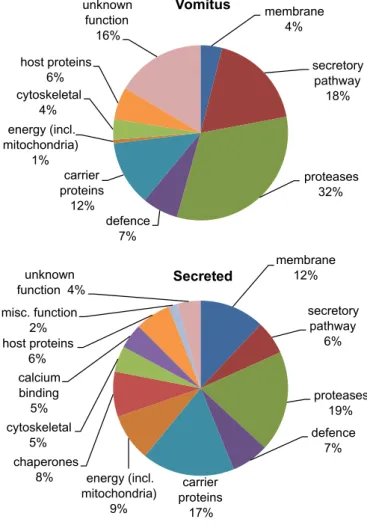
gut contents before collecting the secretions. After recovery from bovine livers, transport to the laboratory and extensive washing, a 30-min pre-incubation step was incorporated in the protocol to facilitate expulsion of the gut contents. This released vomitus was analysed by MS/MS to provide a baseline reference against which to detect potential tegument proteins (Table 3, Supplemen-tary Tables S2–S6). A total of 34 proteins was identified in the vom-itus, the largest group being proteases (32% of the total emPAI score of 13.52; Fig. 5). These were predominantly isoforms of cathepsin L but asparaginyl endopeptidase, prolidase, leucine ami-no peptidase, dipeptidyl-peptidase and cathepsin B were also iden-tified, all apparently of lysosomal origin. The second most abundant group (18%) comprising other secretory proteins, pre-dominantly the Kunitz-type protease inhibitor with one of the highest emPAI scores (2.37) in the entire study, and a lysosomal phospholipase B. Unknown function and carrier proteins were ranked third (16%) and fourth (12%), one of the former category (FhB03836) being especially abundant. The carrier category com-prised two isoforms of the cholesterol transporter NPC-2, and three variants of Fasciola myoglobin (one annotated as haemoglobin). The vomitus also contained four proteins of host origin (6%), the
alpha and beta chains of haemoglobin being the most abundant, but the heavy chain of immunoglobulin A and serum albumin were also identified. The vomitus was notable for the scarcity of fluke cytosolic (<1%; aldolase) and cytoskeletal proteins (4%; calponin, fimbrin, tubulin) and the absence of chaperones, revealing that up to 2 h ex-host, in RPMI-1640 medium, the parasites had in-curred minimal cell damage leading to protein leakage. This obser-vation may indicate that the presence of the defence protein thioredoxin peroxidase (peroxiredoxin) in the vomitus, also noted byRobinson et al. (2009)represents a true release of into the gut lumen, not leakage due to damage. It is also possible that the phos-pholipase A and D detected in the vomitus are of lysosomal origin (i.e. secretory), although placed here in the membrane-associated category.
Subsequent to the collection of vomitus, selected flukes were incubated for a further hour in a secretion experiment and the re-leased proteins subjected to MS/MS analysis. This extra incubation virtually doubled the number of proteins; two aspects are apparent from their identities (Table 3;Fig. 5). Firstly, the flukes continued to regurgitate vomitus into the medium as evidenced by the detec-tion of certain gut proteases (18.6% of the higher total emPAI score
Table 3
of 20.15;Fig. 5). Secondly, there was a marked increase in cytosolic proteins, particularly chaperones (8%) and those involved in energy metabolism (9%) and defence (9%). Host haemoglobins alpha and beta as well as the IgA heavy chain were also present in the secre-tion sample (7%).
In the membrane category, three annexins, a phospholipase and carbonic anhydrase were found, the last of these being potentially glycosylphosphatidylinositol (GPI)-anchored (cf. S. mansoni, (Castro-Borges et al., 2011)). The detection of a multi-membrane spanning tetraspanin is suggestive of tegument membrane damage. Of particular note in the secretory category was a second isoform of the von Willebrand factor, a serpin, and a nucleoside diphosphate kinase predicted to encode a signal peptide. In the carrier protein category one of the NPC-2 cholesterol transporters was released, and ferritin presumably involved in iron transport. The presence of the cytosolic fatty acid binding protein (Fh15;
Table 3) may be further evidence of cellular damage.
4. Discussion
The pioneering investigations of the trematode tegument in the early 1960s that defined its cellular organisation at the ultrastruc-tural level were undertaken onF. hepatica(Threadgold, 1963, 1967; Bjoerkman and Thorsell, 1964). However, since that time there has been much less fundamental work on isolation of the tegument or analysis of its protein composition compared with schistosomes so there are few pointers in the literature to sample preparation for
proteomics. The principal objective of the present study was to adapt techniques used in recent proteomic analyses of the tegu-ment surface of the blood fluke S. mansoni (van Balkom et al., 2005; Braschi et al., 2006; Braschi and Wilson, 2006; Castro-Borges et al., 2011) to theF. hepaticategument.The recent activity in the sequencing of adultF. hepatica transcripts by ourselves (unpub-lished data) and others (Young et al., 2010) meant that an exten-sive EST database was available to aid identification of peptide mass spectra. By cross-species sequence comparisons we were able to show that many tegument membrane and membrane-associ-ated proteins identified inS. mansoniby proteomics were repre-sented by putative orthologues in one or more of the three F. hepaticatranscript databases available.
The FTV protocol developed for theS. mansonitegument has an advantage in relying on physical methods for the initial isolation and enrichment stages, rather than on extractions involving deter-gents. It also depends on the integrity of the meshwork of sub-tegumental circular and longitudinal muscle layers to retain the organelles of the internal tissues. However, it does not prevent leakage of cytosolic proteins from those internal tissues and these must be removed before membrane analysis (Braschi et al., 2006). While the cellular organisation of theF. hepaticaandS. mansoni
teguments is similar, with the machinery for protein synthesis and export residing in cell bodies located beneath the muscle lay-ers (Threadgold, 1967; Morris and Threadgold, 1968), there are important differences. TheF. hepaticategument is much thicker (15
l
m versus 3–4l
m inS. mansoni), it contains many moremito-chondria, the secretory vesicles are quite distinct in appearance and, instead of the secreted membranocalyx that covers the schis-tosome tegument (Wilson and Barnes, 1977), there is a prominent glycocalyx (Threadgold, 1967). All of these factors are likely to im-pact on the techniques for tegument isolation and on the proteins identified. In fact, interrogation of theF. hepaticatranscriptome for orthologues encoding known S. mansoni tegument proteins re-vealed many common components. In consequence we might anticipate that similar biological processes occur in the tegument of the two flukes. Indeed, the detection of orthologues for Sm200 and LMWP (with no known functions) may indicate processes un-ique to, but widespread, in trematodes. In contrast, the identifica-tion of 38 proteins in the FTV experiment that have no homology outside the Trematoda, 10 found in other flukes and 10 possessing signal peptides and/or transmembrane domains, suggests the occurrence ofFasciola-specific tegumental processes. This observa-tion is important and justifies further detailed analysis of the teg-umental proteome to clarify tegument biological functions.
The tegument preparation released by FTV proved not to be as clean as the one forS. mansonimade by the same method (Roberts et al., 1983). The most obvious difference was the presence of numerous mitochondria and expanded secretory vesicles, but also the observation of sperm and a single egg in the S2P fraction, prior to UTCS extraction. We interpret the presence of sperm and egg to indicate there is continuous activity by the fluke reproductive sys-tem right up to the instant of freezing, rather than their release by rupture of the fluke body wall, since vitelline cells or shell granules were not evident in electron micrographs of the pellet. It should be noted thatF. hepaticahas a much greater reproductive capacity per parasite (up to 50,000 eggs/day;Moxon et al., 2010) thanS. man-soni(200–300 eggs/day;Loker, 1983). Despite this contamination, the extraction scheme that produced a final pellet enriched in membrane proteins can be considered a qualified success. The ini-tial wash step with buffer to yield the S2SS fraction contained lar-gely cytosolic proteins. The extraction of the resultant pellet with a combination of chaotropic agents and non-ionic detergents (UTCS) to remove membrane-associated proteins, produced a FP differing markedly in composition from its predecessors. Indeed, the in-crease in combined membrane and secretory pathway proteins
membrane 4%
secretory pathway
18%
proteases 32%
defence 7% carrier proteins 12% energy (incl. mitochondria)
1% cytoskeletal
4% host proteins
6%
unknown function
16%
Vomitus
membrane 12%
secretory pathway
6%
proteases 19%
defence 7%
carrier proteins 17% energy (incl.
mitochondria) 9% chaperones
8% cytoskeletal
5% calcium binding
5% host proteins
6% misc. function
2% unknown function 4%
Secreted
Fig. 5.Pie chart showing the percentage distribution of proteins identified in Vomitus (A) and Secretion (B) preparations on the basis of the exponentially modified Protein Abundance Index (emPAI), classified by biological function.
from 4% to 21% to 29% in the FP whilst protease and inhibitor con-tamination from the gut reduced from 7% to 0%, is a testament to this difference. Mitochondrial and nuclear fractions were also en-riched in the final pellet, which was only to be expected given the morphological observations. In future studies on theFasciola
tegument it will be important to introduce a step early in the prep-aration procedure, which excludes organelles in order to enrich further the surface membrane contribution to the final pellet.
In the final pellet we identified a number of membrane struc-tural proteins (two annexins, a tetraspanin, three ferlins) that are prominent features of theS. mansonitegument surface. We also identified three transporters (two ion channels and a glucose transporter) and three enzymes (two calpains and carbonic anhy-drase) that the tegument surfaces of two flukes have in common. In contrast we did not find any of the phosphohydrolases that are exposed at theS. mansonitegument surface, although the rele-vant genes are represented in F. hepatica cDNA databases. One GTPase and a series of GTP-binding proteins were extracted by the UTCS treatment. Such proteins are usually associated with plasma membranes as components of signalling pathways but no receptors were detected; proteomic studies on theS. mansoni teg-ument have also failed to identify receptors. Finally, a putative orthologue of human CD59 was identified in the final pellet. This aspect is of considerable interest since human CD59 acts as an inhibitor of the complement pathway, blocking formation of the membrane attack complex after binding of C3 (Huang et al., 2006). At least six CD59-like proteins are encoded in theS. mansoni
genome, two of which are GPI-anchored at the surface of the schis-tosome tegument (Castro-Borges et al., 2011); one of these pro-teins (Smp_019350) is the closest homologue (37% identical, 52% conserved amino acids) of the F. hepatica protein we identified. OurF. hepaticadatabases contain transcripts for at least four genes encoding the CD59-like proteins, all of which possess the diagnos-tic CCxxDxCN sequence near the C-terminus (data not shown), and representatives are also present inS. japonicum andSchistosoma haematobium. Their presence in the tegument surface may point to a mechanism, widespread in trematodes, for defence against im-mune attack that would repay further investigation.
The proteins of the secretory pathway that we identified are also informative about processes occurring in the tegument or at its surface. (The Kunitz-type inhibitor and the proteases of gut ori-gin are dealt with in the discussion of vomitus composition below.) One obvious feature is the identification of four potential glycan-binding lectins. All four genes have orthologues in theS. mansoni
andS. japonicum genomes but no lectins have been reported in the tegument of these two blood flukes. Although theF. hepatica
cDNA sequences may be incomplete, at least two (FhA00116 and FhB01221) encode a signal peptide suggesting their secretion to the exterior. It is plausible that these predicted lectins play a role in the binding of glycoproteins for endocytosis (Kerrigan and Brown, 2009) or that they interact with host leucocytes, potentially to subvert host responses (Vasta, 2009). The group of three pro-teins with little homology in common other than the presence of a CUB domain, is more enigmatic. FhC02219 encodes a signal pep-tide and is likely secreted, and the other two transcripts may lack the 50end. In vertebrates, CUB domains are present in diverse
pro-teins. A conserved domain search on NCBInr reveals that the lon-gest FhC transcripts (Fh06620 and Fh06044) each possess two CUB domains and a heterodimerisation interface. They have the closest homology to CUBN type proteins that function on the brush border of the mammalian gut as components of the receptor that acquires vitamin B12 (Andersen et al., 2010). This suggests that theF. hepaticaCUB proteins may cooperate to bind a protein ligand in the tegument.
Other potential secretory proteins are noteworthy. LMWP, a 8 kDa protein when its putative leader sequence is subtracted,
was first cloned inClonorchis sinensisand is also present in theS. mansonitegument (Castro-Borges et al., 2011). The second is the
F. hepatica orthologue of SmKK7, another small protein with homology to the potassium channel blockers of scorpion venom. This protein was first described from the cercarial secretions ofS. mansoni(Curwen et al., 2006), and has been reported subsequently as released when live adult schistosomes are subjected to mild trypsin treatment (Castro-Borges et al., 2011). Its identification in the S2SS but not the UTCS or FP fractions could be taken as evi-dence that it is a cytoplasmic protein, were it not that the presence of a leader sequence implies export into the endoplasmic reticu-lum. Both proteins appear to be trematode-specific but their pre-cise tegumental location requires confirmation. The final protein in this group (FhC00742), present in the UTCS fraction, contains a von Willebrand factor domain in association with an N-terminal inter-alpha trypsin inhibitor (ITI) domain, and a metal ion-depen-dent adhesion site (MIDAS). In humans the von Willebrand factor is required for normal haemostasis and mediates the adhesion of platelets to sites of vascular damage and exposed connective tis-sue, whereas the ITI domain plays a role in extracellular matrix sta-bilisation (Sadler, 1998). The role of this protein in theF. hepatica
tegument and/or its secretions needs to be investigated.
The experiments in which flukes were incubated in vitro for short periods were intended to provide information about tegu-ment secretions. Unfortunately, these experitegu-ments suffered a sin-gular disadvantage compared with similar studies with S. mansoni. The blood fluke is very reluctant to open its mouth and regurgitate gut contents when placed in vitro so the live flukes can be exposed to biotinylation reagents (Braschi and Wilson, 2006) or tegument shaving enzymes (Castro-Borges et al., 2011) without the presence of competing proteins or large amounts of proteases.Fasciola hepatica, on the other hand, readily regurgitates a continuous stream of vomitus into the medium. Our attempt to solve this problem by performing a 30 min pre-incubation pro-vided a vomitus preparation for analysis. However, when clean flukes lacking gut pigment were selected for further incubation, proteomic analysis of the released material merely confirmed the continued secretion of known gut proteases and the Kunitz inhib-itor. It is notable that the tegument of the adult flukes at the start of the secretion experiment, after approximately 1 h in vitro, had a normal appearance, whereas at the end of the 1 h incubation per-iod in RPMI-1640 medium at 37°C, vesicles much smaller than the
type 2 secretory inclusions were present in the surface pits. Pre-sumably of cytosolic origin, they may explain why more glycolytic enzymes, chaperones, cytoskeletal proteins and calcium binding proteins were identified in the secretions than in the initial vomi-tus. We infer from this information that any component identified in the initial vomitus preparation is likely to be of gut origin.
found in theS. mansonivomitus (Hall et al., 2011). The detection of leucine amino peptidase accords with a recent observation that this enzyme is a component ofF. hepaticaexcretory and secretory products (Marcilla et al., 2008) and it has been proposed as a vac-cine candidate (Piacenza et al., 1999; Acosta et al., 2008). However, according to MEROPS (http://merops.sanger.ac.uk) leucine amino peptidases have an intracellular location and are involved in degra-dation of oligopeptides. In human serum, leucine amino peptidase is used as a marker for hepatocyte damage. Its identification inF. hepaticavomitus may not reflect true secretion but simply an arte-fact due to tissue damage, as it has not been identified inS. mansoni
vomitus (Hall et al., 2011). Our proteomic analysis of the vomitus and secretion preparations expands the list of knownF. hepatica
gut proteases by adding a serine carboxypeptidase, hitherto only inferred from transcript data (Robinson et al., 2009), and a dipep-tidyl peptidase. It is notable that bothS. mansoniandF. hepatica
vomitus contain a prolyl-carboxypeptidase which may be involved in the hydrolysis of bulky serum proteins such as albumin ( Robin-son et al., 2009; Hall et al., 2011). In the case ofF. hepaticathe pro-lidase-type enzymes may additionally hydrolyse proline-rich connective tissue collagens.
The objective of the incubation experiment with washed live flukes was to collect tegument secretions after the contribution to the medium by gut vomitus had diminished. A small number of proteins in the membrane and membrane-associated categories was more abundant in the secretion fraction. Five of these proteins (carbonic anhydrase, three annexins, tetraspanin) were also found in the FTV preparation which provides strong circumstantial evi-dence for a tegument surface location. InS. mansoni, at least one tetraspanin elicits protection against cercarial challenge (Tran et al., 2006) whilst a surface annexin has been proposed as a vac-cine candidate (Tararam et al., 2010). On that basis theF. hepatica
orthologues would repay investigation for protective potential. It is surprising that none of the lectins or CUB-domain proteins identi-fied in the UTCS and/or FP after FTV were detected in the secreted material, as at least three possess signal peptides. Conversely, the three phospholipases (FhB00083; FhB00054; FhB00284) assigned to the membrane/membrane associated or secretory categories may well prove to be of gut origin, especially as all three were found in the primary vomitus, as well as the secretions. Finally, a second ITI/von Willebrand factor type protein was found in the secretion sample (FhC01779, showing 45% identical, 63% con-served amino acids with FhC00742 in the UTCS fraction), empha-sising the possible tegumental role for these unusual proteins.
TheF. hepaticavomitus/secretions samples contained the car-rier proteins NPC-2 and ferritin, in common with the vomitus of
S. mansoni(Hall et al., 2011), but no saposins that are prominent in the blood fluke; this is surprising as a saposin has been identified in excretory–secretory products ofF. hepaticaby Western blotting (Espino and Hillyer, 2003). The detection of the host proteins in the secretion sample indicates that the 30 min pre-incubation did not achieve complete wash-out of the gut contents, while the presence of haemoglobin alpha and beta chains in the vomitus together with albumin (the most abundant plasma protein) confirms the impor-tance of blood in the fluke diet, especially as no bovine epithelial proteins were detected. The IgA heavy chain constant region was the only immunoglobulin identified, perhaps an indication that this class may be the most resistant to proteolysis. This observation suggests that design of a Fasciola vaccine to elicit mucosal re-sponses might be profitable, especially if the IgA class is the most resistant to cathepsin attack.
Characterisation of the F. hepatica tegument surface and its secretions was not as straightforward as anticipated, based on pre-vious experience withS. mansoni. With hindsight, a major factor is that the gut occupies a much greater proportion of the body mass inF. hepaticathan inS. mansoni. ForF. hepaticathe value is 57%
(using surface area as the measure;Dawes, 1968) whereas forS. mansonithe values for male and female worms are 6% and 16%, respectively (mean 11%, using cross-sectional area; unpublished observations). This discrepancy, coupled with the propensity ofF. hepaticato vomit, which does not happen withS. mansoni, floods the medium with large amounts of interfering proteins that in-clude a rich mixture of proteases. It is clear that collection ofF. hepaticategument secretions and enzymatic shaving ( Castro-Bor-ges et al., 2011) or biotinylation (Braschi and Wilson, 2006) of ex-posed proteins on the surface of live flukes, will only be achieved if production of vomitus can be prevented. A second impediment is that the FTV method to investigate surface composition releases not only the tegument membranes, but also its cytoplasm, rich in mitochondria and type 2 secretory vesicles. These inclusions need to be depleted from the preparation before proteomic analysis. A combination of density gradient and differential centrifugation might alleviate the problem, as advocated forS. mansonitegument membranes (Roberts et al., 1983) and tegument discoid granules (MacGregor et al., 1988).
Acknowledgements
This research was supported by a Researcher Exchange award from the ARC/NHMRC Research Network for Parasitology, Australia and the Australian Society for Parasitology, and with funds from Charles Sturt University and La Trobe University, Australia. We acknowledge the assistance of Dr. Naveed Aziz and Ms. Celina Whalley of the Genomics Laboratory, Technology Facility, Depart-ment of Biology, University of York, UK, in performing the Roche454 sequencing ofF. hepaticatranscripts that were assem-bled into the contigs and singlets comprising the FhB dataset. Cur-rent research in the Gasser Laboratory is supported mainly through grants from the Australian Research Council (ARC), the National Health and Medical Research Council (NHMRC) and Melbourne Water Corporation, Australia. Support from the Victorian Life Sciences Computation Initiative (VLSCI) and the IBM ‘Collaborato-ry’ is gratefully acknowledged (RBG).
Appendix A. Supplementary data
Supplementary data associated with this article can be found, in the online version, atdoi:10.1016/j.ijpara.2011.08.003.
References
Acosta, D., Cancela, M., Piacenza, L., Roche, L., Carmona, C., Tort, J.F., 2008.Fasciola hepaticaleucine aminopeptidase, a promising candidate for vaccination against ruminant fasciolosis. Mol. Biochem. Parasitol. 158, 52–64.
Andersen, C.B., Madsen, M., Storm, T., Moestrup, S.K., Andersen, G.R., 2010. Structural basis for receptor recognition of vitamin-B(12)-intrinsic factor complexes. Nature 464, 445–448.
Bjoerkman, N., Thorsell, W., 1964. On the fine structure and resorptive function of the cuticle of the liver fluke,Fasciola hepaticaL.. Exp. Cell Res. 33, 319–329. Bozas, S.E., Panaccio, M., Creaney, J., Dosen, M., Parsons, J.C., Vlasuk, G.V., Walker,
I.D., Spithill, T.W., 1995. Characterisation of a novel Kunitz-type molecule from the trematodeFasciola hepatica. Mol. Biochem. Parasitol. 74, 19–29.
Braschi, S., Curwen, R.S., Ashton, P.D., Verjovski-Almeida, S., Wilson, A., 2006. The tegument surface membranes of the human blood parasite Schistosoma mansoni: a proteomic analysis after differential extraction. Proteomics 6, 1471–1482.
Braschi, S., Wilson, R.A., 2006. Proteins exposed at the adult schistosome surface revealed by biotinylation. Mol. Cell. Proteomics 5, 347–356.
Cardoso, F.C., Macedo, G.C., Gava, E., Kitten, G.T., Mati, V.L., de Melo, A.L., Caliari, M.V., Almeida, G.T., Venancio, T.M., Verjovski-Almeida, S., Oliveira, S.C., 2008. Schistosoma mansonitegument protein Sm29 is able to induce a Th1-type of immune response and protection against parasite infection. PLoS Negl. Trop. Dis. 2, e308.
Castro-Borges, W., Dowle, A., Curwen, R., Thomas-Oates, J., Wilson, R.A., 2011. Mass spectrometric identification of exposed proteins on the surface of the schistosome tegument released by enzymatic shaving: a rational approach for selection of vaccine candidates. PLoS Negl. Trop. Dis. 5, e993.