www.cbpv.org.br/rbpv ISSN 0103-846X (Print) / ISSN 1984-2961 (Electronic)
Doi: http://dx.doi.org/10.1590/S1984-29612016090
Clinical, pathological and molecular evaluations and
CT scan screening of coenurosis (
Coenurus cerebralis
) in
sheep and calves
Avalıação clínıca, patológica e molecular e tomografia computadorizada de cenurose
(
Coenurus cerebralis
) em carneiros e bezerros
Abdullah Gazioglu1; Sami Simsek2*; Omer Kizil3; Ali Osman Ceribasi4; Harun Kaya Kesik2; Haroon Ahmed2,5
1 Department of Veterinary Science, Vocational School of Technical Sciences, University of Bingol, Bingol, Turkey 2 Department of Parasitology, Faculty of Veterinary, University of Firat, Elazig, Turkey
3 Department of Internal Medicine, Faculty of Veterinary, University of Firat, Elazig, Turkey 4 Department of Pathology, Faculty of Veterinary, University of Firat, Elazig, Turkey
5 Department of Biosciences, COMSATS Institute of Information Technology – CIIT, Chakh Shazad, Islamabad, Pakistan
Received April 5, 2016 Accepted June 14, 2016
Abstract
The aims of this study were to diagnose coenurosis by means of computerized tomography (CT) scan imaging and molecular characterization of the CO1 gene using the polymerase chain reaction (PCR). Sheep and calves were necropsied, and CT scans on the cephalic region were performed on the animals. Sections of brain tissue infected with parasites were then stained with hematoxylin and eosin for microscopic examination. Material collected from brain cysts was fixed in 70% ethanol. PCR amplification was carried out using the CO1 mitochondrial gene. A total of 60 calves and 80 sheep were examined clinically and, of these, 15 calves and 38 sheep showed signs of depression, with counterclockwise circling movements and altered head carriage. Four sheep and one calf were necropsied, and C. cerebralis cysts were detected in all of them. A hypodense cyst was monitored in the right cerebellar hemisphere on a CT scan on one sheep. A cyst was found in the left frontal lobe on a CT scan on one calf. Microscopically, C. cerebralis cysts were surrounded by a fibrous or epithelial wall that presented necrosis on cerebral sections of both the sheep and the cattle. The CO1-PCR assay yielded a 446 bp band, which was sequenced and phylogenetically analyzed: the results confirmed the presence of T. multiceps. This study reports the first use of CT imaging on naturally infected calves and sheep for diagnosing coenurosis.
Keywords:Coenurus cerebralis, CT imaging, PCR, sheep, calf, Turkey.
Resumo
Os objetivos deste estudo foram diagnosticar cenurose por tomografia computadorizada (CT) por imagem de digitalização e caracterização molecular do gene CO1, usando a Reação em Cadeia da Polimerase (PCR). Ovelhas e bezerros foram necropsiados, e uma tomografia computadorizada da região cefálica foi realizada nos animais. Em seguida, cortes microscópicos de cérebro infectado com parasitas foram corados com hematoxilina e eosina e posterior avaliação ao microscópio de luz. Em seguida, o material recolhido de cada cisto cerebral foi fixado em etanol a 70%. A amplificação pela PCR foi realizada utilizando-se o gene mitocondrial CO1. Um total de 60 bezerros e 80 ovelhas foram clinicamente examinados e, desses, 15 bezerros e 38 ovelhas apresentaram sinais de depressão, com movimentos circulares em sentido anti-horário, e desvio da cabeça. Quatro carneiros e uma vitela foram necropsiados, e cistos de C. cerebralis foram detectados nos animais. Um cisto hipodenso foi monitorado no hemisfério cerebelar direito por imagem do CT de um carneiro. O cisto foi encontrado no lobo frontal esquerdo por imagem do CT de um bezerro. Microscopicamente, cistos de C. cerebralis foram envolvidos por uma parede fibrosa ou epitelial, apresentando necrose em ambos os cortes cerebrais de ovinos e de bovinos. O ensaio CO1-PCR produziu uma banda de 446 pb, sequenciado e submetido à filogenia, confirmou ser T. multiceps. Este estudo relata a primeira utilização de imagens de CT em bezerros e ovelhas naturalmente infectados para o diagnóstico de coenurosis.
Palavras-chave:Coenurus cerebralis, tomografia computadorizada, PCR, ovelha, bezerro, Turquıa.
*Corresponding author: Sami Simsek. Department of Parasitology, Faculty of Veterinary Medicine, University of Firat, 23119, Elazig-Turkey.
Introduction
Coenurosis is a parasitic disease in livestock animals caused by
Taenia multiceps, which is a taeniid cestode that lives in its adult stage in the small intestine of dogs and other canids. The larval stage of T. multiceps causes coenurosis in the intermediate hosts (VARCASIA et al., 2009). It is a common disease that affects sheep, cattle, goats, pigs and horses but rarely affects humans (SOULSBY, 1982; SHARMA & CHAUHAN, 2006; COLLOMB et al., 2007; AVCIOGLU et al., 2011).The disease typically leads to the death of the infected animals. Taenia multiceps cysts are commonly found in the brains and spinal cords of the intermediate hosts. The symptoms of coenurosis include head deviation, headache, blindness, hypermetria, ataxia, stumbling and paralysis. These symptoms appear to be due to the presence of cysts, causing the mortality of the infested animals. In some animals, coenurosis is diagnosed only after the death of them, as no symptoms were evident throughout their lives. Therefore, coenurosis may cause serious economic damage to sheep farms (SCALA et al., 1992).
Coenurosis has been reported in some areas of Europe, America, Africa, and Asia. The prevalence in cattle was 0.47% (5/1045) in the Erzurum province of Turkey (AVCIOGLU et al., 2011). The infestation rate of coenurosis in sheep was 15.5% (60/387) in the Kars Province of Turkey (GOKCE et al., 2013). A clinical diagnosis of chronic coenurosis can be made following a careful neurological examination by paying special attention to general behavior, conducting postural tests and examining the animals for visual deficits. The proper interpretation of the neurological signs is helpful for determining the areas of the brain that are affected by the lesion. Accurate localization is an essential prerequisite for surgical removal of the parasitic cyst. Other diagnostic methods, such as radiology and ultrasound, have been used to determine the location of the parasitic cyst in the central nervous system. There have been a few reports of using Computed Tomography (CT) for ovine coenurosis but not cattle coenurosis (GONZALO-ORDEN et al., 2000).
Currently, either partial or whole mitochondrial genome sequencing based on polymerase chain reaction (PCR) has been used for molecular characterization (SAIKI et al., 1988) using a small amount of DNA from fresh, frozen or even ethanol-fixed parasite material (MCMANUS, 2006; YAMASAKI et al., 2006). The molecular characterization of T. multiceps in cattle was genetically analyzed and confirmed to be T. multiceps metacestodes using NAD1 and CO1 mitochondrial gene sequence analyses. The comparison of NAD1 and CO1 sequences in the T. multiceps
isolates from Erzurum (Turkey) and samples isolated in other countries (GenBank) showed differences ranging from 0.6 to 2.9% and 0.2 to 2.6%, respectively (AVCIOGLU et al., 2011).
A highly effective vaccine has been developed that can reduce hydatid infection in livestock animals (LIGHTOWLERS et al., 1996). The transmission of a hydatid infection to humans could be reduced by the extensive use of this vaccine. Recently, Gauci et al. (2008) described the development of a vaccine against
T. multiceps. The vaccine utilizes oncosphere antigens that are homologues of known host-protective proteins of other Taenia
species (LIGHTOWLERS, 2006). A vaccine that could reduce the
economic losses incurred due to coenurosis in livestock animals would have economic benefits for livestock owners in endemic regions of Turkey.
The aim of the current study was to evaluate the clinical signs and pathological and molecular parasitological findings and determine how those aspects relate to CT scans of sheep and cattle that were screened for coenurosis in an endemic region of Turkey.
Materials and Methods
This study was conducted on two farms, each containing 60 calves and 80 sheep, in the Bingöl Province of Eastern Turkey. All livestock grazed daily on pastures. Fifteen calves and 38 sheep showed signs of depression, counterclockwise circling, and altered head carriage with their noses pointing upwards. The owner granted permission for only four sheep and one calf to be necropsied, and a CT scan of the cephalic region was carried for each of the necropsied animals. The heads of the animals were placed in dorsal recumbency into the CT machine. Transverse scans that were 5 mm thick were collected from the foramen magnum to the rostral limit of the cranial cavity. The area of the affected region was calculated using the image handling functions of the computer.
Then, the heads of the sheep and calf were collected, followed by skin removal and the careful opening of the skull using a machete to avoid damaging the brain. Incisions were made into the meninges using a scalpel blade to expose the brain tissue. The whole brain of each individual animal was collected and examined for visible evidence of C. cerebralis cysts. The brain tissue specimens were fixed in a 10% neutral formaldehyde solution. Samples were routinely processed by paraffin embedding. Sections that were 5 µm thick were stained with hematoxylin and eosin (HE) for a microscopic examination (LUNA, 1968).
trimmed, and the ambiguities were corrected in FinchTV 1.4.0 (Geospiza Inc., Seattle Washington, USA) (PERKINELMER, 2016).
Multiple sequence alignment and phylogenetic tree construction of the obtained sequence of the mt-CO1 gene were performed with the use of CLC Main Workbench software (KNUDSEN et al., 2007). Unidirectional DNA sequence analysis of the mt-CO1 gene of the samples were performed. Unreliable ends of the raw sequences were trimmed and then the phylogenetic tree was built using the neighbour-joining method (SAITOU & NEI, 1987). Based on pairwise comparisons, sequence differences were calculated using the CLC Main Workbench software. The reliability of the inferred tree was evaluated by bootstrap analysis on 1000 replicates. Reference sequences of T. multiceps (JQ710587; FJ886783) were also included for phylogenetic analysis.
Results
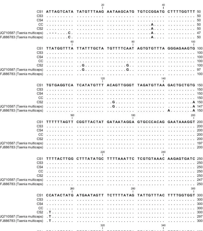
A total of 60 calves and 80 sheep were examined for symptoms of coenurosis, and 13 calves and 38 sheep were found to have signs of the disease. The signs included circling behavior and depression, which are often observed in both sheep and calves with coenurosis. The animals showed signs of apathy, blindness, and nystagmus and intermittently hit their heads against the walls of the stall as they were walking in circles. Except for tachypnea, the other clinical parameters were within normal limits. Four sheep and one calf were necropsied, and C. cerebralis cysts were detected in all the animals. According to the molecular analysis, the CO1-PCR assay yielded a 446 bp band (Figure 1). Genetic alignment (Figure 2) and tree view of the samples was done using Neighbor Joining method of measurement with Kimura 80 (Figure 3).
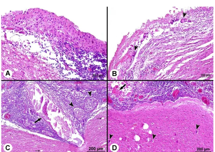
Microscopically, a fibrous or epithelial wall that included necrosis in both sheep and cattle (Figure 4A) surrounded C. cerebralis cysts. The cyst membrane was thinner in the sheep than in the cattle. The areas adjacent to the cyst membrane displayed cellular infiltrate that included many lymphocytes, histiocytes, plasmacytes, epithelioid macrophages and eosinophils. There were many multinucleated
giant cells in the sheep but not in the cattle (Figure 4B). The new cyst formations, including homogenous pink and calcified areas or cellular exudate, were found in the cerebral parenchyma of the cattle. These cysts were closed by dense lymphocytic, histiocytic, plasmacytic and eosinophilic cell infiltrations (Figure 4C). In all cases, severe hyperemia, congestion and perivascular cuffing were detected in the vessels. In addition, gliosis, demyelination, neuronal degeneration and atrophy of the cerebral parenchyma were also detected. However, neuronal dystrophic calcification was found only in the sheep (Figure 4D).
A hypodense cyst was monitored in the right cerebellar hemisphere in the CT images of one of the sheep. It had several millimeters of calcification in the cyst wall. The cyst was compressed in the left cerebellar hemisphere and 4th ventricle. The bilateral
lateral ventricles were dilated (non-communicate hydrocephalus). A cyst was found in the left frontal lobe (51x35x48 mm) of the calf ’s CT image, and the cyst causing cortical thinning, which compressed the anterior horn of the lateral ventricle and caused a rightward shift in the anterior falx cerebri.
Discussion
Coenurosis has been reported in some areas of Europe, America, Africa, and Asia. To the best of our knowledge, there have been only a limited number of previous scientific reports describing the occurrence of coenurosis in sheep and calves in Turkey (AVCIOGLU et al., 2011; GICIK et al., 2007). The post-mortem demonstration of viable C. cerebralis in our study suggests that the disease is prevalent and is likely to constitute a major health problem in sheep and calves with significant economic repercussions. The overall prevalence of C. cerebralis was 3.57% (5% in sheep and 7.69% in calves) in slab-slaughtered sheep and calves based on CT scans and necropsy. Our findings correlate with those obtained by Avcioglu et al. (2011), who reported that the prevalence in cattle was 0.47% (5/1045) in the Erzurum Province of Turkey and 15.5% (60/387) in sheep of the Kars Province of Turkey (GICIK et al., 2007). In contrast, another study found that the prevalence was 36.8% in Turkey (USLU & GUCLU, 2007) and 45.6% in sheep from the Ngorongoro District of Tanzania (CUBEDDU et al., 1990). Reports on coenurosis in some countries in Africa and other tropical to sub-tropical regions revealed a lower prevalence, including 18.65% in Iran (TAVASSOLI et al., 2011), 14.8% in Mozambique (AFONSO et al., 2011), and 5% in Bangladesh (NOORUDDIN et al., 1996). Coenurosis is quite rare in Tanzania (CUBEDDU et al., 1990) but is prevalent in cattle in Greece (GIADINIS et al., 2007). This variation might be due to differences in production systems, levels of natural immunity, relative presence or absence of definitive carnivore hosts and husbandry practices, such as regular deworming.
Currently, either partial or whole mitochondrial genome sequencing based on PCR have been used for the molecular characterization of coenurosis (SAIKI et al., 1988; MCMANUS, 2006; YAMASAKI et al., 2006). The molecular analysis of the CO1-PCR assay from the cysts of these sheep and calves yielded a 446 bp band, which is typical for T. multiceps. Similar findings were reported by Avcioglu et al. (2011), who conducted a molecular
Figure 2. Sequence alignment of samples analyzed in this study using the reference sequences (JQ710587 and FJ886783). CC: Coenurus
characterization of T. multiceps in cattle in Erzurum (Turkey), Iran (AMRABADI et al., 2015) and Italy (VARCASIA et al., 2006) based on CO1 mitochondrial gene sequence analysis.
At necropsy, there was a thickening associated with congestion and turbidity of the meningeal membranes, particularly in the cerebellum. Focal to multifocal necrotic areas and whitish spots were observed in the cortex of the cerebrum and cerebellum. Histopathologically, cross sections of coenurus larvae associated with necrotic suppurative meningoencephalitis were observed. Multiple necrotic areas were also observed in the gray matter of the cerebellum due to the migration of the larvae along with an extensive infiltration of eosinophils and neutrophils. Based on the gross and histopathologic lesions, a diagnosis of acute coenurosis is very rare in sheep (FARJANI KISH et al., 2015). At necropsy,
the cysts were found in the telencephalic portion of the right cranial lobe (ACHENEF et al., 1999).
Several researchers (BIYIKOGLU & DOGANAY, 1998; AVCIOGLU et al., 2012; DESOUKY et al., 2011) reported central nervous system lesions caused by chronic coenurosis. The common properties of the histopathological lesions in chronic coenurosis were inflammatory cell infiltration (lymphocytic-histiocytic-plasmacytic-eosinophilic), congestion, hyperemia, demyelination, perivascular cuffing, necrosis and gliosis (GIADINIS et al., 2007; SHARMA et al., 1998). In the present study, the histopathological lesions of coenurosis were very similar to those of previous reports. However, neuronal dystrophic calcification was observed in samples from the sheep, and multinucleated cells were not detected in the cattle in the current study.
Figure 3. Genetic tree view of the aligned sequences.
A hypodense cyst was monitored in the right cerebellar hemisphere at CT imaging of a sheep. This calcified cyst was compressed to the left cerebellar hemisphere and 4th ventricle. The bilateral lateral ventricles were dilated (non-communicate hydrocephalus). Similarly, the cysts were reported in the caudal region of the cerebral hemisphere of the sheep (ANJOS et al., 2015). Similar observations were found in two sheep cysts that affected the cerebellum, whereas the other eighteen sheep cysts were located in the cerebrum (cortex, parenchyma or lateral ventricles) (GONZALO-ORDEN et al., 1999). The coenurus cysts were reported in the right hemisphere of the infested sheep (BATISTA et al., 2010). The circling behavior, depression, apathy, blindness, nystagmus and intermittently hitting their heads against the walls of the stall while walking in circles are common symptoms that were observed in both sheep and calf infested with coenurosis. The parasitic cysts located in one cerebral hemisphere usually produce contralateral visual and postural deficits. Affected sheep have a tendency to circle toward the side of the cyst. Some authors have suggested that the head tilt is usually directed toward the side with the cyst and that the direction of the circling is not a very reliable guide for locating a cerebral cyst. The location of the cyst causes the visual, behavioral and posture impairments that are the main clinical manifestations of coenurosis (SHARMA & CHAUHAN, 2006; RISSI et al., 2008). The results showed that the cyst was present in the left frontal lobe of the calf ’s CT image, which caused cortical thinning and compressed the anterior horn of the lateral ventricle, causing a rightward shift in the anterior falx cerebri. Varcasia et al. (2013), who found two coenurus cysts in the right hemisphere of cattle by necropsy but not with a CT scan, reported similar observations. Therefore, the present CT scan results were the first to be reported in calves.
Conclusion
Our results suggest that CT imaging is a useful tool for diagnosing ovine coenurosis in sheep and calves in Turkey. CT imaging was used for the first time in calves for diagnosing coenurosis. CT imaging allowed the accurate localization of the cyst and overcame the difficulties of interpreting neurological signs in cattle and sheep.
Acknowledgements
We are very thankful to TUBITAK (2216-research fellowship program for international researchers) to provide an opportunity and funding to work for Dr. Haroon Ahmed.
References
Achenef M, Markos T, Feseha G, Hibret A, Tembely S. Coenurus cerebralis infection in Ethiopian highland sheep: incidence and observations on pathogenesis and clinical signs. Trop Anim Health Prod 1999; 31(1): 15-24. PMid:10399813. http://dx.doi.org/10.1023/A:1005125316275.
Afonso SMS, Mukaratirwa S, Hajovska K, Capece BPS, Cristòfol C, Arboix M, et al. Prevalence and morphological characteristics of Taenia
multiceps Cysts (Coenurus cerebralis) from Abattoir-Slaughtered and experimentally infected goats. J Neuroparasitol 2011; 2: 1-5. http:// dx.doi.org/10.4303/jnp/235532.
Amrabadi O, Oryan A, Moazeni M, Sharifiyazdi H, Akbari M. Comparison of cerebral and non-cerebral coenurosis by genetic markers of glycolytic enzyme (enolase) and mitochondrial sequences in sheep and goats. Vet Parasitol 2015; 214(3-4): 333-336. PMid:26527237. http://dx.doi. org/10.1016/j.vetpar.2015.10.021.
Anjos BL, Fagundes MZ, Gonçalves MA, Rissi DR. Pathology in practice. cerebellar coenurosis in a sheep. J Am Vet Med Assoc 2015; 247(8): 893-895. PMid:26421400. http://dx.doi.org/10.2460/javma.247.8.893.
Avcioglu H, Terim Kapakin KA, Yildirim A. Clinical, morphological and histopathological features of bovine coenurosis: case reports. Rev Méd Vét 2012; 163(6): 295-298.
Avcioglu H, Yıldırım A, Duzlu O, Inci A, Kapakin Terim KA, Balkaya I. Prevalence and molecular characterization of bovine coenurosis from Eastern Anatolian region of Turkey. Vet Parasitol 2011; 176(1): 59-64. PMid:21074326. http://dx.doi.org/10.1016/j.vetpar.2010.10.033.
Batista FA, Pizzigatti D, Martins CF, Nunes MM, Megda TT, Ribeiro OC, et al. First report of coenurosis in sheep in the State of Mato Grosso do Sul, Brazil. Rev Bras Parasitol Vet 2010; 19(4): 265-267. PMid:21184708. http://dx.doi.org/10.1590/S1984-29612010000400016.
Biyikoglu G, Doganay A. Effects of praziquantel and albendazole on Coenurus cerebralis in experimentally infected lambs. Turk J Vet Anim Sci 1998; 22(1): 43-48.
Bowles J, Blair D, McManus DP. Genetic variants within the genus Echinococcus identified by mitochondrial DNA sequencing. Mol Biochem Parasitol 1992; 54(2): 165-173. PMid:1435857. http://dx.doi. org/10.1016/0166-6851(92)90109-W.
Collomb J, Machouart M, Biava MF, Brizion M, Montagne K, Plénat F, et al. Contribution of NADH Dehydrogenase subunit I and cytochrome C oxidase subunit I sequences toward identifying a case of human coenuriasis in France. J Parasitol 2007; 93(4): 934-937. PMid:17918379. http://dx.doi.org/10.1645/GE-1160R.1.
Cubeddu GM, Pintori G, Muzzetto P, Lepori S. La cenurosi cerebellare del bovino. Atti della Società Italiana di Buiatria 1990; 22: 343-348.
Desouky EA, Badawy AI, Refaat RA. Survey on coenurosis in sheep and goats in Egypt. Vet Ital 2011; 47(3): 333-340. PMid:21947971.
Farjani Kish G, Khodakaram-Tafti A, Hajimohammadi A, Ahmadi N. Clinical and morphopathological characteristics of an enzootic occurrence of acute coenurosis (Coenurus cerebralis) in a sheep herd. J Parasit Dis 2015; 39(2): 280-283. PMid:26064018. http://dx.doi.org/10.1007/ s12639-013-0344-z.
Gauci C, Vural G, Oncel T, Varcasia A, Damian V, Kyngdon CT, et al. Vaccination with recombinant oncosphere antigens reduces the susceptibility of sheep to infection with Taenia multiceps. Int J Parasitol 2008; 38(8-9): 1041-1050. PMid:18160069. http://dx.doi.org/10.1016/j. ijpara.2007.11.006.
Giadinis ND, Brellou G, Pourliotis K, Papazahariadou M, Sofianidis G, Poutahidis T, et al. Coenurosis in a beef cattle herd in Greece. Vet Rec 2007; 161(20): 697-698. PMid:18024927. http://dx.doi.org/10.1136/ vr.161.20.697.
Gokce E, Beytut E, Tasci GT, Uzlu E, Kirmızıgul AH, Erdogan HM. An outbreak of coenurosis in a cattle farm. Kafkas Univ Vet Fak Derg 2013; 19(Suppl-A): A199-A202.
Gonzalo-Orden JM, Altónaga JR, Díez A, Gonzalo JM, Asunción Orden M. Correlation between MRI, computed tomographic findings and clinical signs in a case of ovine coenurosis. Vet Rec 2000; 146(12): 352-353. PMid:10777046. http://dx.doi.org/10.1136/vr.146.12.352.
Gonzalo-Orden JM, Díez A, Altónaga JR, Gonzalo JM, Orden MA. Computed tomographic fındings in ovine coenurosis. Vet Radiol Ultrasound 1999; 40(5): 441-444. PMid:10528835. http://dx.doi. org/10.1111/j.1740-8261.1999.tb00372.x.
Knudsen B, Knudsen T, Flensborg M, Sandmann H, Heltzen M, Andersen A, et al. CLC main workbench: version 5.5. Aarhus: CLC Bio; 2007.
Lightowlers MW. Cestode vaccines: origins, current status and future prospects. Parasitology 2006;133(S2 Suppl): S27-S42. PMid:17274847. http://dx.doi.org/10.1017/S003118200600179X.
Lightowlers MW, Lawrence SB, Gauci CG, Young J, Ralston MJ, Maas D, et al. Vaccination against hydatidosis using a defined recombinant antigen. Parasite Immunol 1996; 18(9): 457-462. PMid:9226681. http:// dx.doi.org/10.1111/j.1365-3024.1996.tb01029.x.
Luna LG. Manual of histologic staining methods of the Armed Forces Institute of Pathology. New York: McGraw-Hill; 1968.
McManus DP. Molecular discrimination of taeniid cestodes. Parasitol Int 2006; 55(Suppl): S31-S37. PMid:16337179. http://dx.doi.org/10.1016/j. parint.2005.11.004.
Nooruddin M, Dey AS, Ali MA. Coenuriasis in Bengal goats of Bangladesh. Small Rumin Res 1996; 19(1): 77-81. http://dx.doi.org/10.1016/0921-4488(95)00734-2.
PerkinElmer. [online]. Seattle; 2016 [cited 2016 Apr 5]. Available from: http://www.geospiza.com
Rissi DR, Rech RR, Pierezan F, Gabriel AL, Trost ME, Barros CSL. Coenurosis of sheep in southern Brazil: 16 cases. Cienc Rural 2008; 38(4): 1044-1049. http://dx.doi.org/10.1590/S0103-84782008000400021.
Saiki RK, Gelfand DH, Stoffel S, Scharf SJ, Higuchi R, Horn GT, et al. Primer-directed enzymatic amplification of DNA with a thermostable
DNA polymerase. Science 1988; 239(4839): 487-491. PMid:2448875. http://dx.doi.org/10.1126/science.2448875.
Saitou N, Nei M. The neighbor-joining method: a new method for reconstructing phylogenetic trees. Mol Biol Evol 1987; 4(4): 406-425. PMid:3447015.
Scala A, Ligios C, Leoni A, Nieddu AM. Cenurosi degli ovini in Sardegna: rilievi epidemiologici, parassitologici ed anatomoistopatologici. Atti Società Italiana Scienze Vet 1992; 46(2): 1435-1440. [SISVet]
Sharma DK, Chauhan PPS. Coenurosis status in Afro-Asian region: a review. Small Rumin Res 2006; 64(3): 197-202. http://dx.doi.org/10.1016/j. smallrumres.2005.05.021.
Sharma DK, Singh N, Tiwari HA. Prevalence and pathology of coenurosis in organized goat farms. J Vet Parasitol 1998; 12: 30-32.
Soulsby EL. Helminths, arthropods and protozoa of domesticated animals. 2nd ed. London: Bailliere-Tindall; 1982.
Tavassoli M, Malekifard F, Soleimanzadeh A, Tajik H. Prevalence of Coenurus cerebralis in sheep in Northwest of Iran. Vet Res Forum 2011; 2(4): 274-276.
Uslu U, Guclu F. Prevalence of Coenurus cerebralis in sheep in Turkey. Med Welt 2007; 63(6): 678-680.
Varcasia A, Lightowlers MW, Cattoli G, Cancedda GM, Canu S, Garippa G, et al. Genetic variation within Taenia multiceps in Sardinia, Western Mediterranean (Italy). Parasitol Res 2006; 99(5): 622-626. PMid:16614827. http://dx.doi.org/10.1007/s00436-006-0179-y.
Varcasia A, Pipia AP, Arru D, Pes AM, Tamponi C, Dore F, et al. Morphological and molecular characterization of bovine coenurosis in Sardinia, Italy. Parasitol Res 2013; 112(5): 2079-2082. PMid:23274489. http://dx.doi.org/10.1007/s00436-012-3257-3.
Varcasia A, Tosciri G, Coccone GN, Pipia AP, Garippa G, Scala A, et al. Preliminary field trial of a vaccine against coenurosis caused by Taenia multiceps. Vet Parasitol 2009; 162(3-4): 285-289. PMid:19345506. http:// dx.doi.org/10.1016/j.vetpar.2009.03.008.