TOTAL PROTEIN ELECTROPHORESIS AND RAPD FINGERPRINTING ANALYSIS FOR THE IDENTIFICATION OF AEROMONAS AT THE SPECIES LEVEL.
Ana Paula Longaray Delamare1,2; Liane de Oliveira Artico1; Felipe Gobbi Grazziotin1; Sergio Echeverrigaray1*;
Sérgio Olavo Pinto da Costa1,2
1Instituto de Biotecnologia, Universidade de Caxias do Sul, Caxias do Sul, RS, Brasil. 2Instituto de Ciências Biomedicas, Universidade de São Paulo, São Paulo, SP, Brasil
Submitted: June 20, 2001; Returned to authors for corrections: April 04, 2002; Approved: December 05, 2002
SHORT COMMUNICATION
ABSTRACT
Fifteen well-defined strains of Aeromonas of thirteen species were analyzed by SDS protein electrophoretic analysis (SDS-PAGE) and random amplified polymorphic DNA analysis (RAPD). The comparison between the patterns obtained by both methods allowed differentiating all the strains. Clusters formed by the unweighted pair group method with arithmetic averages applied to protein data correlates with the genetic and biochemical information about the species. The results show that protein fingerprinting has the potential to differentiate
Aeromonas species, but the low qualitative variation indicates that this technique is not efficient for the characterization of strains within a species. Conversely, RAPD fingerprinting allows the identification of strains but the high variability limits its potential as an aiding method for species identification.
Key words: Aeromonas, molecular markers, species identification
The genus Aeromonas comprises several species of oxidase negative and catalase positive, glucose-fermenting, facultative anaerobic, gram-negative, rod-shaped, motile and non-motile bacteria. They are widely distributed in nature, especially in aquatic environments, and have been isolated from a variety of raw foods. Several species of the genera have been associated with several diseases in both warm and cold blood animals (fishes, reptiles, etc.) (6). In humans, they are opportunistic pathogens causing gastroenteritis, and less commonly, cellulitis, wound infections, meningitis, otitis, peritonitis, endocarditis and septicemia (10).
The taxonomy of the genus Aeromonas is confuse and controversial (4). The need of a system for the identification and classification of Aeromonas isolates is justified by their ecological and clinical importance. Different methods as biotyping (22), isozyme electrophoretic analysis (21), DNA hybridization (23), lipopolysaccharide analysis (27), serotyping
* Corresponding author. Mailing Address: Instituto de Biotecnologia, Universidade Caxias do Sul, Caixa Postal 1532. 95001-970, Caxias do Sul, RS, Brasil. Telephone/Fax: (+5554) 212-1133 ext. 2075. E-mail: selaguna@yahoo.com
(7), ribosomal DNA typing (1), SDS-PAGE analysis of cell proteins (14,15), RAPD markers (16,17), AFLP fingerprinting (8), and PCR (18,19) have been used to type isolates. However, these methods are not generally accepted as standard systems for the evaluation of Aeromonas isolates, as a standard method should be simple, rapid, inexpensive, reliable, and applicable in any kind of routine laboratory. SDS-PAGE analysis of cell proteins and RAPD analysis, two methods that have most of these characteristics, have been tested for isolates identification (14, 15, 16, 17). However, few attempts have been made to evaluate their usefulness for the characterization of Aeromonas at the species level. Therefore, the purpose of this study was to compare the efficiency of SDS-PAGE and RAPD analysis for the differentiation of Aeromonas species.
A. caviae (ATCC 15468), A. ichtiosmia (ATCC 49904), A. euchrenophila (ATCC 23309), A. enteropelogenes (ATCC 49803), A. trota (ATCC 49657), A. salmonicida var. salmonicida (ATCC 33658), A. media (ATCC 33907), A. veronii (ATCC 35624),
A. encheleia (CECT 4341), A. sobria (ATCC 43979), A. hydrophyla var. punctata (ATCC 14486), and clinical isolates of
Escherichia coli, Pseudomonas aeruginosa, Enterobacter cloacae, Salmonella typhimurium, Staphylococcus epidermidis, and Citrobacter spp.
For SDS-PAGE analysis, cultures were grown over-night on LB broth (1.0 ml) and centrifuged on microtubes. The pellets were washed with water, and suspended in sample buffer. The proteins were dissociated by immersion for 5 min in boiling water. The samples were centrifuged to eliminate cell debris and used directly for electrophoretic separation (11).
Sodium-dodecyl sulphate polyacrilamide-gel electrophoresis (SDS-PAGE) was performed according to Laemmli (11) with a stacking gel containing 4.5% acrilamide and a resolving gel containing 12% acrilamide. Samples with 120 to 150 µg of proteins, as determined by mini Bradford method (2), were loaded in each track. Electrophoresis was performed at constant voltage of 80V for stacking gels and 150V for resolving gels. The gels were fixed for 15 min in an aqueous solution containing 7% glacial acetic acid and 30% methanol and stained over-night in 0.1% (w/v) Coomassie Brilliant Blue R-250 solution (3). All samples were prepared and examined in triplicate on different gels. After several destaining steps, the gels were photographed on a high intensity light box. The protein profiles were compared by eye. The proteins (bands) were listed as discrete character states per strain (presence/absence). Bands were considered identical only when their width, intensity and position were the same.
For randomly amplified polymorphic DNA analysis (RAPD), bacterial cultures were grown in 1.0 ml of LB broth at 18ºC for 24h, centrifuged at 15000xg for 5 min to pellet the cells. Total DNA was isolated by the method described by Pan et al. (20). DNA content of all samples was measured using spectrophotometer at 260 nm. DNA purity was evaluated by the 260/280 ratio and gel electrophoresis. All extracts were diluted to working solution of 10ng µl-1.
The polymerase chain reaction DNA amplification protocol was a variation of that reported by Williams et al. (29). Reactions were performed in 25 µl volume containing 50 mM KCl; 10 mM Tris-HCl (pH 8.3); 3 mM MgCl2; 0.25% Triton-X-100; 1.25 mM of dNTP (Pharmacia LKB Biotechn.); 30 ng of single decamer primer (40 primers of kits A and B of Operon Techn.); 60 to 80 ng of genomic DNA; and 1.5 units of Taq DNA polymerase (Pharmacia LKB Biotechn.). DNA amplification was performed using a thermal cycler (model PTC100, MJ Research, Watertown, Mass.). The thermal cycle used was 94ºC for 1 min; then 45 cycles of 94ºC (1 min), 35ºC (1 min) and 72ºC (2 min), and finally 72º for 3 min. A negative control including all components except genomic DNA was included in all thermal cycle runs.
Following amplification, the RAPD products (20 µl) were loaded in 1.5% agarose gels in TBE buffer (89 mM Trisma-base, 89 mM boric acid and 8 mM EDTA) and resolved by electrophoresis. After electophoresis the amplification products were stained with ethidium bromide (0.5 µg ml-1) and photographed under UV light. The size of amplification products were determined by comparison with Lambda DNA digested with EcoRI and HindIII restriction enzymes.
Bands were scored as present or absent. Bands that were not well defined were not included in the data set as these were assumed to be unreliable markers.
Total protein and RAPD data were analyzed using NTSYS-pc package, version 1.5 (26). Similarities were computed using the Jaccard´s coefficient, and strains were clustered by the unweighted pair-group method using arithmetic averages (UPGMA) in order to present the results in the form of dendrograms.
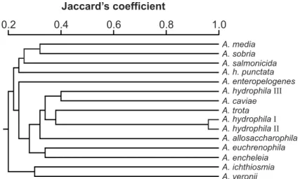
The fifteen Aeromonas strains evaluated yielded similar protein electrophoretic patterns with a high number of bands (>50 bands). However, based on the reproducibility and the criteria adopted for the analysis of the gels, the presence or absence of a total of 24 protein bands with molecular weights between 14000 and 65000 daltons were scored. The patterns obtained in three independent gels were very similar confirming the high reproducibility of protein fingerprinting analysis (14). Protein profiles were very similar among the strains, and several strains exhibited characteristic proteins that may be useful markers for the identification at the species level: A. allosaccharophila (35.5 kDa), A. sobria (39.0 kDa), and A. trota
(40.0, 27.0, and 24.5 kDa).
Using the unweighted pair group method with arithmetic averages for clustering, we identified a total of four clusters at the 70% hierarchical level (Fig. 1). A. salmonicida and A.sobria
formed independent clusters, groupS 1 and 2, respectively. A. salmonicida, a non motile species included in the hybridization group 3 (24), and A. sobria, hybridization group 7, have been separated by different methods as immunoblotted SDS-PAGE gels (15,28), 16S rDNA sequencing (13), RAPD (17), and AFLP (9) A. enteropelogenes and A. trota cluster together within group 3. These data confirm previous results that considered these species as identical or similar by comparing their 16S rRNA sequence (5), and their AFLP profiles (8).
from the “Coleção de Culturas Tropicais”, and the other one from the Animal Pathology Department, University of León, confirmed by their protein fingerprinting as identical, and A. allosaccharophila ATCC 51208, a new species proposed by Martinez-Murcia et al. (13).
For RAPD analysis an initial screening of primers was performed. DNA of A. hydrophila CECT 839 and A. trota ATCC 49657 were amplified using the 40 decamer primers of the kits A and B of Operon Technologies. The results obtained in these first experiments were used to select 14 primers that gave at least four intense amplification products for each species: 02, 04, 09, 11, OPA-19, 04, 06, 07, 10, 12, OPB-13, OPB-15, OPB-16 and OPB-17.
The selected primers were used to analyze the 15 strains of Aeromonas. Using the 14 selected primers 290 RAPD bands were scored (4 to 18 bands per primer), ranging in size from 100 to 2700 base pairs. Of the total bands scored, 280 (95%) were polymorphic. This percentage of polymorphic bands (95%) is extremely high when compared with the variation reported at the species level for other organisms, but is comparable to that obtained Oakey
et al. (17) when comparing the RAPD patterns for some species of Aeromonas. Despite the high variation observed, RAPD profiles exhibited ten bands that were common to all the Aeromonas
species, and absent in clinical isolates of Escherichia coli, Pseudomonas aeruginosa, Enterobacter cloacae, Salmonella typhimurium, Staphylococcus epidermidis, and Citrobacter spp. These bands can be cloned and sequenced to design PCR primers that may be useful for the rapid detection of aeromonads. This approach has been previously used to design PCR primers for the identification of A. salmonicida
(18) and A. hydrophila (19).
The ability to differentiate all tested strains by RAPD suggests that this technique may be practically applied for the identification of
Aeromonas isolates, being necessary for this purpose, the amplification with one or two selected primers. Even showing very different profiles, ten bands were common to all the strains.
Comparing the RAPD patterns of the 15 Aeromonas
strains evaluated, we construct a matrix that was used to calculate the Jaccard´s similarity between strains. As expected, the highest similarity (96.77%) was obtained between the two representatives of
A. hydrophila ATCC 7966. The lowest similarity was observed between A. enteropelogenes and A. hydrophila punctata (20.17%).
The unweighted pair group method with arithmetic averages applied to the RAPD data, did not allow the clear separation of clusters among the tested strains. Similar results were obtained by Huy et al. (8) using AFLP analysis. These results support
0.5 0.6 0.7 0.8 0.9 1.0
A. hydrophilaI A. hydrophilaII
A. caviae A. h. punctata A. ichthiosmia A. media A. veronii A. hydrophilaIII A. euchrenophila A. encheleia A. enteropelogenes A. trota
A. sobria A. salmonicida A. allosaccharophila Jaccard´s coefficient
A. media A. sobria A. salmonicida A. h. punctata A. enteropelogenes A. hydrophilaIII
A. caviae A. trota A. hydrophilaI
A. hydrophilaII
A. allosaccharophila A. euchrenophila A. encheleia A. ichthiosmia A. veronii
0.2 0.4 0.6 0.8 1.0
Jaccard’s coefficient
Figure 2. Dendrogram based on UPGMA analysis of the phenetic similarity between fifteen Aeromonas strains as determined by 290 RAPD bands. (A. hydrophila I and II – ATCC 7966 A and B, respectively; A. hydrophila III- CECT 398).
the existing classification of Aeromonas in several species, since each type strain gave different RAPD patterns.
The present results shows that RAPD analysis in
Aeromonas, with the set of primers tested, even efficient for the discrimination among isolates (17) is not useful for the characterization of strains at the species level and the evaluation of relationships among Aeromonas. Conversely, the results obtained by the analysis of cell protein profiles correlates with the genetic and biochemical data previously reported by other authors, using different analytical methods, and can be used as a rapid, inexpensive and reliable system to help in identification and taxonomy of Aeromonas isolates.
ACKNOWLEDGEMENTS
This work was supported by FAPERGS and Universidade de Caxias do Sul. A.P.L.D. was supported by a grant of CAPES and L.O.A. by a grant of CNPq. The authors would like to thank Dr. Naharro, form the Animal Pathology Department, University of León, Spain, for the donation of some of the bacterial strains.
RESUMO
Análise eletroforética de proteínas totais e marcadores de RAPD na identificação de Aeromonas
ao nível de espécie
Quinze linhagens de Aeromonas pertencentes a treze espécies foram avaliadas através de eletroforese de proteínas totais (SDS-PAGE) e segmentos de DNA amplificados ao acaso (RAPD). A comparação entre os padrões obtidos por ambos métodos permitiu diferenciar todas as linhagens. Agrupamentos formados com base nos dados protéicos mostraram relação com informações bioquímicas e genéticas a respeito das espécies. Os resultados mostraram que análises protéicas têm potencial para diferenciar espécies de Aeromonas, mas a baixa variação qualitativa indica que esta técnica não é eficiente para a caracterização entre linhagens dentro de espécies. Ao contrário, marcadores de RAPD permitem identificar linhagens, mas a alta variabilidade limita seu potencial como método auxiliar na identificação de espécies.
Palavras-chave: Aeromonas, marcadores moleculares, identificação de espécies.
REFERENCES
1. Altwegg, M.; Altwegg-Bissig, R.; Demarta, R.; Peduzzi, A.; Reeves, M.W.; Swaminathan, B. Comparison of four typing methods for
Aeromonas species. J. Diarrh. Dis. Res., 6: 88-94, 1988.
2. Bradford, M. A rapid and sensitive method for the quantitation of microgram quantities of protein utilizing the principle of protein-dye binding . Anal. Biochem., 72: 248-254, 1976.
3. Brune, W.; Alfenas, A.C. Identificação de proteínas em géis. In
Alfenas, A.C. (Ed.) Eletroforese de isoenzimas e proteínas afins. Editora UFV, Viçosa, MG, 1998, p.183-197.
4. Carnaham, A.M.; Altwegg M. Taxonomy. In Austin, B.; Altwegg, M.; Gosling, P.J.; Joseph, S.W. (Ed.) The Genus Aeromonas. John Wiley & Sons Ltd., Chichester, England, 1995, p.1-38.
5. Collins, M.D.; Martinez-Murcia, A.J.; Cai, J. Aeromonas enteropelogenes and Aeromonas ichtiosmia are identical to
Aeromonas trota and Areomonas veronii, respectively, as revealed by small-subunit rRNA sequence analysis. Int. J. Syst. Bacteriol., 43: 855-856, 1993.
6. Gosling, P.J. Aeromonas species in disease of animals. In Austin, B.; Altwegg, M.; Gosling, P.J.; Joseph, S.W. (Ed.) The Genus Aeromonas. John Wiley & Sons Ltd., Chichester, England, 1995, p.175-196. 7. Havelaar, A.H.; Schets, F.M.; Van Silfhout, A.; Janssen, W.H.;
Wieten, G.; Van der Kooij, D. Typing of Aeromonas strains from patients with diarrhoea and from drinking water. J. Appl. Bacteriol., 72: 435-444, 1992.
8. Huys, G.; Coopman, R.; Janssen, P.; Kersters, K. High-resolution genotypic analysis of the genus Aeromonas by AFLP fingerprinting.
Int. J. Syst. Bacteriol., 46: 572-580, 1996.
9. Huys, G.; Kampfer, P.; Alwtwegg, M.; Coopman, R.; Janseen, P.; Gillis, M.; Kersters, K. Inclusion of Aeromonas DNA hybridization group 11 in Aeromonas encheleia and extended descriptions of the species Aeromonas eucrenophila and A. encheleia. Int. J. Syst. Bacteriol., 47: 1157-1164, 1997.
10. Janda, J.M.; Abbott, S.L. Human Pathogens. In Austin, B.; Altwegg, M.; Gosling, P.J.; Joseph, S.W. (Ed.) The Genus Aeromonas. John Wiley & Sons Ltd., Chichester, England, 1995, p.151-174. 11. Laemmli, U.K. Cleavage of structural proteins during the assembly
of the head of bacteriophage T4. Nature, 227: 680-685, 1970. 12. Lee, J.V. Identification of Aeromonas in the routine laboratory.
Experientia, 43: 355-356, 1987.
13. Martinez-Murcia, A.J.; Benlloch, S.; Collins, M.D. Phylogenetic interrelationships of members of the genera Aeromonas and
Plesiomonas as determined by 16S ribosomal DNA sequencing: lack of congruence with results of DNA-DNA hybridizations. Int. J. Syst. Bacteriol., 42: 412-421, 1992.
14. Millership, S.E.; Want, S.V. Characterization of strains of Aeromonas
spp. by phenotype and whole-cell protein fingerprint. J. Med. Microbiol., 39: 107-113, 1993.
15. Muller, R.; Millership, S.E. Typing of Aeromonas spp. by numerical analysis of immunoblotted SDS-PAGE gels. J. Med. Microbiol., 39: 325-333, 1993.
16. Myata, M.; Aoki, T.; Inglis, V.; Yoshida, T.; Endo, M. RAPD analysis of Aeromonas salmonicida and Aeromonas hydrophila. J. Appl. Bacteriol., 79: 181-185, 1995.
17. Oakey, H.J.; Ellis, J.T.; Gibson, L.F. Differentiation of Aeromonas
genomospecies using random amplified polymorphic DNA polymerase chain reaction (RAPD-PCR). J. Appl. Bacteriol., 80: 402-410, 1996.
18. Oakey, H.J.; Ellis, J.T.; Gibson, L.F. The development of random DNA probes specific for Aeromonas salmonicida. J. Appl. Microbiol., 84: 37-46, 1998.
19. Oakey, H.J.; Gibson, L.F.; George, A.M. DNA probes specific for
Aeromonas hydrophila. J. Appl. Microbiol., 86: 187-193, 1999. 20. Pan, Y.B.; Grisham, M.P.; Burner, D.M. A polymerase chain reaction
protocol for the detection of Xanthomonas albilineans, the causal agent of sugarcane leaf scald disease. Plant Disease, 81: 189-194, 1997. 21. Picard, B.; Goullet, Ph. Comparative electrophoretic profiles of esterases, and of glutamate, lactate and malate deshydrogenases, from Aeromonas hydrophila, A. caviae and A. sobria. J. Gen. Microbiol., 131: 3385-3391, 1985.
22. Popoff, M.; Veron, M. A taxonomic study of the Aeromonas hydrophila-Aeromonas punctata group. J. Gen. Microbiol., 94: 11-22, 1976.
24. Popoff, M. Genus III. Aeromonas. In Krieg N.R.; Holt, J.G. (Ed.)
Berger´s Manual of Systematic Bacteriology, Vol. 1. Williams & Wilkins, Baltimore, MD, 1984, p.545-548.
2 5 . Janda, J.M.; Abbott, S.L. Human Pathogens. In Austin, B.; Altwegg, M.; Gosling, P.J.; Joseph, S.W. (Ed.) The Genus Aeromonas. John Wiley & Sons Ltd., Chichester, England, 1995, p . 1 5 1 - 1 7 4 .
26. Rohlf, F.J. NTSYS-pc numerical taxonomy and multivariate analysis system, version 1.70. Exeter Software, Setauket, NY, 1989.
27. Shaw, D.; Hodder, H.J. Lipopolysaccharides of the motile aeromonads; core oligosaccharide analysis as an aid to taxonomic classification. Can. J. Microbiol., 24: 864-868, 1978.
28. Stephenson, J.R.; Millership, S.E.; Tabaqchali, S. Typing of