Prognostic Value of Dipyridamole Stress Echocardiography in
Women
Maria Celita de Almeida
1e Brivaldo Markman Filho
1,2Procárdio Diagnósticos1; Universidade Federal de Pernambuco2, Recife, PE - Brazil
Mailing address: Brivaldo Markman Filho •
Av. Visconde de Jequitinhonha, 2544/1902 - 51130-020 - Recife, PE - Brazil E-mail: [email protected]
Manuscript received August 03, 2009; revised manuscript received Marh 04, 2010; accepted May 31, 2010.
Abstract
Background: Stress echocardiography is an important diagnostic and prognostic tool in ischemic heart disease.
Objective: To evaluate the role of dipyridamole stress echocardiography (DSE) in the investigation of myocardial ischemia in women and its ability to predict combined events (cardiovascular death, acute myocardial infarction [AMI], unstable angina, coronary artery bypass grafting [surgery or percutaneous intervention] at an average follow-up of 16 months.
Methods: A prospective study using the protocol of dipyridamole at 0.84 mg in 10 minutes, associated with atropine (0.25 mg/min up to 1.0 mg).
Results: This study evaluated 147 women. DSE was positive in 14 patients (9.5%), negative in 128 (87.1%) and inconclusive in 5 (3.4%). Events occurred in 8 patients, 7 had positive DSE. The other 138 did not present any events. Our of these, 128 had negative DSE. The sensitivity, specificity, accuracy, the positive and negative predictive values of the test before the events were respectively: 83%, 95%, 94%, 42% and 99%. The event-free survival for patients with negative DSE was 99.2% compared with 58% for positive DSE (p < 0.001). Univariate analysis identified the DSE result, basal electrocardiogram (ECG), LV ejection fraction, dyslipidemia, wall motion score index at rest and peak, history of AMI, coronary artery bypass grafting, as prognostic predictors related to outcomes. The results of DSE and ECG remained significantly associated with outcomes in the multivariate analysis (p < 0.001).
Conclusion: The baseline ECG and positive DSE were independent variables for the occurrence of outcomes. The DSE showed excellent negative predictive value, confirming its usefulness in evaluating prognosis in such patients. (Arq Bras Cardiol 2011; 96(1): 31-37)
Keywords: Echocardiography, stress; myocardial ischemia; diagnostic techniques and procedures; dipyridamole; prognostic; women.
Introduction
In developed countries, atherothrombotic disease, including coronary artery disease (CAD) and cerebrovascular accident (CVA), is the leading cause of death among women1.
However, in women, CAD is presented differently related to men. The onset of symptoms is late (5-10 years), the group of symptoms is more atypical and there is a greater prevalence of comorbidities such as hypertension (HBP), diabetes mellitus (DM) and dyslipidemia (DLP).Besides this, there is a poor specificity of routine diagnostic tests, and increased mortality during percutaneous coronary intervention (PCI) and coronary artery bypass grafting (CABG)².
The treadmill test (TT) is associated with a high rate of false-positive results and a huge number of variables may change the accuracy of such test in women. The combination of imaging
tests significantly increases the diagnostic accuracy for CAD and several authors have used stress echocardiography (EE) in this subgroup patients3-5.
In dipyridamole stress echocardiography (DSE), this drug works by causing myocardial ischemia through the mechanism of coronary “flow steal”, as previously explained6, 7. Concomitant administration of atropine, increasing heart
rate, enhances the effects described.
This study was intended to evaluate the prognostic value of DSE in women with suspected myocardial ischemia.
Methods
Prospective observational study conducted from Mar/2005-Jun/2007 in women with some contraindication to ET or ET positive for ST-segment alteration without clinical manifestation of CAD or inconclusive ET, who underwent DSE for evaluation of myocardial ischemia. The DSE was performed without discontinuation of drug therapy.
Patients with improper echocardiographic window, previously known contraindication to dipyridamole or those who had used xanthine products in the 24 hours preceding the examination were excluded.
The protocol used consisted of intravenous administration of a total dipyridamole dose of 0.84 mg/kg in 10 minutes, as follows: 0.56 mg/kg in four minutes, four minutes of observation, and if no criterion of positivity was evidenced, an additional dose of 0.28 mg/kg were infused in two minutes8.
If no criteria for test interruption arouse, atropine was administered at a dose of 0.25 mg every minute, totaling 1.0 mg. The test was completed with the infusion of aminophylline, to reverse the effects of dipyridamole (maximum dose of 240 mg for three minutes) in the 16th minute.
D u r i n g t h e p r o c e d u r e , b l o o d p r e s s u r e a n d echocardiographic images were recorded at each stage. Criteria for test interruption were: onset of segmental abnormalities in left ventricular contraction, chest pain of at least moderate intensity, ventricular arrhythmias or side effects deemed important by the physician who performed the examination.
The equipment used was a Vivid 3 Pro – GE Medical Systemsâ. According to current recommendations, the left
ventricle (LV) was divided into 17 segments9. A score of 4
points was given for each segment, as follows: 1 = normal, 2 = hypokinesia, 3 = akinesia, 4 = dyskinesia7,10.
Then, the LV wall motion score index (LVWMSI) was calculated, considering the sum of the points of the 17 LV segments divided by the number of segments analyzed. Values greater than one were considered abnormal. For each patient, the scores at rest (pre) and at the peak of drug infusion were determined.
The test was considered positive for ischemia with the onset of a change in the LV segmental contractility (hypokinesia, akinesia or dyskinesia) or worsening of a pre-existing contractile change. The studies were evaluated off-line by two independent examiners.
Follow-up was through medical records, telephone interview or an interview with the attending physician.
The clinical events assessed during follow-up were: death of cardiac origin, acute myocardial infarction (AMI), unstable angina (UA) and coronary artery bypass surgery (CABG) or percutaneous intervention (PCI), occurring three months after the DSE.
Death was attributed to cardiac origin when the following was documented: significant arrhythmia, congestive heart failure (CHF) or AMI11. The diagnosis of AMI and UA followed
the diagnostic criteria defined by current guidelines12,13.
Clinical follow-up of each patient was closed after the occurrence of any event. Therefore, only the first event was considered.
Statistical analysis
The results were expressed as mean, median and standard deviation for quantitative variables. Qualitative variables were expressed in absolute and relative frequencies. Faced with clinical outcomes, we calculated the sensitivity, specificity,
accuracy, positive predictive value (PPV) and negative predictive value (NPV) of the DSE.
The cumulative probabilities of events were estimated by the Kaplan-Meier curves and the presence of differences by the log rank test. The COX proportional hazards model was used to determine the variables with independent prognostic value for occurrence of events. For the model, we considered all variables that, in the univariate analysis, showed statistical significance (p < 0.20).
The significance level was 0.05. Data were analyzed with SPSS (Statistical Package for Social Science) - IBM™ for Windows version 12.0.
Results
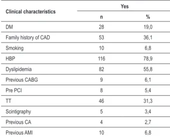
A total of 147 patients whose mean age was 62 years (37-87 years) was assessed by DSE. Five tests were stopped early due to disabling symptoms (two cases of intense dyspnea, two cases of important chest pain with no significant alteration of myocardial contractility and one case of bronchospasm), which were excluded from the analysis of results. Clinical characteristics of patients are shown in Table 1. Noteworthy is the high number of patients with hypertension and dyslipidemia. On the other hand, the number of coronary events characterized by history of AMI was low, as well as the number of coronary revascularisation procedures.
Out of 147 women, 66.0% had atypical chest pain, 7.5% typical chest pain (angina) and 26.8% were asymptomatic, of which 51.3% with TT characterized by: ischemic ST-T segment changes or angina. In this sample, 72.8% presented normal basal electrocardiogram (ECG) and 27.2%, ECG with abnormalities. Abnormalities were considered: changes in left ventricular repolarization, left bundle branch block (LBBB), electrically inactive area and left ventricular hypertrophy. Regarding ventricular function, only 08 patients (5%) had lowered EF
Table 1 - Distribution of sampling frequency according to clinical characteristics
Clinical characteristics Yes
n %
DM 28 19,0
Family history of CAD 53 36,1
Smoking 10 6,8
HBP 116 78,9
Dyslipidemia 82 55,8
Previous CABG 9 6,1
Pre PCI 8 5,4
TT 46 31,3
Scintigraphy 5 3,4
Previous CA 4 2,7
Previous AMI 10 6,8
Table 2 - Distribution of sampling frequency according to event
n %
Event 8 5,4
CABG 4 2,7
PCI 3 2,0
UA 1 0,7
CABG - coronary artery bypass grafting, PCI - percutaneous coronary intervention;
UA - unstable angina.
(<0.56) at baseline Doppler echocardiography. History of CAD, previous AMI, CABG or PCI, or prior coronary angiography indicating the presence of obstructive coronary artery lesion of at least 50.0% of the vessel lumen was present in 27 patients. As for medications, 42.0% were using beta-blockers, 22% used calcium blocker, 16% nitrate, 39% angiotensin-converting enzyme inhibitors, 35% aspirin and 32% statins. There were 5.0% of patients using beta-blocker and nitrate combination, 5% with beta-blocker and calcium blocker and only 3% under combined use of beta-blocker, calcium blocker and nitrate.
The average follow-up for events was 16 months (minimum of two months and a maximum of 27 months).
The DSE showed myocardial ischemia in 14 (9.5%) patients. The result was negative in 128 (87.1%) and inconclusive in 5 (3.4%) patients. Out of 147 patients analyzed, eight (5.4%) had cardiac events during follow-up period. Out of the 8 events, 7 occurred among the 14 positive tests for myocardial ischemia, of which two events occurred earlier than three months of the examination and, therefore, were excluded from the analysis, and one among patients with negative test. Among the 7 patients with positive test without events, 4 were patients with CAD.
Regarding the number of events, there was significant difference between positive and negative tests. The probability of occurrence of events during follow-up for women with positive test was 42% and for women with negative test was 0.8% (p < 0.0001) (Chart 1).
We observed 4 CRM (2.7%), three PCI (2.0%) and one patient (0.7%) had UA. No AMI or death occurred during follow-up (Table 2).
Thus, the sensitivity, specificity, accuracy, positive predictive value and negative predictive value of the test related to clinical outcomes were respectively 83.0%, 95.0%, 94.0%, 42.0% and 99.0%.
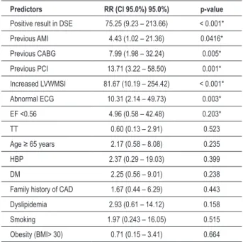
Regarding the survival analysis of all clinical, electrocardiographic and echocardiographic variables analyzed, DSE positive for previous ischemia, acute myocardial infarction, PCI and CABG, increased LVWMSI, abnormal ECG and ejection fraction (EF) <0.56 presented statistically significant association with the occurrence of events (Table 3).
With the Cox regression at a significance level of 5.0%, only the variables DSE and abnormal baseline ECG were significant. It was found that patients with positive DSE for ischemia were roughly 57 times more likely to have the event than patients with negative DSE. Patients with abnormal baseline ECG were 13 times more likely to present the event than those with normal ECG (Table 4).
Chart 1 - Survival free of events according to the DSE result.
P
rob. s
urvi
va
l fre
e
of e
ve
nt
Table 3 -Results of univariate analysis of Cox - Predictors for event
Predictors RR (CI 95.0%) 95.0%) p-value
Positive result in DSE 75.25 (9.23 – 213.66) < 0.001*
Previous AMI 4.43 (1.02 – 21.36) 0.0416*
Previous CABG 7.99 (1.98 – 32.24) 0.005*
Previous PCI 13.71 (3.22 – 58.50) 0.001*
Increased LVWMSI 81.67 (10.19 – 254.42) < 0.001*
Abnormal ECG 10.31 (2.14 – 49.73) 0.003*
EF <0.56 4.96 (0.58 – 42.48) 0.203*
TT 0.60 (0.13 – 2.91) 0.523
Age ≥ 65 years 2.17 (0.58 – 8.08) 0.235
HBP 2.37 (0.29 – 19.03) 0.399
DM 2.25 (0.56 – 9.01) 0.238
Family history of CAD 1.67 (0.44 – 6.29) 0.443
Dyslipidemia 2.93 (0.61 – 14.12) 0.158
Smoking 1.97 (0.243 – 16.05) 0.515
Obesity (BMI> 30) 0.71 (0.15 – 3.41) 0.664 * Statistically signiicant (p <0.005), RR - relative risk, CI - conidence interval,
AMI - acute myocardial infarction, CABG - coronary artery bypass grafting; PCI - percutaneous coronary intervention; LVWMSI - left ventricular wall motion score index, ECG - electrocardiogram; EF - ejection fraction; TT - treadmill test; HBP - high blood pressure, DM - diabetes mellitus, BMI - body mass index, CAD - coronary artery disease.
Table 4 - Results of the adjustment of the Cox regression model
Predictors RR (CI 95%) p-value
Positive DSE 56.59 (6.74 – 174.87) < 0.001
Abnormal baseline ECG 12.68 (1.51 – 86.42) 0.019 RR - relative risk; CI - conidence interval; DSE - dipyridamole stress
echocardiography; ECG - electrocardiogram.
Table 5 - Distribution of sampling frequency of side effects
Side effect n %
Ventricular extrasystole 8 5.4
Headache 7 4.8
Chest pain 4 2.7
Dyspnea 2 1.4
Bronchospasm 1 0.7
factors were associated with more than 90.0% of the risk attributable to a first AMI. Dyslipidemia, smoking, diabetes and hypertension were the most important ones. In our study, more than three quarters of the patients were hypertensive, and approximately half of them had dyslipidemia.
Among the clinical variables of interest, history of AMI, CABG and PCI proved to be independent predictors for cardiac events15. In a study using the pharmacological stress
echocardiography to stratify the risk of AMI, previous AMI, CABG or PCI were adverse prognostic factors in univariate analysis16. History of AMI, CABG or PCI were predictive risk
factors by univariate analysis, but did not reach statistical significance in multivariate analysis due to the size of the sample.
The LV systolic function is a determinant factor of prognosis in patients with ischemic heart disease17. Left ventricular
ejection fraction (LVEF) is the measure most used in clinical practice, and is extremely valuable for risk stratification18.
Only 5.0% of women analyzed presented LVEF <0.56. Out of these patients, 20.0% had events. There was a statistically significant association between LVEF and events, although the magnitude of such association has been jeopardized by the small number of patients with reduced EF.
Abnormal baseline ECG was significantly associated with future events. Literature data show that normal ECG is a favorable prognostic factor in patients with suspected CAD or CAD defined, as it suggests preserved left ventricular function19. On the other hand, conduction disturbances,
most often BRE and left anterior fascicular block, may occur in patients with chronic CAD, are more associated with left ventricular dysfunction and may reflect multiarterial disease and previous myocardial damage20. Left ventricular
hypertrophy on ECG is an independent predictor of events in patients with chronic CAD21.
Regarding the results of DSE, significant difference was observed between patients with negative and positive tests in relation to the occurrence of cardiac events during follow-up. Cortigiani et al22 reported that DSE on women has specificity
of 93.0% and diagnostic accuracy of 87.0%, better than the TT for diagnosis of CAD. These results were also observed in two reviews: an American review23 and an English one24.
This study showed that out of eight events, only one happened among 128 women with negative tests for ischemia (0.8%), highlighting the difference.
This sample of women with negative tests shows the high negative predictive value of the test in the study group (99.0%). The positive predictive value found (42.0%) is consistent with the sample studied, especially when compared with patients with higher cardiovascular risk25. Consistently with our results,
Marwick et al26 reported that normal stress echocardiography
(SE) corresponds to low risk for cardiac events (<1.0% per annum) under analyses of 4-5 year of follow-up. Similar data were also found in the American recommendations of non-invasive tests in the clinical evaluation of women with suspected CAD27.
The negative test in which an event occurred was of a diabetic and smoker patient. Some studies show that false negatives are more frequent in submaximal tests, single-vessel lesions or moderate lesions (stenosis between 50 As to the DSE safety profile, the side effects observed
in the sample are shown in Table 5. There were no major complications and all the side effects were reversed with the administration of aminophylline.
Discussion
to 70.0%)28,29. Diabetic patients may present more rapid
progression of CAD30 and the patient underwent coronary
angiography after 6 months of testing. This fact could explain the false-negative result.
Among the patients with positive tests with no events during the follow-up period (7 patients), 4 had proven CAD. This indicates the possibility that, at the option of the medical team, clinical treatment has been enhanced, rather than proposed coronary artery revascularisation procedures (surgical or percutaneous). One patient had normal coronary arteries and had her test considered ischemic due to abnormalities found on the inferior wall contractility, a common cause of false-positive tests31. The presence of typical chest pain may
suggest microcirculation disease, more common in women than in men32. From the other two patients with positive test,
one had a coronary spasm during angiography and coronary arteries free of significant atheromatous disease and the other presented aneurysmal segment in left anterior descending artery, which may have contributed to the positive test33.
The analysis of clinical, electrocardiographic and echocardiographic variables by logistic regression analysis showed that positive DSE for ischemia and abnormal baseline ECG were independent predictors for cardiac events during follow-up. We observed a clear superiority of the DSE over the electrocardiography variable in the multivariate analysis, which is consistent with other studies34,35.These data are corroborated by the findings
reported by Shaw et al, who evaluated 4,234 women who underwent Dobutamine Stress Echocardiography (DoSE) or stress echocardiography with exercise (SEE) for five years, concluding that the SE is a strong independent predictor of long-term cardiac events in women36.
Biagini et al evaluated the overall mortality in women with suspected CAD or with known CAD undergoing DoSE, and observed that the presence of myocardial ischemia during DoSE had an independent association with increased overall mortality risk after adjustment with clinical data37. This study,
once again, further demonstrates the superiority of SE on the ECG findings as a prognostic factor.
Some studies have assessed the safety of DSE in patients with chronic coronary artery disease or under investigation, delivering good results38,39. Mathias Jr et al40, evaluating
the safety profile of DoSE in a prospective study in 4,033 patients, found significant adverse effects related to the examination in 10 patients (0.25%), with no record of death. A multicenter study evaluated the safety profile of DSE in 10,451 examinations. Significant side effects occurred in 113 patients (1.2%), including 7 major adverse events (0.07%): one death, one cardiac arrest of short duration, readily reversed, two AMI, one pulmonary edema and an episode of prolonged ventricular tachycardia41.
In our study, the occurrence of side effects was small, which were promptly reversed after the infusion of
aminophylline, with no record of serious complications, corroborating the literature data.
Study limitations
This study was observational and there was no interference in the conduct of assistant physicians.
The post-test bias cannot be eliminated, since the results of DSE were available to the assistant physicians. Positive tests for ischemia may have influenced the indication for coronary angiography and therefore the decision for revascularization, either surgical or percutaneous. This fact contributed for patients with higher risk to have reduced chances of death and AMI. Similarly, the positive test result may have influenced the enhancement of the clinical treatment, reducing the possibility of occurrence of events.
On the other hand, failure to suspend antianginal medications prior to testing may have contributed to the decrease in sensitivity even in terms of outcome. However, as to the prognosis, based on the excellent negative predictive value against clinical outcomes at follow-up, the impact was not relevant.
Even in the realm of hypotheses, only patients with more extensive coronary disease could have positive DSE, and this fact could have influenced the positive predictive value of the test.
Conclusions
Abnormal baseline ECG and a positive DSE for myocardial ischemia were independent predictors for the occurrence of clinical outcomes combined. DSE is a safe, feasible and effective method in the evaluation of women with clinical suspicion of myocardial ischemia. The test presented excellent negative predictive value, confirming its usefulness in prognostic evaluation in this group of patients.
Acknowledgements
The authors thank Camila Sarteschi for the careful statistical analysis of data.
Potential Conflict of Interest
No potential conflict of interest relevant to this article was reported.
Sources of Funding
There were no external funding sources for this study.
Study Association
References
1. Diercks DB, Kirl JD. Chest pain units: management of special populations. Cardiol Clin. 2005; 23 (4): 549-57.
2. Canto JG, Shlipak MG, Rogers WJ, Malmgren JA, Frederick PD, Lambrew CT, et al. Prevalence, clinical characteristics, and mortality among patients with myocardial infarction presenting without chest pain. JAMA. 2000; 283 (24): 3223-9.
3. Merz NB, Johnson BD, Kelsey PSF, Reis SE, Lewis JF, Reichek N, et al. Diagnostic, prognostic and cost assessment of coronary artery disease in women. Am J Manag Care. 2001; 7 (10): 959-65.
4. Picano E, Bedetti G, Varga A, Cseh E. The comparable diagnostic accuracies of dobutamine-stress and dipyridamole-stress echocardiographies: a meta-analysis. Coron Artery Dis. 2000; 11 (2): 151-9.
5. Pingitore A, Picano E, Varga A, Gigli G, Cortigiani L, Previtali M, et al. Prognosis value of pharmacological stress echocardiography in patients with known or suspected coronary artery disease. J Am Coll Cardiol. 1999; 34 (6): 1769-77.
6. Picano E, Lattanzi F. Dipyridamole echocardiography a new diagnostic window on coronary artery disease. Circulation. 1991; 83 (5 Suppl): III19-26.
7. Sicari R, Nihoyannopoulos P, Evangelista A, Kasprzak J, Lancellotti P, Poldermans D, et al. Stress echocardiography expert consensus statement: European Association of Echocardiography (EAE) (a registered branch of the ESC). Eur J Echocardiogr. 2008; 9 (4): 415-37.
8. Picano E, Pingitore A, Conti U, Kozàkovà M, Boem A, Cabani E, et al. Enhanced sensitivity for detection of coronary artery disease by addition of atropine to dipyridamole echocardiography. Eur Heart J. 1993; 14 (9): 1216-22.
9. Cerqueira MD, Weissman NJ, Dilsizian V, Jacobs AK, Kaul S, Laskey WK, et al. Standardized myocardial segmentation and nomenclature for tomographic imaging of the heart: a statement for healthcare professionals from the Cardiac Imaging Committee of the Council on Clinical Cardiology of the American Heart Association. Circulation. 2002; 105 (4): 539-42.
10. Bach DS, Armstrong WF. Dobutamine stress echocardiography. Am J Cardiol. 1992; 69 (20): 90H-6H.
11. Cortigiani L, Dodi C, Paolini EA, Bernardi D, Bruno G, Nannini E. Prognostic value of pharmacological stress echocardiography in women with chest pain and unknown artery disease. J Am Coll Cardiol. 1998; 32 (7): 1975-81.
12. Piegas LS, Timerman A, Nicolau JC, Mattos LA, Rossi Neto JM, Feitosa, G. Sociedade Brasileira de Cardiologia. III Diretriz sobre tratamento do infarto agudo do miocárdio. Arq Bras Cardiol. 2004; 83 ( supl 4): 7-87.
13. Nicolau JC, Timerman A, Piegas LS, Marim-Neto JA, Rassi A Jr.: Guidelines for unstable angina and non-ST segment elevation myocardial infarction of the Brazilian Society of Cardiology (II Edition, 2007). Arq Bras Cardiol 2007 Abr; 89(4): e89-e131
14. Yusuf S, Hawken S, Ounpuu S. INTERHEART Study Investigations. Effects of potentially modifiable risk factors associated with myocardial infarction in 52 countries (the interheart study): Case-control study. Lancet. 2004; 364 (9438): 937-52.
15. Sitges M, Azqueta M, Paré C. Dobutamine stress echocardiography and exercise eletrocardiography for risk stratification in medically treated unstable angina. J Am Soc Echocardiogr. 2000;13 (12): 1084-90.
16. Sicari R, Picano E, Landi P, Pasanisi E, Venneri L. Pharmacologic stress echocardiography predicts total mortality early after acute myocardial infarction. J Am Soc Echocardiogr. 2004; 17 (2): 114-20.
17. Allman KC, Shaw LJ, Hachamovitch R, Udelson JE. Myocardial viability testing and impact of revascularization on prognosis in patients with coronary artery disease and left ventricular dysfunction: a meta-analysis. J Am Coll Cardiol. 2002; 39 (7): 1151-8.
18. Multicenter Posinfarction Research Group: Risk stratification and survival after myocardial infarction. N Engl J Med. 1983; 309 (6): 331-6.
19. Morrow DA, Gersh BJ, Braunwald E. Chronic coronary artery disease. In: Zipes DP, Libby P, Bonow RO, Braunwald E, (eds.). Braunwald´s heart disease. 7th ed. Philadelphia: Elsevier Saunders; 2005. p.1285-354.
20. Crenshaw JH, Mirvis DM, el-Zeky F, van der Zwaag R, Ramanathan KB, Maddock V, et al. Interactive effects of ST-T wave abnormalities on survival of
patients with coronary artery disease. J Am Coll Cardiol. 1991; 18 (2): 413-20.
21. Fragasso G, Lu C, Dabrowski P, Pagnotta P, Sheiban I, Chierchia S. Comparison of stress/rest myocardial perfusion tomography, dipyridamole and dobutamine stress echocardiography for the detection of coronary disease in hypertensive patients with chest pain and positive exercise test. J Am Coll Cardiol. 1999; 34 (2): 441-7.
22. Cortigiani L, Desideri A, Bigi R. Noninvasive assessment of coronary artery disease: the role of stress echocardiography. Ital Heart J. 2001; 2 (4): 250-5.
23. Gibbons RJ, Chatterjee K, Daley J, Deedwania PC, Douglas JS, Ferguson TB, et al. ACC/AHA 2002 guideline uptade for the management of patients with chronic stable angina: a report of the American College of Cardiology/ American Heart Association Task Force on Practice Guidelines (Committee to Update the 1999 Guidelines for the Management of Patients with Chronic Stable Angina). 2002. [Acessed on 2008 Aug 21]. Available from http://www. acc.org/clinical/guidelines/stable/stable.pdf
24. Senior R, Monaghan M, Becher H, Mayet J, Nihoyannopoulos P. Stress echocardiography for the diagnosis and risk stratification of patients with suspected or known coronary artery disease: a critical appraisal. Supported by the British Society of Echocardiography. Heart. 2005; 91 (4): 427-36.
25. Markman Filho B, Almeida MC, Markman M, Chaves A, Moretti MA, Ramires JAF, et al. Estratificando o risco na angina instável com a ecocardiografia sob estresse com dobutamina. Arq Bras Cardiol. 2006; 87 (3): 294-9.
26. Marwick T, D’Hondt AM, Baudhuin T, Willemart B, Wijns W, Detry JM, et al. Optimal use of dobutamine stress for the detection and evaluation of coronary artery disease: combination with echocardiography or scintigraphy, or both? J Am Coll Cardiol. 1993; 22 (1): 159-67.
27. Mieres JH, Shaw LJ, Arai A, Budoff MJ, Flamm SD, Hundley WG, et al. Role of noninvasive testing in the clinical evaluation of women with suspected coronary artery disease. Circulation. 2005; 111 (5): 682-96.
28. Marwick TH, Nemec JJ, Pashkow FJ, Stewart WJ, Salcedo EE. Accuracy and limitations of exercise echocardiography in a routine clinical setting. J Am Coll Cardiol. 1992; 19 (1): 74-81.
29. Geleijnse ML, Krenning BJ, Soliman OI, Nemes A, Galema TW, ten Cate FJ. Dobutamine stress echocardiography for the detection of coronary artery disease in women. Am J Cardiol. 2007; 99 (5): 714-7.
30. Smanio P. Cardiovascular disease in diabetic women without cardiac symptoms. Arq Bras Endocrinol Metab. 2007; 51 (2): 305-11.
31. Borges AC, Pingitore A, Cordovil A, Sicari R, Baumann G, Picano E. Heterogeneity of left ventricular regional wall thickening following dobutamine infusion in normal human subjects. Eur Heart J. 1995; 16 (11): 1726-30.
32. Bugiardini R, Manfrini O, Pizzi C, Fontana F, Morgagni G. Endothelial function predicts future development of coronary artery disease: a study of women with chest pain and normal coronary angiograms. Circulation. 2004; 109 (21): 2518-23.
33. Sicari R, Palinkas A, Pasanisi EG, Venneri L, Picano E. Long-term survival of patients with chest pain syndrome and angiographically normal or near-normal coronary arteries: the additional prognostic value of dipyridamole echocardiography test (DET). Eur Heart J. 2005; 26 (20): 2136-41.
34. Dodi C, Cortigiani L, Masini M, Olivotto I, Azzarelli A, Nannini E. The incremental prognostic value of pharmacological stress echo over exercise electrocardiography in women with chest pain of unknown origin. Eur Heart J. 2001; 22 (2): 145-52.
35. Mahenthiran J, Bangalore S, Yao SS, Chaudhry FA. Comparison of prognostic value of stress echocardiography versus stress electrocardiography in patients with suspected coronary artery disease. Am J Cardiol. 2005; 96 (5): 628-34.
36. Shaw LJ, Vasey C, Sawada S, Rimmerman C, Marwick TH. Impact of gender on risk stratification by exercise and dobutamine stress echocardiography: long-term mortality in 4234 women and 6898 men. Eur Heart J. 2005; 26 (5): 447-56.
38. Minardi G, Manzara CC, Pulignano G, Carmenini E, Gaudio C, Giovannini E. Safety and diagnostic accuracy of intravenous accelerated high-dose dipyridamole-atropine stress echocardiography. Ital Heart J. 2002; 3 (12): 726-9.
39. Cortigiani L, Picano E, Coletta C, Chiarella F, Mathias W, Gandolfo N, et al. Safety, feasibility, and prognostic implications of pharmacologic stress echocardiography in 1482 patients evaluated in an ambulatory setting. Am
Heart J. 2001; 141 (4): 621-9.
40. Mathias Jr W, Arruda A, Santos FC, Arruda AL, Mattos E, Osório A, et al. Safety of dobutamine-atropine stress echocardiography: a prospective experience of 4033 consecutive studies. J Am Soc Echocardiogr. 1999; 12 (10): 785-91.