Prognostic Value of Tc-99m Tetrofosmin Myocardial Perfusion Gated
SPECT in Patients with Diabetes Mellitus and Suspected Coronary
Artery Disease
Márcia Maria Sales dos Santos, Mauricio da Rocha Pantoja, Eduardo Cwajg Universidade Federal do Rio de Janeiro, Cintilab, Rio de Janeiro, RJ - Brazil
Summary
Background: The cardiovascular disease is the main cause of death among diabetic patients, which makes it crucial to identify the individuals at higher risk of cardiovascular events.
Objective: To evaluate the prognostic value of scintigraphy with gated single photon emission computed tomography
(SPECT) in patients with diabetes mellitus (DM) and suspected coronary artery disease.
Methods: Retrospective study with 232 diabetic patients submitted to scintigraphy with gated SPECT. Perfusion Gated SPECT (scores and number of altered segments) as well as ventricular function parameters (ejection fraction, left ventricle volume and contractility) were evaluated. Cardiac death, acute ischemic coronary syndrome, revascularization procedures or encephalic vascular accident were considered future cardiovascular events. The uni- and multivariate analyses were carried out by the multiple logistic regression model (p< 0.05).
Results: At the univariate analysis, age (p=0.02), chest angina (p=0.01), insulin therapy (p=0.02), myocardial perfusion abnormalities (p<0.0001), the number of segments involved (p=0.0001), the perfusion scores (p=0.0001), the ejection fraction (p=0.004), the final systolic volume (p=0.03) and the finding of segmental alteration at the LV contractility (p<0.0001) were associated with future events at the univariate analysis. At the multivariate analysis, the male sex S DJHS DQJLQDS LQVXOLQWKHUDS\S DQGWKH6'6S DQGWKHQXPEHURI DOWHUHGVHJPHQWVS ZHUHSUHGLFWRUVRIFDUGLRYDVFXODUHYHQWV
Conclusion: The myocardial scintigraphy with gated SPECT adds independent information to the stratification of the
risk of future cardiovascular events in patients with DM and suspected coronary artery disease. (Arq Bras Cardiol 2008;90(1):2-10)
Key words: Diabetes Mellitus; coronary arteriosclerosis; tomography, emission-computed; prognosis.
Mailing address: Márcia Maria Sales dos Santos •
Rua Barão de Lucena, 43/102 - 22260020 - Botafogo, Rio de Janeiro, RJ - Brazil E-mail: [email protected]
Manuscript received October 24, 2006; revised manuscript received May 22, 2007; accepted August 16, 2007.
Introduction
There is a current worldwide epidemic of diabetes mellitus (DM), affecting around 200 million people and this number tends to increase1,2.
The American Heart Association (AHA) considers diabetes a higher risk factor for cardiovascular disease3,4.
The cardiovascular disease, especially the coronary artery disease (CAD), is the main cause of death among diabetic individuals2,5. Additionally, some studies state that the risk of
cardiac death in patients with diabetes mellitus in the absence of known cardiovascular disease is similar to that of the non-diabetic individuals with established CAD2,3-6-8.
The adverse scenario of this disease supports the need for early detection and stratification of the presence of coronary
artery disease. There are several non-invasive methods for the stratification of CAD and among them, the myocardial perfusion scintigraphy.
Nuclear Cardiology, throughout its thirty years of experience in clinical use, has become a safe and effective tool for the diagnostic and prognostic evaluation of coronary artery disease.
However, there are scarce available data in literature on the role of myocardial perfusion gated SPECT (single photon emission computed tomography) in diabetic individuals.
The aim of the present study was to evaluate the prognostic value of myocardial scintigraphy with gated SPECT using Tc-99m Tetrofosmin in a cohort of Brazilian patients with diabetes mellitus suspected to have CAD.
Methods
generating three planes of tomographic slice images of the left ventricle: the short axis, the long vertical axis and the long horizontal axis9.
The image of the stress test was divided in 4 pictures based on the R-R interval of the ECG. The images from each picture were added and reconstructed using filtered retroprojection and a Butterworth filter order 5 for the images synchronized with the ECG. The Cedars Quantitative “Gated SPECT”® program was then applied to the
reconstructed image.
The interpretation of the perfusion scintigraphy images was carried out quantitative and qualitatively, by more than one experienced observer, according to the recommendations of the American Society of Nuclear Cardiology (ASNC)9.
For the quantification of the perfusion scintigraphy, a numerical value was subjectively (visually) assigned to each of the 17 segments in both phases that varied from 0 (homogenous uptake); 1 (mild hypouptake); 2 (moderate hypouptake); 3 (accentuated hypouptake) and 4 (absence of uptake). The addition of the scores attributed to the 17 segments at the stress phase (SSS) and resting (SRS) allows the semi-quantitative evaluation of the intensity and extension of the coronary disease9. The difference between the stress
and resting scores (SDS) represents the degree of reversibility of the uptake defect.
In order to quantify the extension of hypouptake in relation to the left ventricle (LV) volume (Perfusion Defect Size- PDS) a program called CEqual® was used9.
At the end of the processing of the gated SPECT, the results of the left ventricle final diastolic volume (FDV), final systolic volume (FSV) and ejection fraction (LVEF) were presented. The values admitted as being within the normal range were: FDV up to 140 ml, FSV up to 70 ml and LVEF > 45%10.
The images synchronized to the ECG were evaluated subjectively regarding their contractility (systolic movement and thickening) and each segment was classified qualitatively regarding its movement as: normal, hypokinetic, akinetic or dyskinetic.
Results that were considered normal were those that showed a homogenous distribution of the radiotracer throughout the LV myocardium at the stress and resting images and with normal systolic movement and thickening. The fixed perfusion defects, present in both images and with a segmental contractile deficit and systolic thickening were interpreted as fibrosis. The so-called transient perfusion defects, present at the stress phase and absent at the resting phase, with normal range of movement and thickening, were considered to be ischemia. When the recovery of these defects was only partial, with a contractile deficit, it configured the simultaneous existence of fibrosis and ischemia.
In order to rule out the possible influence of the test results on the clinical procedures, the start of the follow-up occurred six months after the inclusion of the last patient. The patients were followed according to the recommendations made by the physician in charge of the patients, with no interference on the adopted therapeutics or test performance.
The demographic, clinical history and scintigraphy data It is a retrospective study, formulated from the database of
the Nuclear Cardiology Laboratory, Cintilab, Rio de Janeiro. A total of 5,967 scintigraphies were performed from February 2000 to April 2002, of which 583 were carried out in diabetic individuals with no previous diagnosis of CAD.
At the moment of the examination, data such as: date of the examination (scintigraphy), age, sex, weight, height, body mass index (BMI), history of risk factors for CAD, history of cardiac symptoms, type of stress performed, scintigraphy results, clinical history data, laboratory assessment and use of medications, were collected and recorded for each patient.
The exclusion criteria included: third-degree left bundle block; history of acute or chronic ischemic coronary syndrome; percutaneous coronary intervention or myocardial revascularization surgery; physical stress with maximum stress heart rate < 85% of the predicted heart rate for the age range; unsatisfactory technical results at the ECG-gated perfusion scintigraphy using Thallium-201 and/or Tc-99m sestamibi. These criteria were established to minimize possible interferences at the scintigraphy image assessment.
All patients were submitted to a stress myocardial perfusion gated SPECT (physical stress or pharmacological stress with dipiridamol) and at rest, in separate days, with 99mTc-tetrofosmin.
The stress phase was carried out with physical stress or pharmacological stress with dipiridamol, according to the assistant physician’s recommendation. The patients were requested to avoid caffeine and cardiovascular action medications in the 48 hours prior to the test.
Initially, a peripheral venipuncture was carried out in one of the upper limbs. The heart rate, blood pressure and electrocardiogram (ECG) were continuously monitored. For each phase of the test, the patients received a dose of 555 to 740 MBq of 99mTc-tetrofosmin and the imaging capture was initiated 45-90 minutes later.
The physical stress was attained by the Bruce-protocol stress test. The criteria for test interruption were: muscular exhaustion, onset of chest angina or angina equivalent symptoms, presence of severe cardiac arrhythmia or systemic arterial pressure decline > 10 mmHg from a test phase to the subsequent one. The electrocardiographic findings of the ergometric test were classified as negative (without ST-segment
DOWHUDWLRQSRVLWLYH67VHJPHQWGHSUHVVLRQPPDWPV
from J point) and inconclusive (altered basal ECG).
The pharmacological stress was attained with dipiridamol, 0.56 mg/Kg i.v., for 4 minutes, with injection of the radiotracer 4 minutes after the end of the stress agent infusion. If the patient presented an adverse reaction to dipiridamol, 240 mg of aminophilin was administered.
The scintigraphic images were carried out in two tomographic gamma-chambers Starcam 3200 and Millennium VG GE Medical Systems with a rotation arch of 1800. The
patients were randomly distributed between the two equipments available at the Service. The stress images were acquired in synchronization with the patient’s ECG.
were obtained retrospectively, through the review of the Service database.
Aiming at evaluating the prognosis of this population, the following were considered as cardiovascular events: history of sudden cardiac death or not; acute ischemic coronary syndrome (AICS) with or without ST-segment depression; myocardial revascularization surgery (MRS) or percutaneous coronary intervention (PCI) and encephalic vascular accident (EVA).
The follow-up of the sample was carried out by telephone contact with the patient, family member or the assistant physician. During the contact, a questionnaire was used to verify the occurrence of cardiovascular events after the scintigraphy.
The categorical variables were expressed through percentages and compared using the Chi-square test or Fisher’s exact test. Age was expressed by the mean and the median. Student’st test was used to compare the age mean between the groups with normal or altered scintigraphy results.
At the univariate analysis, variables with a p value <0.05 were considered statistically significant and a trend value of
S 7KH PXOWLYDULDWH DQDO\VLV ZDV SHUIRUPHG
by the multiple logistic regression model. The selection of the model co-variables was carried out according to the statistical significance, from a p value obtained at the univariate analysis. For the logistic regression analysis model, a statistical significance level of 10% was considered. The coefficient, standard error, p value, chance ratio and confidence interval (95%) were determined for each variable. Considering these objectives, a p value < 0.05 was statistically significant and
SZDVFRQVLGHUHGDVWUHQG
The sensitivity, specificity and positive and negative predictive values of the scintigraphy were calculated in order to predict cardiac events.
An actuarial survival curve was compiled through the Kaplan-Meier method. The difference between the survival
curves for the different subgroups was statistically based on the log rank evaluation.
Results
Of the 583 diabetic patients, 310 were excluded from the study. Of the final sample of 273 diabetic patients with suspected coronary artery disease submitted to myocardial scintigraphy, follow-up was attained in 232 (85%) of them.
The contacted population that was in fact studied consisted of 121 women (52.2%) and 111 men (47.8%), with mean and median age of 62 and 63 years, respectively, ranging from 36 to 90 years, as shown in Table 1.
The risk factors for the cardiovascular disease were distributed as follows: 74.1% of systemic arterial hypertension; 59.2% of dyslipidemia; 10.4% of cigarette-smoking and 61.5% of family history for coronary history.
At the time of the scintigraphy, 34 patients had a history of typical angina, 63 of atypical angina, and 135 had no angina. Only 29 patients (12.5%) used insulin.
The stress phase was carried out through physical stress in 138 patients (59.5%) and through pharmacological stress with dipiridamol in 94 patients (40.5%). Of the 138 patients submitted to the ergometric stress test, 50 patients had a stress ECG result that was negative for ischemia, 69 had a positive result and 19 had an inconclusive result.
The scintigraphy result was abnormal in 18.1% of the patients, with only 22 of them being asymptomatic (16.4%). The image aspect observed was that of ischemia in 30 patients, ischemia plus fibrosis in 11 and fibrosis in 1 patient.
The left ventricle ejection fraction (LVEF) varied from 19 to 94%, with a mean of 61.1%. Global and segmental contractility of LV was considered normal in 90.9% of the assessments.
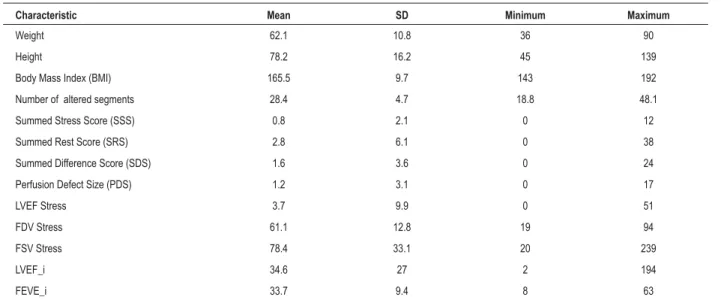
Tables 1 and 2 show the profile of the main numerical and categorical variables analyzed in this population.
Table 1 - Descriptive analysis of the numerical variables of 232 patients
Characteristic Mean SD Minimum Maximum
Weight 62.1 10.8 36 90
Height 78.2 16.2 45 139
Body Mass Index (BMI) 165.5 9.7 143 192
Number of altered segments 28.4 4.7 18.8 48.1
Summed Stress Score (SSS) 0.8 2.1 0 12
Summed Rest Score (SRS) 2.8 6.1 0 38
Summed Difference Score (SDS) 1.6 3.6 0 24
Perfusion Defect Size (PDS) 1.2 3.1 0 17
LVEF Stress 3.7 9.9 0 51
FDV Stress 61.1 12.8 19 94
FSV Stress 78.4 33.1 20 239
LVEF_i 34.6 27 2 194
FEVE_i 33.7 9.4 8 63
The mean follow-up duration was 28.9 ± 6.9 months (ranging from 10.9 to 45.9 months).
When the scintigraphy results are analyzed (normal x abnormal) it was observed that the group with abnormal perfusion presented a significantly higher mean age (p=0.04) when compared to the group with normal perfusion.
Thirty-two cardiovascular events occurred (14% of the sample), which are shown in Graphic 1: 1 cardiac death, 6 acute ischemic coronary syndromes, 12 percutaneous coronary interventions, 11 myocardial revascularization surgeries and 2 encephalic vascular accidents.
Of the 32 patients with cardiovascular events, 20 (62.5%) were males (p=0.07) and 12 (37.5 %) were females. The mean age of the group with cardiac events was 66 years (p= 0.02). In the group of patients with events only 37.5% of the patients were asymptomatic and 62.5% had angina complaints (p=0.01). The presence of angina (p=0.01) and insulin use (p=0.01) were associated with cardiac event (Graphic 2).
The proportion of abnormal scintigraphies in the group with cardiac events was significantly higher than in the group without them. Of the 190 patients with normal scintigraphy, only 14 presented cardiac events. It was observed that in the group with cardiac events, the perfusion scintigraphy parameters were compatible with more severe and extensive coronary disease, as shown in Graphic 2.
The proportion of alterations in the ventricular contractility and in the LV volumes was significantly higher in the group of patients with cardiovascular event as described in Table 3 (both with p<0.0001).
The independent predictors of cardiovascular events were:
6'6WKHSUHVHQFHRIDQJLQDPDOHVH[LQVXOLQXVHDQG
older age. Based on the information of clinical rationale,
WKHQXPEHURIDOWHUHGVHJPHQWVDWWKHSHUIXVLRQLPDJH
segments was included in the model for the logistic regression analysis. Hence, it was observed that the number of altered
VHJPHQWVLQVXOLQXVHSUHVHQFHRIDQJLQDPDOHVH[DQG
older age were also statistically significant to predict cardiac events. The event-free survival curves according to the SDS
VH[QXPEHURIDOWHUHGVHJPHQWVLQVXOLQXVHDQG
presence of angina are shown in Graphics 3, 4 and 5. The sensitivity, specificity, positive and negative predictive value and accuracy for the occurrence of cardiovascular events were calculated and were 56.3%, 88.4%, 43.9%, 92.6% and 84%, respectively. In this sample, it was noteworthy the high negative predictive value of the perfusion myocardial scintigraphy with gated SPECT.
Discussion
Diabetes mellitus is a systemic metabolic disease that affects approximately 5 to 8% of the world’s population11,12.
Table 2 - Descriptive analysis of the categorical variables of the sample
Characteristics Sample n (%)
Male sex 111(47.8)
Angina 97(41.9)
SAH 172(74.1)
Dyslipidemia 129(59.2)
Family history 139(61.5)
Smoking 24(10.4)
Negative perfusion 190(81.9)
Positive perfusion
Ischemia 30(13)
Fibrosis+Ischemia 11(4.8)
Fibrosis 1(0.4)
Attenuation (breast) 50(21.6)
Attenuation (diaphragm) 54(23.3)
Attenuation (musculature) 4(1.7)
Apical thinning 28(12.1)
N. of altered segments 42(18.1)
LV increase 10(4.3)
Myocardial contractility
Seg. height 12(5.2)
Diffuse hypokinesis 9(3.9)
SAH - Sistemic arterial hypertension; LV - left ventricle;
Graph 1 - *CD - cardiac death; EVA – encephalic vascular accident; AICS – acute ischemic coronary syndrome; MRS- myocardial revascularization surgery; PCI- percutaneous coronary intervention.
There is a current worldwide epidemic of this disease and it is estimated that by the end of 2030, there will be 360 million diabetics in the world 12,14.
Diabetes mellitus can be defined as a set of metabolic alterations characterized by hyperglycemia caused by a deficit in insulin secretion associated or not to resistance to insulin action12.
Many etiopathogenic processes have been described for the development of the disease, from the autoimmune, infectious or drug-induced destruction of the insulin-producing pancreatic cells to the decreased tissue response to insulin action.
Frequently, the insulin production and action deficits co-exist, impairing the identification of the primary cause of hyperglycemia12.
Around 65 to 70% of the deaths among diabetics are due to cardiovascular disease. Diabetes mellitus increases up to 4-fold the risk of developing coronary artery disease 15,16. The
diabetic individuals without coronary artery disease have the same future risk of cardiac death that a non-diabetic individual with a previous infarction has 8,17. Among the patients with
established coronary disease, diabetes also increases the risk of ischemic cardiac events and sudden death8,15.
These data allow us to state that diabetes is a cardiovascular disease4.
The genesis of the atherosclerotic disease in diabetics is multifactorial, comprehending endothelium, smooth muscle cell and platelet abnormalities. The main mechanisms include
metabolism disorders, oxidative stress, endothelial function, coagulation and inflammatory response3.
Nuclear Cardiology has renowned experience and has contributed to the diagnostic and prognostic evaluation of the well-established coronary artery disease. The main reason for carrying out the present study arose upon verifying the scarceness of publications involving Nuclear Cardiology, especially the gated SPECT technique, and the diabetic patient’s prognosis. The literature data on the subject comprehend studies in patients with several different characteristics and this fact hinders the comparability among them. Some studies included patients with type-1 or type-2 diabetes, populations with or without diabetes, with or without known coronary disease, with or without symptoms. Most of the studies that included scintigraphic assessment were performed with different radiotracers, techniques and stress protocols.
The present retrospective study evaluated 232 diabetic patients, with or without cardiac symptoms without known coronary disease, through stress myocardial scintigraphy (physical stress or dipiridamol-induced pharmacological stress), synchronized to the ECG. During the follow-up period (between 10 and 46 months), the total rate of cardiovascular events found in this study was 14%.
It is known that the population distribution of CAD predominates in the male sex and from the sixth decade of life18.
In this sample, it was observed that the age median was 63 years
Table 3 - Statistical analysis of the numerical variables according to the cardiovascular event
Characteristic Event Mean SD P Value
Age yes 66.2 10.8 0.020
no 61.4 10.7
Number of altered segments yes 3.2 3.6 0.0001
no 0.4 1.4
Summed Stress Score (SSS) yes 9.3 11.0 0.0001
no 1.8 4.0
Summed Rest Score (SRS) yes 4.2 6.2 0.0009
no 1.2 2.8
Summed Difference Score (SDS) yes 5.2 5.6 0.0001
no 0.6 1.8
Perfusion Defect Size (PDS) yes 14.9 17.3 0.0001
no 1.9 6.7
LV* ejection fraction at stress yes 53.7 15.2 0.004
no 62.3 12
/9¿QDOGLDVWROLFYROXPHDWVWUHVV yes 91.5 40.6 0.050
no 76.3 31.4
/9¿QDOV\VWROLFYROXPHDWVWUHVV yes 47.3 35.74 0.030
no 32.5 24.8
LV ejection fraction at stress_i yes 29.8 10 0.011
no 34.3 9.2
and there was a higher rate of events among males (62.5%). The multivariate analysis showed that both older age and male sex were independent variables of cardiovascular events.
The diagnostic evaluation of the CAD in the diabetic patients is complex19. The increase in cardiovascular mortality
in diabetic patients is due not only to the diabetic status, but also to the aggregation of several cardiovascular risk factors, such as obesity, systemic arterial hypertension (SAH) and dyslipidemia, among others6,7. SAH is two-fold more
frequent among diabetics than in the general population. The diabetic patient commonly has dyslipidemia. The most frequent lipidic alterations are hypertriglyceridemia and low HDL-cholesterol1,6,7.
Another relevant aspect is that the coronary disease in the diabetic patient can present as atypical or “silent”, which makes it difficult to manage the disease clinically.
Much is discussed about the possible denervation and lower pain sensitivity observed in the diabetic patient, but there are no definite conclusions about their existence. Diabetic individuals, especially those with neuropathy, have lower pain sensitivity and can present atypical manifestations of AMI without referring angina16,18. The scarcity of typical angina
symptoms can hinder the ischemic disease diagnosis20.
The mechanism of silent ischemia in diabetic individuals is still unknown, but the main hypothesis is that of attenuation of the sensory impulses of myocardial ischemia due to the autonomic neuropathy20. The chest angina results from the
stimulation of the afferent fibers of the cardiac sympathetic nerves20. The variation of the intensity of myocardial ischemia,
Graph 3 - Actuarial curve of event-free survival according to sex, insulin use and presence of angina. *NIDDM - insulin dependent diabetes mellitus; *Non-NIDDM - non-insulin dependent diabetes mellitus.
Graph 4 -Actuarial curve of event-free survival according to the number of altered segments and the summed difference score (SDS); * SDS-summed difference score.
pain threshold and destruction of the nociceptive fibers can explain the variations in pain perception. It is likely that the autonomic diabetic neuropathy interferes in the transmission of the afferent cardiac sensory impulses.
Hence, it is crucial, when performing a cardiologic evaluation in the diabetic patient, to carry out a detailed anamnesis and consider the presence of typical and atypical symptoms in the clinical history, even if they are minor.
The strategy to investigate the existence of coronary disease in all diabetic patients is not cost-effective. The American Diabetes Association (ADA) recommends the stress test in asymptomatic diabetic patients in the presence of peripheral or cerebral vascular disease, minor alterations at the ECG or the presence of two or more risk factors. The ADA recommends the myocardial scintigraphy if there is any evidence of ischemia or infarction at the ECG12.
To date, the real efficacy of clinical event prevention and treatment of coronary disease in asymptomatic individuals is still unknown6,12. In the literature, the prevalence of silent
ischemia in diabetic individuals varies from 9 to 48%12. This
variation is due to differences among the studied populations, as well as selection and diagnosis criteria.
The “Milan Study on Atherosclerosis and Diabetes Group”21
(MiSAD), evaluated the prevalence of silent ischemia in non-insulin dependent diabetic individuals; the prevalence of ischemia detected by ergometry and by scintigraphy was 12.1% and 6.4%, respectively.
Our study disclosed an incidence of 16.4% of silent ischemia, whereas the available evidence reports an incidence of 4 to 57% of silent ischemia at the scintigraphy. These variations are due to the differences in sample selection. It is likely that this population did not have the same degree of disease severity, considering that these were patients with no previous history of CAD and a small percentage of positive scintigraphies.
In the present study, 41.8% of the patients referred to the laboratory for a scintigraphy presented precordial pain, which was considered typical in only 14.7% of them. The presence of chest angina was an independent variable for the occurrence of cardiovascular events (p=0.001). These results reinforce the importance of the detailed clinical investigation and taking symptoms into account when managing the diabetic patient.
The diabetes mellitus treated with insulin reflects the presence of a more advanced and more severe disease12. In
this sample, this variable presented statistical significance as a marker of worse prognosis (p=0.02).
In this cohort, the statistical analysis clearly shows that the presence of a normal scintigraphy for the diagnosis of CAD positively influences the event-free survival. The association between the presence of an abnormal scintigraphy result (ischemia, fibrosis or fibrosis associated to ischemia) for the diagnosis of CAD and the occurrence of cardiac events presented statistical significance at the univariate analysis (p < 0.0001). Giri and cols.22 carried out a multicenter
study in a large cohort and concluded that the presence of an abnormal scintigraphy result and the extension of the perfusion defect were the main predictors of cardiac events among diabetic women.
Other authors have stated23,28 that the score of perfusion at
the stress phase (SSS) and that of the reversibility (SDS) as well as the number of altered segments are important predictors of cardiac events. As described in the literature, this study confirmed through the uni- and multivariate analyses that the extension of the scintigraphic alterations of perfusion and the presence of myocardial ischemia were correlated with cardiac events. At the multivariate analysis, the number of
VHJPHQWVDWWKHSHUIXVLRQDQG6'6ZHUHLQGHSHQGHQW
predictors of complications during the follow-up (p=0.0001). Some reports refer to the ischemia scintigraphic pattern as a marker of survival reduction and a determinant for myocardial ischemic event 29,30.
In 1995, a new methodology, called “gated SPECT”, was introduced in clinical practice29. The acquisition of the
myocardial perfusion scintigraphy images synchronized with the cardiac cycle (“gated SPECT”) through an electrocardiographic signal, would allow a single study to simultaneously evaluate myocardial perfusion, the global and segmental function and the left ventricle (LV) volumes29,31,32. One of the main benefits
of the gated SPECT is to help the differentiation between attenuation artifacts and real fixed perfusion defects (fibrosis), thus increasing the specificity and accuracy of the results of the perfusion study10,30,33-36.
The gated SPECT has been proven to have an additional diagnostic value to the clinical one and to the stress test10.
Additionally, the calculation of the left ventricle ejection fraction (LVEF) obtained through the gated SPECT is useful in the risk stratification for future cardiac events36.
The importance of the LV function as a determinant of survival is unquestionable37,38. In this sample, the presence of
alterations in the ventricular contractility was associated with cardiac events, as well as the values of LVEF, FDV and FSV. These variables were submitted to the multivariate analysis and did not show statistical significance in the model. Perhaps in larger populations and with a higher rate of events, these parameters would be statistically significant.
In the present study, it was not possible to reproduce, specifically, the sensitivity, specificity, positive and negative predictive value mentioned in the literature29,30. Possibly, the
discordance with the previous publications might be due to the differences regarding the sample selection and the inherent limitations of the study. Another relevant aspect is the fact that, it is believed that the myocardial scintigraphy sensitivity is overestimated in many publications39,41.
Some factors that are specific of the diabetic patient can interfere with the non-invasive assessment and accuracy of the myocardial scintigraphy. The frequent association between diabetes mellitus and risk factors for CAD can make it difficult to take certain image aspects into account. Such is the case of LV hypertrophy secondary to systemic arterial hypertension, which can cause a false-positive result at the scintigraphy 42. The diabetic cardiomyopathy can lead to
myocardial perfusion and ventricular contraction alterations that are similar to ischemic disease.
The endothelia dysfunction can interfere in the vasodilation capacity of the vessel, leading to an altered scintigraphy, without anatomical alteration (“false-positive” result).
There are some limitations to the present study. The fact that it was a retrospective study is itself a limitation. It was not possible to evaluate with details the severity of the diabetes in this cohort. The presence of complications of the underlying disease and the degree of glycemia control are important factors that can influence the results. Another limitation was the impossibility to obtain information on the coronary circulation anatomy.
Therefore, it seems that the great dilemma involving CAD in the diabetic patient is the differentiation between the low and very high-risk individuals, as the presence of diabetes itself constitutes an intermediate risk situation for cardiovascular complications.
Based on the results of the present study, we consider that gated SPECT can significantly contribute to the clinical management of the diabetic patient.
Conclusion
The present study demonstrates that the myocardial perfusion gated SPECT has an additional value in the risk stratification of future complications in patients with diabetes
mellitus and suspected coronary artery disease.
Acknowledgements
To the patients that participated in this study, to Doctors Elizabeth Costa, Luiz Cláudio Baldi, Gustavo Gavina, Sergio Doedge Gaspar, who collaborated in data collection and to the staff of Cintilab Laboratory.
To Professors Fátima Lucia Conceição, Aristarco Siqueira, Ivan da Costa Barros and João Manoel Pedrosa, who have greatly contributed to this manuscript.
Potential Conflict of Interest
No potential conflict of interest relevant to this article was reported.
Sources of Funding
There were no external funding sources for this study.
Study Association
This article is part of the thesis of doctoral submitted by Márcia Maria Sales dos Santos, from Universidade Federal do Rio de Janeiro.
References
1. Smith S, Greenland P, Grundy S. AHA Conference Proceedings. Prevention conference V: Beyond secondary prevention: identifying the high risk patient for primary prevention: executive summary. Circulation. 2000; 101: 111-6.
2. Creager M, Luscher T, Cosentino F, Beckman JA. Diabetes and vascular disease- pathophysiology, clinical consequences and medical therapy: Part I. Circulation. 2003;108 (12): 1527-32.
3. Clark C, Perry C. Type 2 diabetes and macrovascular disease: epidemiology and etiology. Am Heart J. 1999; 138: S330-3.
4. Grundy S, Benjamin I, Burke G, Chait A, Eckel RH, Howard BV, et al. Diabetes and cardiovascular disease: a statement for healthcare professionals from the American Heart Association. Circulation. 1999; 100: 1134-46.
5. Luscher T, Creager M, Beckman JA, Cosentino F. Diabetes and vascular disease- pathophysiology, clinical consequences and medical therapy: Part II. Circulation. 2003;108 (13): 1655-61.
6. Grundy S, Howard B, Smith S Jr, Eckel R, Redberg R, Bonow RO. Prevention Conference VI Diabetes and Cardiovascular Disease: executive summary: conference proceeding for healthcare professionals from a special writing group of the American Heart Association. Circulation. 2002; 105: 2231-9.
7. Taegtmeyer H, McNulty P, Young M. Adaptation and maladaptation of the heart in diabetes: part I. Circulation. 2002; 105 (pt I): 1727-33.
8. Cho E, Rimm E, Stampfer M, Willet WC, Hu FB. The impact of diabetes mellitus and prior myocardial infarction on mortality from all causes and from coronary heart disease in men. J Am Coll Cardiol. 2002; 40: 954-60.
9. Imaging guidelines for nuclear cardiology procedures. Part 2. American Society of Nuclear Cardiology. J Nucl Cardiol. 1999; 6 (2): G47-84.
10. Sharir T, Germano G, Kavanagh P, Lai S, Cohen I, Lewin HC, et al. Incremental prognostic value of post-stress left ventricular ejection fraction and volume by gated myocardial perfusion single photon emission computed tomography. Circulation. 1999; 100: 1035-42.
11. The Expert Committee on the diagnosis and classification of diabetes melito. Report of The Expert Committee on the diagnosis and classification of diabetes mellitus. Diabetes Care. 2003; 26 (Suppl I): 5-20.
12. American Diabetes Association. Standards of medical care in diabetes. Diabetes Care. 2006; 29: (Suppl I) 4-48.
13. Bax J, Van der Wall E. Assessment of coronary artery disease in patients with (a)symptomatic diabetes. Eur Heart J. 2006; 27: 631-2.
14. Anand D, Lim E, Lahiri A, Bax JJ. The role of non- invasive imaging in the risk stratification of asymptomatic diabetes subjects. Eur Heart J. 2006; 27: 905-12.
15. Mooradian A. Cardiovascular disease in type 2 diabetes mellitus. Arch Intern Med. 2003; 163: 33-40.
16. Solomon C. Reducing cardiovascular risk in type 2 diabetes. N Engl J Med. 2003; 348: 457-9.
17. Genest J, Pedersen T. Prevention of cardiovascular ischemic events: high–risk and secondary prevention. Circulation. 2003; 107: 2059-65.
18. Fazzini P, Prati P, Rovelli F. Epidemiology of silent myocardial ischemia in asymptomatic middle-aged men. Am J Cardiol. 1993; 72: 1383-8.
19. Alberts A, Krichavsky M, Balady G. Stress testing in patients with diabetes mellitus: diagnostic and prognostic value. Circulation. 2006; 113: 583-92.
20. Sheifer S, Manolio T, Gersh B. Unrecognized myocardial infarction. Ann Intern Med. 2001; 135: 801-11.
21. Milan Study on Atherosclerosis and Diabetes Group: prevalence of unrecognized silent myocardial ischemia and its association with atherosclerotic risk factors in non insulin-dependent diabetes melito. Am J Cardiol. 1997; 79: 134-9.
symptoms suggestive of coronary artery disease. Circulation. 2002; 105: 32-40.
23. Navare S, Noble G, Ahmed A. Interaction of age and gender on risk stratification of diabetic patients with rest / stress ECG-Gated Tc-99m sestamibi SPECT imaging [abstract]. In: 53rd Annual Scientific Sessions, Mar 7-10; New Orleans. J Am Coll Cardiol. 2004; 43: 339A.
24. Berman D, Kang X, Hayes S, Friedman JD, Cohen I, Abidov V, et al. Adenosine myocardial perfusion single photon emission computed tomography in women compared with men. J Am Coll Cardiol. 2003; 41: 1125-33.
25. Kang X, Berman DS, Lewin H, Cohen I, Friedman JD, Germano G, et al. Incremental prognostic value of myocardial perfusion single photon emission computed tomography in patients with diabetes mellitus. Am Heart J. 1999; 138: 1025-32.
26. Janand-Delenne B, Savin B, Habib G, Bory M, Vague P, Lassman-Vague V. Silent myocardial ischemia in patients with diabetes: who to screen. Diabetes Care. 1999, 22: 1396-400.
27. Vanzetto G, Halimi S, Hammoud T, Fagret D, Benhamou PY, Cordonnier D, et al. Prediction of cardiovascular events in clinically selected high- risk NIDDM patients: prognostic value of exercise stress test and thallium-201 single- photon emission computed tomography. Diabetes Care. 1999; 22: 19-26.
28. Schinckel A, Elhendy A, van Domburg R, Bax JJ, Vourvouri EC, Sozzi FB, et al. Prognostic value of dobutamine-atropine stress myocardial perfusion imaging in patients with diabetes. Diabetes Care. 2002; 25: 1637-43.
29. Germano G, Kiat H, Kavanagh P, Moriel M, Mazzanti M, Su HT, et al. Automatic quantification of ejection fraction from gated myocardial perfusion SPECT. J Nucl Med. 1995; 36: 2138-47.
30. Smanio P, Watson D, Segalla D, Vinson EL, Smith WH, Beller GA. Value of gating of technetium-99m sestamibi single photon emission computed tomographic imaging. J Am Coll Cardiol. 1997; 30: 1687-92.
31. Cwajg E, Cwajg J, He Z, Hwang WS, Keng F, Nagueh SF, et al. Gated myocardial perfusion tomography for the assessment of left ventricular
function and volumes: comparison with echocardiography. J Nucl Med. 1999; 40: 1857-65.
32. Cwajg E. Tomografia de perfusão miocárdica sincronizada ao ciclo cardíaco (gated SPECT) na avaliação funcional do ventrículo esquerdo: estudo comparativo com a ecocardiograma bidimensional [Tese]. Rio de Janeiro: Universidade Federal do Rio de Janeiro; 2001.
33. Bonow R. Gated myocardial perfusion imaging for measuring left ventricular function. J Am Coll Cardiol. 1997; 30: 1649-50.
34. Fleischmann S, Koepfti P, Namdar M, Wyss CA, Jenni R, Kaufmann PA. Gated 99m Tc-tetrofosmin SPECT for discriminating infarct from artifact in fixed myocardial perfusion defects. J Nucl Med. 2004; 45: 754-9.
35. Bavelaar-Croon C, Pauwels E, van der Wall E. Gated single-photon emission computed tomographic myocardial imaging: a new tool in clínical cardiology. Am Heart J. 2001; 141: 383-90.
36. Shaw L, Iskandrian A. Prognostic value of gated myocardial perfusion single photon emission computed tomography. J Nucl Cardiol. 2004; 11: 171-85.
37. Zaret B., Beller G. Nuclear cardiology: state of the art and future directions. 2nd ed. Philadelphia: Mosby; 1999. 640 p.
38. Iskandrian A, Verani M. Nuclear cardiac imaging: principles and applications. 2nd ed. Philadelphia: F.A. Davis Company; 1996. 451 p.
39. Chol BC. Sensitivity and specificity of a single diagnostic test in the presence of work up bias. J Clin Epidemiol. 1992; 45: 581-6.
40. Miller TD, Hodge DO, Christian TF, Milavetz JJ, Balley KR, Gibbons RJ. Effects of adjustment for referral bias on the sensitivity and specificity of single photon emission computed tomography for the diagnosis of coronary artery disease. Am J Med. 2002; 112: 290-7.
41. Lauer MS. Coronary artery disease in diabetes: Wich (if any) test is best. Cleve Clin J Med. 2005; 72 (1): 6, 8-9.